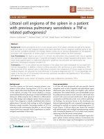
báo cáo khoa học: "Chest pain with ST segment elevation in a patient with prosthetic aortic valve infective endocarditis: a case report" ppsx
Bạn đang xem bản rút gọn của tài liệu. Xem và tải ngay bản đầy đủ của tài liệu tại đây (913.44 KB, 4 trang )
JOURNAL OF MEDICAL
CASE REPORTS
Chest pain with ST segment elevation in a
patient with prosthetic aortic valve infective
endocarditis: a case report
Luther et al.
Luther et al. Journal of Medical Case Reports 2011, 5:408
(24 August 2011)
CAS E REP O R T Open Access
Chest pain with ST segment elevation in a
patient with prosthetic aortic valve infective
endocarditis: a case report
Vishal Luther
1*
, Refai Showkathali
2
and Reto Gamma
2
Abstract
Introduction: Acute ST-segment elevation myoc ardial infarction secondary to atherosclerotic plaque rupture is a
common medical emergency. This condition is effectively managed with percutaneous coro nary intervention or
thrombolysis. We report a rare case of acute myocardial infarction secondary to coronary embolisation of valvular
vegetation in a patient with infective endocarditis, and we highlight how the management of this phenomenon
may not be the same.
Case presentation: A 73-year-old British Caucasian man with previous tissue aortic valve replacement was
diagnosed with and treated for infective endocarditis of his native mitr al valve. His condition deteriorated in
hospital and repeat echocardiography reveale d migration of vegetation to his aortic valve. Whilst waiting for
surgery, our patient developed severe central crushing chest pain with associated anterior ST segment elevation on
his electrocardiogram. Our patient had no history or risk factors for ischaemic heart disease. It was likely that
coronary embolisation of part of the vegetati on had occurred. Thrombolysis or percutaneous coronary intervention
treatments were not performed in this setting and a plan was made for urgent surgical intervention. However, our
patient deteriorated rapidly and unfortunately died.
Conclusion: Clinicians need to be aware that atherosclerotic plaque rupture is not the only cause of acute myocardial
infarction. In the case of septic vegetation embolisation, case report evidence reveals that adopting the current
strategies used in the treatment of myocardial infarction can be dangerous. Thrombolysis risks intra-cerebral
hemorrhage from mycotic aneurysm rupture. Percutaneous coronary intervention risks coronary mycotic aneurysm
formation, stent infections as well as distal septic embolisation. As yet, there remains no defined treatment modality
and we feel all cases should be referred to specialist cardiac centers to consider how best to proceed.
Introduction
Atheroscleroticplaquerupturewithinacoronaryvessel
can lead to rapid vessel occlusion and subsequent myo-
cardial ischaemia and necrosis [1]. Risk factors for the
development of a therosclerosis include hypertension,
diabetes mellitus, high cholesterol, a history of smoking,
and a family history of ather osclerotic disease [2]. Cur-
rent treatment involves either percutaneous coronary
intervention (PCI) to relieve the occlusion, or thrombo-
lysis to dissolve the occlusion [3].
There are more rare causes of acute myocardial
infarction (AMI). We present and discuss the case of a
patient with AMI secondary to embolisation of vegeta-
tion sitting on a prosthetic aortic valve in a patient with
confirmed aortic valve infective endocarditis (IE).
Case presentation
A 73-year-old British Cauca sian man who had under-
goneatissueaorticvalvereplacement five years pre-
viously was admitted to his lo cal hospital with a two-
week history of breathlessness, general malaise and
night sweats. On examination, he was found to have an
ejection systolic murmur in the aortic area a nd a pan-
systolic murmur in t he mitral area radiating to the
axilla. His white cell count was elevated (15.1 × 10
9
* Correspondence:
1
Department of Medicine, Whittington Hospital NHS Trust, Magdala Avenue,
London, N19 5NF, UK
Full list of author information is available at the end of the article
Luther et al. Journal of Medical Case Reports 2011, 5:408
/>JOURNAL OF MEDICAL
CASE REPORTS
© 2011 Luther et al; licensee BioMed Central Ltd. This is an Open Access article distr ibuted under the terms of the Creativ e Commons
Attribution License ( which permits unr estri cted use, distribution, and reproduction in
any medium, provided the original work is properly cited.
cells/L, neutrophils 10.7 × 10
9
cells/L) and he had a
raised C-reactive protein level of 101 mg/dL. The results
of three consecutive blood cultures samples were nega-
tive even after five days in th e culture media. His trans-
thoracic and trans-oesophageal echocardiogram (ECG)
results demonstrated vegetation involving the native
posterior m itral valve leaflet (Figure 1) with moderate
mitral regurgitation and a moderately stenosed tissu e
aortic valve. Vancomycin, Gentamicin and Rifampicin
were given under microbiology guidance. Five days later,
our patient became more unwell, and was found to be
in worsening cardiac failure. A repeat echocardiogram
showed the known vegetation on the mitral valve and
new v egetation on the aortic valve of 1.5 cm (Figure 2)
causing moderate aortic regurgitation. Our patient was
subsequently transferred to our center for valve surgery.
Whilst awaiting surgery, our patient developed severe
central crushing chest pain with associat ed anterior seg-
ment ST elevation on his ECG (Figure 3). Our patient
had no previous history of angina, and was a non-smo-
ker with no other cardiac risk factors. A coronary angio-
gram performed five years ago prior to his valv e surgery
revealed unobstructed coronaries. The most likely expla-
nation for this ST segment elevation myocardial infarc-
tion (STEMI) was coronary embolisation of either part
of the vegetation or thrombus atta ched to the vegeta-
tion. Thrombolysis is relatively contraindicated in this
scenario. PCI risked mycotic aneurysm formation and
either further systemic or coronary embolisation. There-
fore, ur gent surgical intervention was planned; howev er,
our patient deteriorated rapidly and unfortunately died.
Discussion
Coronary embolisation is a rare cause of AMI and needs
to be considered in patients with atrial fibrillation,
prosthetic heart valves, dilated cardiomyopathy, and IE,
where either thrombus or vegetation can embolize into
the coronary circulat ion. Although systemic embolisa-
tion can occur in up to 50% of cases of IE [4], coronary
embolisation rate is ab out 0.3% [5]. The re appears to be
an increased risk of embolisation with vegetations that
are > 1 cm in diameter, as in our patient’s case [6]. Suc-
cessful strategies that have been used to manage coron-
ary embolisation i n non-endocarditic patients include
thrombolytics [7], PCI and thrombus aspiration [8].
There is no clear evidence available about the best
treatment option for patients with coronary embolisa-
tion in the setting of acute IE [9]. Thrombolytic treat-
ment of septic coronary embolisation is associated
with an increased risk of cerebral vascular hemorrhage
due to bleeding from silent cerebral microinfarctions
or mycotic aneurysms [10]. Indeed AMI caused by sep-
tic embolisation is a relative contraindication to the
use of thrombolytic agents. PCI involves coronary bal-
loon angioplasty and stent deployment, and this risks
mycotic aneurysm forma tion at the dilatation site. This
occurs as the balloon crushes vegetation against the
vessel wall [11]. Implanting foreign stent material into
an infective site can lead to stent infection, and this
can require stent excision and debridement [12]. In
addition, PCI risks further distal vegetation embolisa-
tion [13]. As reported in a previous case report, ‘ the
impulse to follow conventional strategies for coronary
reperfusion should be tempered by thoughts of possi-
ble consequences’ [11].
Surgical intervention in left-sided IE is in fact recom-
mended in the contex t of systemic embolisation [14].
However, evidence of successf ul surgical intervention in
the context of coronary embolisation is scarce, with a
Figure 1 Echocardiogram (apical view) showing vegetation in
the native posterior mitral valve leaflet (white arrow). LA = left
atrium; LV = left ventricle; RA = right atrium; RV = right ventricle.
Figure 2 Echocardiogram (parasternal long axis view) showing
large vegetation in the tissue prosthetic aortic valve (white
arrow). LA = left atrium; LV = left ventricle; MV = mitral valve; RV =
right ventricle.
Luther et al. Journal of Medical Case Reports 2011, 5:408
/>Page 2 of 3
few case reports demonstrating success through coron-
ary embolectomy [15].
Conclusions
This case report prese nts a common condition seen in
an uncommon setting. AMI is common, and the man-
agement is well defined and performed by acute physi-
cians and cardiologists. However, in the absence of risk
factors for ischaemic heart disease, clinicians need to
consider alternate causes of AMI.
This is especially important in the case of septic cor-
onary embolisation in patients with IE, as adopting the
current strategies used in the management of myocar-
dial infarction can be dangerous. Where suspicion is
high, care shou ld be urgent ly transferred to specialist
cardiac centers where both interventional and surgical
skills are available to decide on how best to proceed.
Consent
Written informed consent was obtained from the
patient’s next-of-kin for publication of this case report
and any accompanying images. A copy of the written
consent is available for review by the Editor-in-Chief of
this journal.
Author details
1
Department of Medicine, Whittington Hospital NHS Trust, Magdala Avenue,
London, N19 5NF, UK.
2
Department of Cardiology, The Essex Cardiothoracic
Centre, Nethermayne, Basildon, Essex, UK, SS16 5NL, UK.
Authors’ contributions
VL wrote the initial draft of the case report. RS edited the case report and
selected all the images to use. RG was our patient’s consultant. All authors
read and approved the final manuscript.
Competing interests
The authors declare that they have no competing interests.
Received: 11 April 2011 Accepted: 24 August 2011
Published: 24 August 2011
References
1. Rozenman Y, Rosenheck S, Nassar H, Welber S, Sapoznikov D, Lotan C,
Mosseri M, Weiss AT, Gotsman MS: Acute myocardial infarction–the
angiographic picture: new insights into the pathogenesis of myocardial
infarction. Int J Cardiol 1995, 49 :s11-6.
2. Virmani R, Farb A, Burke AP: Risk factors in the pathogenesis of coronary
artery disease. Compr Ther 1998, 24:519-529.
3. Cohen M: High-risk acute coronary syndrome patients with non-ST-
elevation myocardial infarction: definition and treatment. Cardiovasc
Drugs Ther 2008, 22:407-418.
4. Kraus PA, Lipman J: Coronary embolism causing myocardial infarction.
Intensive Care Med 1990, 16:215-216.
5. Fabri J Jr, Issa VS, Pomerantzeff PM, Grinberg M, Barretto AC, Mansur AJ:
Time-related distribution, risk factors and prognostic influence of
embolism in patients with left-sided infective endocarditis. Int J Cardiol
2006, 110:334-339.
6. Sanfilippo AJ, Picard MH, Newell JB, Rosas E, Davidoff R, Thomas JD,
Weyman AE: Echocardiographic assessment of patients with infectious
endocarditis: prediction of risk for complications. J Am Coll Cardiol 1991,
18:1191-1199.
7. Quinn EG, Fergusson DJG: Coronary embolism following aortic and mitral
valve replacement: successful management with abciximab and
urokinase. Cathet Cardiovasc Diagn 1998, 43 :457-459.
8. Kiernan TJ, Flynn AMO, Kearney P: Coronary embolism causing myocardial
infarction in a patient with mechanical aortic valve prosthesis. Int J
Cardiol 2006, 112:E14-E16.
9. Glazier JJ: Interventional treatment of septic coronary embolism:
Sailing into uncharted and dangerous waters. J Interv Cardiol 2002,
15:305-307.
10. Hunter AJ, Girard DE: Thrombolytics in infectious endocarditis associated
myocardial infarction. J Emerg Med 2001, 21:401-406.
11. Herzog CA, Henry TD, Zimmer SD: Bacterial endocarditis presenting as
acute myocardial infarction: a cautionary note for the era of reperfusion.
Am J Med 1991, 90:392-397.
12. Dieter RS: Coronary artery stent infection. Clin Cardiol 2000, 23:800-810.
13. Ural E, Bildirici U, Kahraman G, Komsuoğlu B: Coronary embolism
complicating aortic valve endocarditis: treatment with successful
coronary angioplasty. Int J Cardiol 2007, 119:377-379.
14. Chopra T, Kaatz GW: Treatment strategies for infective endocarditis. Exp
Opin Pharmacother
2010, 11:345-360.
15. Baek MJ, Kim HK, Yu CW, Na CY: Mitral valve surgery with surgical
embolectomy for mitral valve endocarditis complicated by septic
coronary embolism. Eur J Cardiothorac Surg 2008, 33:116-118.
doi:10.1186/1752-1947-5-408
Cite this article as: Luther et al.: Chest pain with ST segment elevation
in a patient with prosthetic aortic valve infective endocarditis: a case
report. Journal of Medical Case Reports 2011 5:408.
Figure 3 Electrocardiogram showing ST elevation in V1 to V4 leads.
Luther et al. Journal of Medical Case Reports 2011, 5:408
/>Page 3 of 3