báo cáo khoa học: "Unusual exanthema combined with cerebral vasculitis in pneumococcal meningitis: a case report" pdf
Bạn đang xem bản rút gọn của tài liệu. Xem và tải ngay bản đầy đủ của tài liệu tại đây (889.81 KB, 4 trang )
CAS E REP O R T Open Access
Unusual exanthema combined with cerebral
vasculitis in pneumococcal meningitis: a case
report
Theonimfi Tavladaki
1
, Anna-Maria Spanaki
1
, Stavroula Ilia
1
, Elisabeth Geromarkaki
1
, Maria Raissaki
2
and
George Briassoulis
1*
Abstract
Introduction: Bacterial meningitis is a complex, rapidly progressive disease in which neurological injury is caused
in part by the causative organism and in part by the host’s own inflammatory responses.
Case presentation: We present the case of a two-year-old Greek girl with pneumococcal meningitis and an
atypical curvilinear-like skin eruption, chronologically associated with cerebral vasculitis. A diffusion-weighted MRI
scan showed lesions with restricted diffusion, reflecting local areas of immunologically mediated necrotizing
vasculitis.
Conclusions: Atypical presentations of bacterial meningitis may occur, and they can be accompanied by serious
unexpected complications.
Introduction
Neurological injury in Streptococcus pneumoniae menin-
gitis can be due to meningeal inflammation, cerebral
edema, necrosis and intracranial hemorrhage. There is a
widely held belief that cerebral infarction after bacterial
meningitis is alw ays caused by vasculitis; however, evi-
dence for this is weak. Vergouwen et al.hypothesized
that diffuse cerebral intravascular coagulation is an addi-
tional explanation for cerebral infarction in patients with
pneumococcal meningitis [1]. At the molecular level, S.
pneumoniae cell walls have been shown to induce cere-
brovascular endothelial cells, microglia, and meningeal
inflammatory cells to release cytokines, chemokines and
reactive oxygen species [2]. These include tumor necro-
sis factor a, int erleukins 1 and 6, platelet-activating fac-
tor, peroxynitrites, matrix metalloproteinases and
urokinase plasminogen activator. Release of such biopro-
ducts is believed to play a role in the development of
disseminated intravascular coagulation in the setting of
pneumococcal sepsis. To the best of our knowledge, we
present a previously-unreported association of an
exaggerated host response, as shown b y the develop-
ment of vasculitis, with an unusual rash in a child with
pneumococcal meningitis.
Case presentation
A two-year-old Greek girl was referred to our Pediatric
Intensive Care Unit (PICU) with a two-day history of
fever (39.3°C), vomiting, reduced appetite for feeding
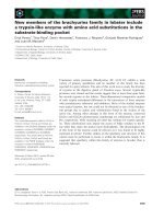
and seizures. A physical examination showed nuchal
rigidity, a decreased level of consciousness and multiple
erythematous, flat macules present on her hands and
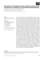
the dorsal and plantar aspects of her feet (Figure 1), tak-
ing a curvilinear appearance (Figure 2A, B). Our patient
had an unremarkable medical history; she had not been
vaccinated for S. pneumoniae.
A complete blood cell count revealed 18,000 cells/μL
white blood cells (neutrophil s 80%, leukocytes 17%), her
C-reactive protein serum level was 28.87 mg/dL, and
pronounced coagulation disturbances were detected
(prothrombin time: 15.4 seconds; activated partial
thromboplastin time: 33 seconds; international normal-
ized ratio: 1.38; fibrinogen: 375 mg/dL, D-dimers: 91.63
mg/dL). Results of a lumbar puncture showed white
blood cells at 4 0 cells/mm
3
, a total protein content of
169 mg/dL and hypoglycorrhachia of 2 mg/dL. Gram-
* Correspondence:
1
Paediatric Intensive Care Unit, University Hospital of Heraklion, University of
Crete, Heraklion, Crete, Greece
Full list of author information is available at the end of the article
Tavladaki et al. Journal of Medical Case Reports 2011, 5:410
/>JOURNAL OF MEDICAL
CASE REPORTS
© 2011 Tavladaki et al; lice nsee BioMed Central Ltd. This is an Open Access article distributed under the terms o f the Creative
Commons Attribution License ( g/licenses/by/2.0), which permits unrestricted use, distribution, and
reproduction in any medium, provided the origi nal work is properly cited.
staining results revealed the presence of Gram-positive
cocci in pair s. Two days after admission, blood and cer-
ebrospinal fluid cultures yielded pure growth of vanco-
mycin susceptible (MIC # 1 μg/mL, 25 mm) and
penicillin susceptible (MIC # 0.12 μg/mL) Streptococcus
pneumoniae. Serotype 23F was identified by PCR from
two blood samples and in the first sample of cerebros p-
inal fluid (CSF). The same isolate was also cultured
from our patient’s throat. IgG subclasses were normal
and the results of an HIV test were negative. Due to the
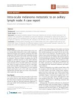
lack of clinical improvement, an urgent diffusion-
weighted MRI scan was performed six days after admis-
sion. The MRI showed ill-defined hyperintense lesions
at the peri-ventricular a nd white matter, exhibiting
restricted diffusion (Figure 3).
Boluses of intravenous fluids, fresh frozen plasma
and intravenous dexamethasone (0.15 mg/kg) were
given, immediately followed by systematic administra-
tion of ceftr iaxone (100 mg/kg/day) and vancomy cin
(60 mg/kg/day). Due to persistent drowsiness and
further clinical deterioration, a second lumbar punc-
ture was taken. The results of this were 90 leukocytes/
mm
3
, a glucose level of 36 mg/dL, and protei n 124
mg/dL, whereas a further CSF culture did not reveal
any isolation. Aiming at bett er permeability through
the blood brain barrier, intravenous rifampicin (40 mg/
kg/day, MIC # 1 μg/mL, 27 mm) was added. Although
the responsible isolate was sensitive to the antibiotics
administered, our patient showed a slow clinical
response; consequently the combined antibiotic regi-
men was administered for a total of 14 days after ther-
apy initiation. Her fever and atypical rash started
resolving after the first week. Our patient made a full
neurological recovery, apart from bilateral hearing
impairment confirmed by brain stem response.
Figure 1 Multiple non-hemorrhagic erythematous flat macules
on the dorsal and plantar aspects of feet.
B
A
Figure 2 Confluent elongated skin lesions (A, arrows) with
curvilinear projections (B, arrows) at the time of isolation of
Streptococcus pneumoniae in blood and cerebrospinal fluid.
Figure 3 MRI scan showing i ll-defined hyperi ntense les ions at
the peri-ventricular and subcortical white matter (arrows) that
were identified shortly after the skin eruption and the
Streptococcus pneumoniae growth.
Tavladaki et al. Journal of Medical Case Reports 2011, 5:410
/>Page 2 of 4
Discussion
Following usage of the pneumococcal conjugate vaccine
in children, the incidence of invasive pneumococcal dis-
ease (IPD) has declined in both children and adults
(reflecting herd immunity). Since our patient’ s responsi-
ble serotype is included in all types of current S. pneu-
moniae vaccines, her life-threatening atypical bacterial
infection could have been avoided if the child had been
appropriately vaccinated . (Following the introduction of
heptavalent pneumococcal conjugate vaccine (PCV7),
the incidence rates of IPD caused by vaccine serotypes
declined across all age groups [3,4].)
Although atypical presentations of bacterial meningitis
still occur, emergency or community physicians are
rarely involved [5]. Only an atypical exanthema
(erythema nodosum) associated with meningitis (due to
Chlamydia pneumonia) has been reported in the litera-
ture [6]; t o the best of our knowledge such an unusual
exanthema, presented in clusters of curvilinear skin
lesions and associated with severe pneumococcal infec-
tion, has never been described previously. Absence of
hemorrhagic rash has been recently reported as one of
the most significant clinical predictors of childhood
pneumococcal meningitis [7]. Regardless, such an atypi-
cal skin er uption, chronologically associated with cere-
bral vasculitis, has not been described in a child with
pneumococcal meningitis to date. However, a low CSF
glucose level, which was profoundly low (2 mg/dL) in
our patient, is an established significant risk factor for
hearing loss after pneumococcal meningitis [8,9].
As in our patient, in adult patients with meningoence-
phalitis caused by S. pneumoniae, diffusion-weighted
MRI may show lesions with restricted diffusion, reflect-
ing local areas of ischemia with cytotoxic edema second-
ary to immunological ly mediated necrotizing vasculitis
and thrombosis [10]. Conventional angiography and
magnetic resonance angiography may show tapering and
stenosis of arteries [11]. Importantly, in a series in
adults, pneumococcal meningitis-associated arterial
(21.8%) or venous (9.2%) cerebrovascular complications
have been shown to develop more frequently than pre-
viously reported [12]. Other reported findings from the
same study were subarachnoid hemorrhages in associa-
tion with angiographic evidence of vasculitis (9.2%) and
acute spinal cord dysfunction due to myelitis (2.3%).
Delayed cerebral thrombosis has also been described in
adult patients with pneumococcal meningitis, with
pathology suggesting an immunological reaction target-
ing cerebral blood vessels [13].
S. pneumoniae bacteria do not readily penetrate the
pia and invade the brain. However, the interaction
between S. pneumoniae and the host results in menin-
geal inflammation, vascular injury, disruption of the
blood-brain barrier, vasogenic, interstitial and cytotoxic
edema, and disruption of normal CSF flow. Many of th e
neurological and systemic conditions that contribute to
morbidity and mortali ty in pneumococcal mening itis, in
particular vascular injury and cerebral edema, have
already been set in motion by the time antibiotics are
administered. So even if antibiotic treatment is started
early and the bacteria are drug sensitive, as in our
patient’s case, unfavorable outcomes and severe neurolo-
gical sequelae of bacterial meningitis frequently still
occur. Treatment options to suppress the inf lammatory
cascade causing neuronal injury include corticosteroids,
as they exert various immunomodulatory actions.
Although previously controversial, as steroids reduce
antibiotic penetration into the CSF, meta-analysis of
trial data now support treatment with a short course of
adjunctive therapy with the corticosteroid dexametha-
sone to improve outcome and partially prevent neurolo-
gical sequelae from bacterial meningitis in adults and
children [14]; this is however only achieved when given
earlyinthediseasecourseandwhenstartedwithor
before parenteral antibiotics [14]. It has been recently
suggested that d examethasone inhibits increase of CSF
soluble tumor necrosis factor 1 levels after antibiotic
therapy in bacterial meningitis, an important indicator
of neurological sequelae in bacterial meningitis [15].
Conclusions
The interaction between S. pneumoniae bacteria and the
host results not only in meningeal inflammation but
also in vascular injury. Early administration of dexa-
methasone and empiric antibiotic treatment should
begin in all cases to prevent neurological sequel and
hearing loss associated with low CSF glucose levels.
Accordingly, the pre sence of an atypical rash should not
deter the physician from a clinical suspicion of this
potentially fatal pneumococcal infection. Brain MRI
scans and/or angiography, as well as CSF findings in
conjunction with the clinical course of this life-threaten-
ing disease, may d ictate appropriate treatment adjust-
ments. Importantly, to the best of our knowledge, an
atypical skin eruption chronologically associated with
cerebral vasculitis has not been described previously.
However, with routine effective use of pneumococcal
conjugate vaccines a general decline in IPD, antibiotic
non-susceptibility and vaccine serotypes was observed.
Consent
Written informed consent was obtained from the
patient’ s legal guardian for publication of this case
report and any accompanying images. A copy of the
written consent is available for review by the E ditor-in-
Chief of this journal.
Tavladaki et al. Journal of Medical Case Reports 2011, 5:410
/>Page 3 of 4
Author details
1
Paediatric Intensive Care Unit, University Hospital of Heraklion, University of
Crete, Heraklion, Crete, Greece.
2
Department of Radiology, University Hospital
of Heraklion, University of Crete, Heraklion, Crete, Greece.
Authors’ contributions
GB, TT, SI, EG, and AMS were responsible for the management of our
patient; MR performed the MRI, and interpreted and discussed findings; GB,
TT and AMS participated in the study design and coordination and helped
draft the manuscript. All authors read and approved the final manuscript.
Competing interests
The authors declare that they have no competing interests.
Received: 4 March 2011 Accepted: 24 August 2011
Published: 24 August 2011
References
1. Vergouwen MD, Schut ES, Troost D, van de Beek D: Diffuse cerebral
intravascular coagulation and cerebral infarction in pneumococcal
meningitis. Neurocrit Care 2010, 13:217-227.
2. Scheld WM, Koedel U, Nathan B, Pfister H-W: Pathophysiology of bacterial
meningitis: mechanism(s) of neuronal injury. J Infect Dis 2002, 186(Suppl
2):S225-233.
3. Vestrheim DF, Høiby EA, Bergsaker MR, Rønning K, Aaberge IS, Caugant DA:
Indirect effect of conjugate pneumococcal vaccination in a 2+1 dose
schedule. Vaccine 2010, 28:2214-2221.
4. Harboe ZB, Valentiner-Branth P, Benfield TL, Christensen JJ, Andersen PH,
Howitz M, Krogfelt KA, Lambertsen L, Konradsen HB: Early effectiveness of
heptavalent conjugate pneumococcal vaccination on invasive
pneumococcal disease after the introduction in the Danish Childhood
Immunization Programme. Vaccine 2010, 28:2642-2647.
5. Fisher JD: Insidious presentation of pediatric pneumococcal meningitis:
alive and well in the post vaccine era. Am J Emerg Med 2009, 27 :1173.
e5-7.
6. Sundelöf B, Gnarpe H, Gnarpe J: An unusual manifestation of Chlamydia
pneumoniae infection: meningitis, hepatitis, iritis and atypical erythema
nodosum. Scand J Infect Dis 1993, 25:259-261.
7. Karanika M, Vasilopoulou VA, Katsioulis AT, Papastergiou P,
Theodoridou MN, Hadjichristodoulou CS: Diagnostic clinical and
laboratory findings in response to predetermining bacterial pathogen:
data from the Meningitis Registry. PLoS One 2009, 4:e6426.
8. Kutz JW, Simon LM, Chennupati SK, Giannoni CM, Manolidis S: Clinical
predictors for hearing loss in children with bacterial meningitis. Arch
Otolaryngol Head Neck Surg 2006, 132:941-945.
9. Worsøe L, Cayé-Thomasen P, Brandt CT, Thomsen J, Østergaard C: Factors
associated with the occurrence of hearing loss after pneumococcal
meningitis. Clin Infect Dis 2010, 51:917-924.
10. Jorens PG, Parizel PM, Demey HE, Smets K, Jadoul K, Verbeek MM,
Wevers RA, Cras P: Meningoencephalitis caused by Streptococcus
pneumoniae: a diagnostic and therapeutic challenge. Diagnosis with
diffusion-weighted MRI leading to treatment with corticosteroids.
Neuroradiology 2005, 47:758-764.
11. Appenzeller S, Faria AV, Zanardi VA, Fernandes SR, Costallat LT, Cendes F:
Vascular involvement of the central nervous system and systemic
diseases: etiologies and MRI findings. Rheumatol Int 2008, 28:1229-1237.
12. Kastenbauer S, Pfister HW: Pneumococcal meningitis in adults: spectrum
of complications and prognostic factors in a series of 87 cases. Brain
2003, 126:1015-1025.
13. Schut ES, Brouwer MC, de Gans J, Florquin S, Troost D, van de Beek D:
Delayed cerebral thrombosis after initial good recovery from
pneumococcal meningitis. Neurology 2009, 73
:1988-1995.
14. Fisher JD: Insidious presentation of pediatric pneumococcal meningitis:
alive and well in the post vaccine era. Am J Emerg Med 2009, 27:1173, e5-7.
15. Ichiyama T, Matsushige T, Kajimoto M, Tomochika K, Matsubara T,
Furukawa S: Dexamethasone decreases cerebrospinal fluid soluble tumor
necrosis factor receptor 1 levels in bacterial meningitis. Brain Dev 2008,
30:95-99.
doi:10.1186/1752-1947-5-410
Cite this article as: Tavladaki et al.: Unusual exanthema combined with
cerebral vasculitis in pneumococcal meningitis: a case report. Journal of
Medical Case Reports 2011 5:410.
Submit your next manuscript to BioMed Central
and take full advantage of:
• Convenient online submission
• Thorough peer review
• No space constraints or color figure charges
• Immediate publication on acceptance
• Inclusion in PubMed, CAS, Scopus and Google Scholar
• Research which is freely available for redistribution
Submit your manuscript at
www.biomedcentral.com/submit
Tavladaki et al. Journal of Medical Case Reports 2011, 5:410
/>Page 4 of 4