báo cáo khoa học: " Recurrent takotsubo cardiomyopathy in the setting of transient neurological symptoms: a case report" pptx
Bạn đang xem bản rút gọn của tài liệu. Xem và tải ngay bản đầy đủ của tài liệu tại đây (632.96 KB, 4 trang )
CAS E REP O R T Open Access
Recurrent takotsubo cardiomyopathy in the
setting of transient neurological symptoms: a
case report
Muhammad Rizwan Sardar
1
, Catherine Kuntz
2
, Jeremy A Mazurek
3*
, Naveed Hassan Akhtar
4
, Wajeeha Saeed
1
and
Timothy Shapiro
5
Abstract
Introduction: First described in Japan, takotsubo cardiomyopathy is increasingly beco ming recognized worldwide
as a cause of sudden and reversible diminished left ventricular function characterized by left apical ballooning and
hyperkinesis of the basal segments, often with symptoms mimicking a myocardial infarction. Associated with
physical or emotional stress, its exact pathogenesis has not been established, though evidence supports a
neurohumoral etiology. Additionally, recurrence of this condition is rare. In this report, we present a rare case of
recurrent takotsubo cardiomyopathy in a post-menopausal woman who presented with transient neurological
complaints on both occasions.
Case presentation: We present a rare case of a 76-year-old Caucasian woman with no history of congestive heart
failure who presented to our emergency department twice with transient neurological complaints. On the first
occasion, she was found to have transient aphasia which resolved within 24 hours, yet during that period she also
developed symptoms of congestive heart failure and was noted to have a new, significantly depressed ejection
fraction with apical ak inesis and possible apical thrombus. One month after her presentation a repeat
echocardiogram revealed complete resolution of all wall motion abnormalities and a return to baseline status.
Seven months later she presented with ataxia, was diagnosed with vertebrobasilar insufficiency, and again
developed symptoms and echocardiography findings similar to those of her first presentation. Once again, at her
one-month follow-up examination, all wall motion abnormalities had completely resolved and her ejection fraction
had returned to normal.
Conclusion: Though the exact etiology of takotsubo cardiomyopathy is unclear, a neurohumoral mechanism has
been proposed. Recurrence of this disorder is rare, though it has been reported in patients with structural brain
abnormalities. This report is the first to describe recurrent takotsubo cardiomyopathy in a patient with transient
neurological symptoms. In our patient, as expected in patients with this condition, complete resolution of all left
ventricular abnormalities occurred within a short period of time. It is important for clinicians to be aware of this
increasingly recognized syndrome, including its association with recurrence, especially in the clinical setting of
neurologic dysfunction.
Introduction
Left apical ballooning syndrome, also known as takot-
subo cardiomyopathy (TTC), is a clinical syndrome of
transient diminished left ventricular (LV) apical wall
motion with relative preservation of the basal heart
segment in the presence of normal coronary arteries. It
was first described in Japan in the early 1990s [1] and is
named for ventricles which seem similar in appearance
to a Japanese octopus trap on ventriculography scans.
This syndrome has been linked to emotional or physical
stress, with a high incidence among post-menopausal
women [2]. However, the etiology of TTC is not com-
pletely understood. Several possible mechanisms, includ-
ing microvascular dysfunction, coronary artery
* Correspondence: om
3
Department of Medicine, Jacobi Medical Center, Albert Einstein College of
Medicine, 1400 Pelham Parkway South, Suite 3N1, Bronx, NY 10461, USA
Full list of author information is available at the end of the article
Sardar et al. Journal of Medical Case Reports 2011, 5:412
/>JOURNAL OF MEDICAL
CASE REPORTS
© 2011 Sardar et al; licensee BioMed Central Ltd. This is an Open Access article distributed under the terms of the Creative Commons
Attribution License ( which permits unrestricted use, distribution, and reproduction in
any medium, provided the original work is properly cited .
vasospasm, aborted myocardial infarction, and exc ess
catecholamine stimulation,havebeenproposed[2,3].
Typically, LV function returns to normal within six to
eight weeks. Recurrence, which is increasingly being
reported in the literature [2,4,5], can be related to neu-
rological pathology [6,7]. We present a case of a woman
with recurrent TTC whose presenting symptoms on
both occasions were of neurological origin.
Case presentation
A 76-year-old Caucasian woman presented to our ter-
tiary care hospital on two separate occasions. She had a
medical history significant for hypertension, diabetes
mellitus, dyslipidemia, hypothyroidism, no congestive
heart failure (CHF), no evidence of coronary artery dis-
ease visualized by catheterization two years prior to her
initial presentation, and a normal baseline left ventricu-
lar ejection fraction (LVEF) of 55% to 60%. In each epi-
sode, she presented without evidence of existing
physical or emotional distress.
At her first presentation, she arrived at the emergency
department with the onset of aphasia in the setting of
hypertensive emergency (blood pressure (BP) 226/100
mmHg). Her aphasia was isolated and transient, and her
physical examination was otherwise unremarkable.
Because we were concerned about an acute intracerebral
event in the setting of hypertensive emergency, com-
puted tomography and diffusion-weighted MRI/mag-
netic resonance angiography (MRI/MRA) of the brain
were performed, both of which showed no evidence of
acute ischemia, hemorrhage, or cerebral edema. She was
admitted for work-up of acute stroke and transient
ischemic attack and treatment of her hypertensive emer-
gency. Her aphasia resolved within 24 hours after onset,
and a modest reduction in BP was achieved. Shortly
after admission, h owever, she developed acute onset of
dyspnea, and rales were heard bilaterally during her phy-
sical examination.
In light of these new symptoms, a chest X-ray was
ordered, which revealed bilateral pulmonary vascular
congestion. Her electrocardiogram (ECG) was unre-
markable for ST elevation or depression, though she
was found to have elevated cardiac enzymes on serial
measurements (tr oponin I levels 0.8 ug/L and 0.90 ug/L,
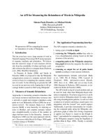
respectively). An echocardiogram obtained at that time
revealed apical akinesis and depressed LV systolic func-
tion, with a LVEF of 25% and a possible apical thrombus
(Figure 1A). Cardiac catheterization was planned to
assess whether the patient had acute CHF secondary to
possible acute myocardial infarction. The patient and
her family refused to allow us to perform the procedure,
however, as she had had a normal cardiac catheteriza-
tion two years earlier.
Given the clinical, laboratory, and echocardiography
data, the patient was started on anti-platelet and anti-
coagulation therapy and optimized on heart failure
treatment for presumed CHF secondary to ischemic car-
diomyopathy. Her symptoms improved, and she was dis-
charged from the hospital with follow-up planned in the
out-patient clinic. At one month post-presentation, an
echocardiogram revealed complete improvement of LV
function back to baseline level (LVEF of 55%) and no
evidence of wall motion abnormalities or thrombus (Fig-
ure 1B).
Seven months after her initial presentation, the patient
returned to the emergency room for ataxia, dizziness,
and associated nausea without focal neurological symp-
toms. MRI/MRA of her brain was inconclusive, and clo-
pidogrel was added to her treatment regimen based on
the suspicion of vertebrobasilar insufficiency. At the
time of presentation, an ECG revealed new T-wave
inversions in the anterolateral leads and QT
Figure 1 Echocardiogram on initial presentation and follow-up. (A) Echocardiogram with contrast enhancement revealing apical ballooning
during the patient’s first presentation to our hospital. (B) Echocardiogram revealing normally contracting apex with complete recovery of
ejection fraction one month after the patient’s initial presentation.
Sardar et al. Journal of Medical Case Reports 2011, 5:412
/>Page 2 of 4
prolongation (QTc 490 milliseconds), along with ele-
vated levels of troponin I on serial measurements (0.50
ug/L,0.89ug/L,and0.91ug/L, respectively) Addition-
ally, during this hospital stay, she again developed symp-
toms of CHF with worsening shortness of breath,
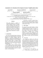
orthopnea, and pedal edema. Her echocardiogram
showed a LVEF of 15% to 20% with apical and septal
akinesis but without evidence of thrombus (Figure 2A).
The patient was treated for CHF and was stable with
reso lution of her symptoms at the time of discharge. As
before, an echocardiogram obtained at her one-month
follow-up examination showed complete reversal of her
LV systolic functio n to her normal baseline without evi-
dence of any wall motion abnormality (Figure 2B).
Discussion
Our patient presented twice with various neurological
complaints, and on each occasion she was found to have
evidence of acute onset severe apical LV dysfunction
that completely resolved within one month of onset.
After her first presentation, we suspected that this
patient had developed TTC. This condition is a syn-
drome of transient diminished LV apical wall motion
with relative preservation of the basal heart segment in
thepresenceofnormalcoronaryarteries[2].First
described in Japan [1], TTC has since been identified
worldwide [8]. Named for the ventricl e’s similar appear-
ance on ventriculograms to a takotsubo, or Japanese
octopus trap, this syndrome, also known as left apical
ballooning syndrome or “broken heart syndrome,” has
been linked to emotional or p hysical stress, with a high
incidence among post-menopausal women. As the syn-
drome is transient, LV function typically normalizes
within six to eight weeks of onset [2].
Our patient is unique in that she had a recurrence of
this syndrome within seven months of h er initial
presentation, and both episodes were preceded by tran-
sient neurological complaints. Recur rence of TTC, once
thought to be rare, has b een increasingly recognize d in
the literature, with a recurrence rate of 5% to 10%
worldwide [2,4,5]. Additionally, the development of TTC
in the setting of fleeting neurological symptoms such as
aphasia and ataxia without structural brain disease has
never been reported. Histological and nuclear imaging
data in humans have shown regional differences in effer-
ent sympathetic innervation where the basal ventricular
wall possesses a greater mean density of nerve endings
and local catecholamine concentrations compared to the
apex, while the apex possesses higher concentrations of
adrenoreceptors. This differential distribution is pro-
posed to induce the LV dysfunction found in patients
with TTC [2,9,10].
Similarly, neurogenic myocardial stunning has also
been proposed as a cause of the findings in patients
with TTC [2,11]. LV dysfunction with a takotsubo-like
state is often seen with intracranial pathology, includ ing
subarachnoid hemorrhage and congenital brain abnorm-
alities in children. In the setting of intracranial injury or
dysfunction, such as the catecholamine toxicity
described in our patient, a surge in sympathetic release
will cause a relative hyperdynamic contraction of the LV
basal segments with a relative stunning, or ballooning,
of the LV apical portion d ue to saturatio n of the adre-
noreceptors in that distribution [2].
Therefore, there appears to be a correlation between
intracranial and LV dysfunction. In fact, recurrence of
TTC in association with underlying chronic neurological
pathology, mainly status epilepticus, has previously been
reported [6,7]. Our present report, however, is the first
to describe a patient with recurrent TTC in the setting
of transient neurological symptoms without structural
evidence of neurologic dysfunction.
Figure 2 Echocardiogram on second presentation and follow-up. (A) Echocardiogram revealing akinesis of the apex during the patient ’ s
second presentation to our hospital. (B) Echocardiogram showing normally contracting apex with complete recovery of ejection fraction after
the patient’s second presentation.
Sardar et al. Journal of Medical Case Reports 2011, 5:412
/>Page 3 of 4
Conclusions
Although no clearly defined etiology for TTC exists,
clinicians should be aware of the possibility of TTC in
patients whose presentation mimics a cute myocardial
infarction, especially in the setting of emotional, physi-
cal, and specifically neurological stress. Additionally, it
has been reported that patients with one episode of
TTC are at increased risk for recurrence [2]. As our
patient’ s two presentations suggest, the development
and recurrence of TTC likely involve a neurocardiogenic
mechanism. Though this condition rarely leads to death,
it is imperative that the clinician be aware of this syn-
drome to ensure the prompt initiation of appropriate
supportive care so that a return of normal LV function
can be achieved [2]
Consent
Written informed consent was obtained from the patient
for publication of this case report and any accompany-
ing images. A copy of the written c onsent is available
for review by the Editor-in-Chief of this journal.
Acknowledgements
We thank Dr Adnan Siddiqui, Dr Brian Triola, and Barbara Schiavi for
obtaining the echocardiographic images and Dr Jerome Santoro and Dr
Cynthia Smith of the Department of Medicine, Lankenau Hospital,
Wynnewood, PA, USA.
Author details
1
Congestive Heart Failure Program, Division of Cardiology, Montefiore
Medical Center, 1825 Eastchester Road, Suite W-195, Bronx, NY 10461, USA.
2
Division of Pulmonology and Critical Care, Lankenau Medical Center,
Lankenau Hospital MOB West, Suite 230, 100 Lancaster Avenue,
Wynnewood, PA 19096, USA.
3
Department of Medicine, Jacobi Medical
Center, Albert Einstein College of Medicine, 1400 Pelham Parkway South,
Suite 3N1, Bronx, NY 10461, USA.
4
Department of Medicine, Weill-Cornell
Medical College, 1300 York Avenue, New York, NY 10065, USA.
5
Division of
Cardiology, Lankenau Medical Center, Campus Chief and Program Director,
Interventional Cardiology, Lankenau Medical Science Building, Suite 380, 100
Lancaster Avenue, Wynnewood, PA 19096, USA.
Authors’ contributions
The patient was admitted to our hospital under the care of MRS, CK, and TS.
JAM, NHA, and WS were major contributors to the researching, writing, and
editing of the manuscript. All authors read and approved the final
manuscript.
Competing interests
The authors declare that they have no competing interests.
Received: 24 January 2011 Accepted: 24 August 2011
Published: 24 August 2011
References
1. Dote K, Sato H, Tateishi H, Uchida T, Ishihara M: [Myocardial stunning due
to simultaneous multivessel coronary spasms: a review of 5 cases] [in
Japanese]. J Cardiol 1991, 21:203-214.
2. Akashi YJ, Goldstein DS, Barbaro G, Ueyama T: Takotsubo cardiomyopathy:
a new form of acute, reversible heart failure. Circulation 2008,
118:2754-2762.
3. Lindsay J, Paixao A, Chao T, Pichard AD: Pathogenesis of the takotsubo
syndrome: a unifying hypothesis. Am J Cardiol 2010, 106:1360-1363.
4. Yamamoto Y, Watari Y, Kobayashi K, Tanaka K: Recurrent takotsubo
cardiomyopathy can appear as transient midventricular ballooning
syndrome. J Cardiol Cases 2009, 1:e141-e143.
5. Sharkey SW, Windenburg DC, Lesser JR, Maron MS, Hauser RG, Lesser JN,
Haas TS, Hodges JS, Maron BJ: Natural history and expansive clinical
profile of stress (tako-tsubo) cardiomyopathy. J Am Coll Cardiol 2010,
55:333-341.
6. Legriel S, Bruneel F, Dalle L, Appere-de-Vecchi C, Georges JL, Abbosh N,
Henry-Lagarrigue M, Revault D’Allonnes L, Ben Mokhtar H, Audibert J,
Guezennec P, Troche G, Bedos JP: Recurrent takotsubo cardiomyopathy
triggered by convulsive status epilepticus. Neurocrit Care 2008, 9:118-121.
7. Lemke DM, Hussain SI, Wolfe TJ, Torbey MA, Lynch JR, Carlin A,
Fitzsimmons BF, Zaidat OO: Takotsubo cardiomyopathy associated with
seizures. Neurocrit Care 2008, 9:112-117.
8. Sharkey SW, Lesser JR, Zenovich AG, Maron MS, Lindberg J, Longe TF,
Maron BJ: Acute and reversible cardiomyopathy provoked by stress in
women from the United States. Circulation 2005, 111:472-479.
9. Tsuchihashi K, Uesima K, Uchida T, Oh-mura N, Kimura K, Owa M,
Yoshiyama M, Miyazaki S, Haze K, Ogawa H, Honda T, Hase M, Kai R, Morii I,
Angina Pectoris-Myocardial Infarction Investigations in Japan: Transient left
ventricular apical ballooning without coronary artery stenosis: a novel
heart syndrome mimicking acute myocardial infarction: Angina Pectoris-
Myocardial Infarction Investigations in Japan. J Am Coll Cardiol 2001,
38:11-18.
10. Kawano H, Okada R, Yano K: Histological study on the distribution of
autonomic nerves in the human heart. Heart Vessels 2003, 18:32-39.
11. Wittstein IS, Thiemann DR, Lima JA, Baughman KL, Schulman SP,
Gerstenblith G, Wu KC, Rade JJ, Bivalacqua TJ, Champion HC:
Neurohumoral features of myocardial stunning due to sudden
emotional stress. N Engl J Med 2005, 352:539-548.
doi:10.1186/1752-1947-5-412
Cite this article as: Sardar et al.: Recurrent takotsubo cardiomyopathy in
the setting of transient neurological symptoms: a case report. Journal of
Medical Case Reports 2011 5:412.
Submit your next manuscript to BioMed Central
and take full advantage of:
• Convenient online submission
• Thorough peer review
• No space constraints or color figure charges
• Immediate publication on acceptance
• Inclusion in PubMed, CAS, Scopus and Google Scholar
• Research which is freely available for redistribution
Submit your manuscript at
www.biomedcentral.com/submit
Sardar et al. Journal of Medical Case Reports 2011, 5:412
/>Page 4 of 4