báo cáo khoa học: "Dysphagia as a manifestation of esophageal tuberculosis: a report of two cases" pps
Bạn đang xem bản rút gọn của tài liệu. Xem và tải ngay bản đầy đủ của tài liệu tại đây (1.62 MB, 5 trang )
CAS E REP O R T Open Access
Dysphagia as a manifestation of esophageal
tuberculosis: a report of two cases
Joana Gomes
*
, Ana Antunes, Aurora Carvalho and Raquel Duarte
Abstract
Introduction: Esophageal involvement by Mycobacterium tuberculosis is rare and the diagnosis is frequently made
by means of an esophageal biopsy during the evaluation of dysphagia. Th ere are few cases reported in the
literature.
Case presentation: We present two cases of esophageal tuberculosis in 85- and 65-year-old male Caucasian
patients with initial complaints of dysphagia and epigastric pain. Upper gastrointestinal endoscopy resulted in the
diagnosis of esophageal tuberculosis following the biopsy of lesions of irregular mucosa in one case and a sessile
polyp in the other. Pulmonary tuberculosis was detected in one patient. In one patient esophageal stricture
developed as a complication. Antituberculous therapy was curative in both patients.
Conclusion: Although rare, esophageal tuberculosis has to be kept in mind in the differential diagnosis of
dysphagia. Pulmonary involveme nt has important implications for contact screening.
Introduction
Tuberculosis of the esophagus is a rare condition, even in
countries with a high incidence of tuberculosis (TB) [1,2],
and studies estimate that it constitutes about 0.3% of gas-
trointestinal TB cases [3]. Involvement o f the gastroin-
testinal tract occurs through ingestion of infected
sputum or hematogenous spread from primary pulmon-
ary TB [4]. Most cases of esophageal tuberculosis are sec-
ondary to direct extension from adjacent structures, such
as mediastinal lymph nodes or pulmonary sites. Primary
esophageal tuberculosis is even rarer [5]. Esophagic invol-
vement by tuberculosis usually affects the middle third of
the esophagus at the carina level [6]. The most common
symptoms are dysphagia or retrosterna l pain, but odyno-
phagia and weight loss may also be present.
We present two case reports of esophageal involve-
ment by Mycobacterium tuberculosis infection in immu-
nocompetent persons.
Case presentations
Case one
A Portuguese Caucasian man, 89 years old, with a his-
tory of hypertension and benign prostatic hypertrophy,
started to experie nce dysphagia, epigastric pai n and
anorexia one month prior to presentation. He was trea-
ted with omeprazole and sucralfate without a ny
improvement. He had a normal blood count, with an
erythrocyte sedimentation rate of 16 mm (normal range:
1-7 mm). An upper gastrointestinal endoscopy was per-
formed. This revealed congestion of the entire esopha-
geal mucosa, mainly i n the proximal portion (20-25 cm
from incisors), with easy bleeding to t he touch, some
irregular mucosa (biopsy one performed) and, in the
lower part of the esophagus, an irregular mucosa with
nodular areas (biopsy two performed). Diagnostic
hypotheses were esophageal cancer and esophagitis. The
histological examination for biopsy one revealed heavy
lymphocytic infiltrate and polymorphonuclear cells, with
ulceration, without lesions of malignancy. The histologi-
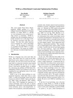
cal examinations of biopsy two showed esophageal
mucosa with extensive ulceration, inflammatory lesions
with epithelioid granulomas, and acid-fast alcohol resis-
tant microorganisms on staining (Figure 1). A chest
radiography showed no lesions. A purified protein deri-
vative skin test was negative. Because of persistent
cough, mycobacteriological sputum examination was
performed; the smear was negative but the culture was
positive on the second month. Human immunodefi-
ciency virus (HIV) -1 and -2 serology was negative.
* Correspondence:
Centro Hospitalar de Gaia/Espinho, EPE, Rua Conceição Fernandes 4434-502
Vila Nova de Gaia, Portugal
Gomes et al. Journal of Medical Case Reports 2011, 5:447
/>JOURNAL OF MEDICAL
CASE REPORTS
© 2011 Gomes et al; licensee BioMed Central Lt d. This is an Open Access article distributed under the terms of the Creative Commons
Attribution License (http://creativecom mons.org/licenses/by/2.0), which permits unrestricted use, distribution, and reproduction in
any medium, provided the original work is properly cited.
Our patient began antituberculous therapy with iso-
niazid, rifampicin, pyrazinamide and ethambutol with
symptomatic improvement. The persistence of dysphagia
to solids led to upper gastrointestinal endoscopy repeti-
tion three months after starting antituberculous treat-
ment, a nd revealed cicatricial stenosis of his esophagus
requiring repeated esophageal dilatations (Figure 2). He
completed treatment with two months of isoniazid,
rifampicin, pyrazinamide and ethambutol, followed by
four additional months of rifampicin and isoniazid. No
further esophageal dilatations were required and our
patient has no gastrointestinal complaints.
Case two
The second patient was a Portuguese Caucasian man, 65
year s old, with a history of hypertension, Ménière’ssyn-
dromeandaknownallergytopenicillin.Hereported
anorexia and weight loss of 8 kg two years earlier. The
examination performed at that time revealed a right
pleural effusion that resolved spontaneously over six
months of follow-up, with no etiological diagnosis made.
He presented with epigastric and left upper abdominal
quadrant pain and had an erythrocyte sedimentation
rate of 15 mm and a C-reactive protein level of 5.8 mg/
dL (normal range: < 0.5 mg/dL). An abdominal ultraso-
nography showed mild polypoid thickening in his gall-
bladder without calculus, with no pain to elective area
compression and no other changes. An upper gastroin-
testinal endoscopy revealed a sessile 6 mm polyp w ith
an irregular surface in the distal third of his esophagus,
located 2 cm above the junction with his stomach
(biopsy performed, see Figure 3), which was removed
through endoscopy. Histology revealed the presence of
epithelioid granulomas with multinucleated giant Lan-
ghans cells, caseous necrosis an d acid-fast bacilli. A
chest radiography showed no relevant changes. A puri-
fied protein derivative skin test was positive after 48
hours.
Our patient started therapy with isoniazid, rifampicin,
pyrazinamide and ethambutol. Serology for HIV1 and 2
was negative. Because of worsening pain in his left
upper abdomen irradiating to the left thoracic region, a
computed tomography scan was done and revealed a
left juxtadiaphragmatic fluid collectio n. After two weeks
of antituberculous therapy, there was a significant
reduction of the effusion (Figure 4 ). Our patient com-
pleted treatment with four drugs during the first two
months and an additional fo ur months of therapy with
isoniazid and rifampicin. The clinical outcome was
good. No drug toxicity or complications were observed.
Discussion
The diagnosis of esophageal tuberculosis is rare, hence
there is a need for a high clinical suspicion. Tuberculo-
sis rarely causes dysphagia, which occurs due to esopha-
geal ulcers, tracheoesophageal fistula or extrinsic
Figure 1 Histology of esophageal mucosa in case one.
Esophageal mucosa with extensive ulceration and inflammatory
lesion with epithelioid granulomas. The acid-fast staining was
positive.
Figure 2 Upper gastrointestinal endoscopy after starting treatment in case one revealed cicatricial stenosis of the esophagus.
Gomes et al. Journal of Medical Case Reports 2011, 5:447
/>Page 2 of 5
compression by the mediastinal or neck lymph nodes
[6,7]. Tuberculosis can involve the esophagus as a pri-
mary infection or as a secondary manifestation of dis-
ease reactivation [8]. Case one was clearly a case of
secondary esophageal tuberculosis, as there w as proven
pulmonary tuberculosis by sputum mycobacteriologica l
examin ation. However, in case two there was a past his-
tory of pleural effusion that resolved spontaneously, as
well as a left juxtadiaphragmatic effusion that resolved
with antituberculous therapy and was most likely caused
by M. tuberculosis. Given these facts, we cannot state
for sure what was the primary focus of infection.
The most f requent symptom reported in esophageal
tuberculosis is dysphagia, which occurs in about 90% of
cases [2,9]. Other symptoms are odynophagia and retro-
sternal pain and the occurrence of symptoms such as
fever, weight loss and anorexia [2,9] is also common. In
our case s, dysphagia and epigastric pain were the cardi-
nal symptoms.
Esophageal tuberculosis lesions can involve any seg-
ment of this organ, but is most often located in the mid-
dle third of the esophagus because of its proximity to
the hilar and mediastinal lymph nodes surrounding the
bifurcation of the trachea [2,9,10]. In the cases pre-
sented, esophageal involvement was at the distal l evel.
The most common macroscopic finding is an esopha-
geal ulcer as observed in case one. Howe ver, hyper-
trophic growth as esophageal polyps may also be
present as in case two [5,11]. Esophageal carcinoma is
part of the differential diagnosis as was the case for both
our patients. Diagnosis is usually made by upper
gastrointestinal endoscopy with histology examination
showing epithelioid granuloma with Langhans cells, cen-
tral necrosis and acid-fast bacilli. This was the method
that allowed the diagnosis in both cases. In secondary
esophageal tuberculosis, diagnosis may be suggested by
confirmation of tuberculosis i nvolving adjacent struc-
tures [5], which was not possible in case two, given the
quick resolution of the abdo minal effusion after starting
antituberculous therapy.
Esophageal tuberculosis treatment is based on che-
motherapy with four drugs (isoniazid, rifampicin, pyra-
zinamide and ethambutol) in a first phase lasting for
two months, followed by a period of four to six
months with two drugs (isoniazid and rifampicin).
There are cases where treatment was successfully car-
ried out with only three drugs for six months, exclud-
ing ethambutol [12,13]. Surgical treatment is reserved
for complications such as esophageal, tracheoesopha-
geal and aortoesophageal fistulas, the latter of which
can lead to death by massive hematemesis [14]. In
both cases presented, six-months of antituberculous
therapy was curative.
Esophageal strictures may result from external com-
pression of the esophagus due to mediastinal or cervical
lymph nodes as well as mediastinal fibrosis induced by
tuberculosis. This condition results in long and narrow
strictures that are difficult to dilate, and in which dila-
tion may be associated with a higher rate of complica-
tions [15].
Esophageal stenosis as a complication of esophageal
tuberculosis is rare and the re are few reports in the
Figure 3 Upper gastrointestinal endoscopy in case two. Sessile polyp with irregular surface in the distal third of the esophagus.
Gomes et al. Journal of Medical Case Reports 2011, 5:447
/>Page 3 of 5
literature [16,17]. The stenosis in case one was probably
the result of the healing process, but was severe and
repeated esophageal dilatation was needed to maintain
esophagogastric transit.
Conclusion
Although rare, esophageal tuberculosis must be kept in
mind in patients with dysphagia, especially in countries
with high prevalence of tube rculosis,eveninimmuno-
competent patients. Active pulmonary tuberculosis
should be ruled out, since early recognition of this infec-
tion is very important for public health. Treatment of
esophageal tuberculosis with antituberculous drugs is
curative, although complications may sometimes occur
as in our case. Finally, esophageal cancer must be
included in the differential diagnosis of the endoscopic
findings in this situation.
Consent
Written informed consent was ob tained from both
patients for publication of this case series and accompa-
nying images. A copy of the written consent is available
for review by the Editor-in-Chief of this journal.
Authors’ contributions
JG was a major contributor in writing and revising the manuscript. RD was
involved in drafting the manuscript and revising it critically for important
intellectual content. AC and AA analyzed and interpreted the patient data
regarding mycobacteriological examination. All authors were responsible for
the diagnosis, treatment and follow-up of the patients whose case reports
were described. All authors read and approved the final manuscript.
Competing interests
The authors declare that they have no competing interests.
Received: 6 January 2011 Accepted: 8 September 2011
Published: 8 September 2011
References
1. Rosario MT, Raso CL, Comer GM: Esophageal tuberculosis. Dig Dis Sci 1989,
34:1281-1284.
2. Jain SK, Jain S, Jain M, Yaduvanshi A: Esophageal tuberculosis: is it so
rare? Report of 12 cases and review of the literature. Am J Gastroenterol
2002, 97(2):287-291.
3. Marshall JB: Tuberculosis of the gastrointestinal tract and peritoneum.
Am J Gastroenterol 1993, 88(7):989-999.
4. Welzel TM, Kawan T, Bohle W, Richter GM, Bosse A, Zoller WG: An unusual
cause of dysphagia: esophageal tuberculosis. J Gastroint Liver Dis 2010,
19(3):321-324.
5. Leung VKS, Chan WH, Chow TL, Luk ISC, Chau TN, Loke TKL: Oesophageal
tuberculosis mimicking oesophageal carcinoma. Hong Kong Med J 2006,
12(6):473-476.
6. Huang YK, Wu YC, Liu YH, Liu HP: Esophageal Tuberculosis mimicking
submucosal tumor. Interact CardioVasc Thorac Surg 2004, 3(2):274-276.
7. Rathinam S, Kanagavel M, Tiruvadanan BS, Santhosam R,
Chandramohan SM: Dysphagia due to tuberculosis. Eur J Cardiothorac
Surg 2006, 30(6):833-836.
8. Grubbs BC, Baldwin DR, Trenkner SW, McCabe RP Jr, Maddaus MA: Distal
oesophageal perforation caused by tuberculosis. J Thorac Cardiovasc Surg
2001, 121(5):1003-1004.
9. Mokoena T, Shama DM, Ngakane H, Bryer JV: Oesophageal tuberculosis: a
review of eleven cases. Postgrad Med J 1992, 68(796):110-115.
10. Lockard LB: Oesophageal tuberculosis: a critical review. Laryngoscope
1913, 23:561-584.
11. Damtew B, Frengley D, Wolinsky E, Spagnuolo PJ: Esophageal tuberculosis:
mimicry of gastrointestinal malignancy. Rev Infect Dis 1987, 9(1):140-146.
12. Pimenta APA, Preto JR, Gouveia AMF, Fonseca E, Pimenta MML: Mediastinal
tuberculous lymphadenitis presenting as an esophageal intramural
tumor: a very rare but important cause for dysphagia. World J
Gastroenterol 2007, 13(45):6104-6108.
13. Hadlich E, Galperim B, Rizzon CF: Esophageal ulcers caused by
reactivation of ganglionary tuberculosis: a case report. Braz J Infect Dis
2007, 11(2):293-296.
14. Devarbhavi HC, Alvares JF, Radhikadevi M: Esophageal tuberculosis
associated with esophagotracheal or esophagomediastinal fistula: report
of 10 cases. Gastrointest Endosc 2003, 57(4):588-592.
15. Management of benign esophageal strictures. [ />contents/management-of-benign-esophageal-strictures].
Figure 4 Thoracic computed tomography scan in ca se two showing a left juxt adiaphragmatic effusion (arrow) which almost
disappeared after two weeks of antituberculous therapy.
Gomes et al. Journal of Medical Case Reports 2011, 5:447
/>Page 4 of 5
16. Prakash K, Kuruvilla K, Lekha V, Venugopal A, Jacob G, Ramesh H: Primary
tuberculous strictures of the oesophagus mimicking carcinoma. Trop
Gastroenterol 2001, 22(3):143-144.
17. Milnes JP, Homes GKT: Recurrent oesophageal stricture due to
tuberculosis. Br Med J (Clin Res Ed) 1983, 286(6382):1977.
doi:10.1186/1752-1947-5-447
Cite this article as: Gomes et al.: Dysphagia as a manifestation of
esophageal tuberculosis: a report of two cases. Journal of Medical Case
Reports 2011 5:447.
Submit your next manuscript to BioMed Central
and take full advantage of:
• Convenient online submission
• Thorough peer review
• No space constraints or color figure charges
• Immediate publication on acceptance
• Inclusion in PubMed, CAS, Scopus and Google Scholar
• Research which is freely available for redistribution
Submit your manuscript at
www.biomedcentral.com/submit
Gomes et al. Journal of Medical Case Reports 2011, 5:447
/>Page 5 of 5