báo cáo khoa học: "Contribution of magnetic resonance imaging in the diagnosis of talus skip metastases of Ewing’s sarcoma of the calcaneus in a child: a case report" pdf
Bạn đang xem bản rút gọn của tài liệu. Xem và tải ngay bản đầy đủ của tài liệu tại đây (909.95 KB, 4 trang )
CAS E REP O R T Open Access
Contribution of magnetic resonance imaging in
the diagnosis of talus skip metastases of Ewing’s
sarcoma of the calcaneus in a child: a case report
Hicham Jalal
1*
, Zoubida Belhadj
1
, Hind Enneddam
1
, Mohammed Madhar
2
, Tarik Fikry
2
, Omar Essadki
1
and
Ahmed Ousehal
1
Abstract
Introduction: Ewing’s sarcoma of the calcaneus is rare. About thirty cases with calcaneus involvement have been
reported in the literature. Talus skip metastases have rarely been described in the available literature
Case presentation: We report a case of a 14-year-old Moroccan boy, who presented with Ewing’s sarcoma of his
right calcaneus, diagnosed by swelling of the calcaneus evolving over a year. Radiography, computed tomo graphy
and magnetic resonance imaging showed an important tumoral process of the calcaneus and talus skip
metastases. The diagnosis was confirmed with histology after a biopsy. In spite of amputati on and postoperative
chemotherapy, our patient died six months later due to secondary respiratory distress after lung metastasis.
Conclusion: Imaging, especially magnetic resonance, is important in the diagnosis of Ewing sarcoma and skeletal
skip metastases. Treatme nt of Ewing’s sarcoma consists of chemotherapy, radiation therapy and surgical resection
depending on the stage and extent of the disease. With the exception of lesions in the calcaneus, the prognosis
for disease-free survival of Ewing’s sarcoma of the foot is excellent.
Introduction
Ewing’s sarcoma is a rare malignant bone tumor that
may affect any bone, usually occurring in long bones,
pelvis and ribs, with only 3-5% of cases in the bones of
the hands and feet [1]. It i s a highly anaplastic round-
cell tumor, primarily arising in the intramedullary por-
tion of the bone.
Case presentation
A 14-year-old Moroccan boy presented with painful
swelling of his right foot of 12 months duration. A gen-
eral examination was unremarkable, while local exami-
nation revealed a diffuse swelling involving his right
ankle joint and foot. The overlying skin was normal.
The swelling was tender and mobility at the joint was
restricted. Hematological and biochemical investigations
revealed a normal hemogram and normal liver and renal
function tests.
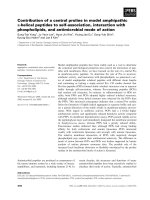
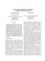
Anteroposterior (Figure 1) and lateral radiographs
(Figure 2 ) showed a condensed le sion in the calcaneus
of his right foot with aggressive periosteal reaction and
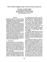
soft-tissue swelling. Computed tomography (CT)
revealed a soft-tissue mass o f the foot originating from
his calcaneus and a sclerotic lesion of the entire bone
with aggressive spiculated periosteal reaction and corti-
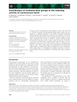
cal destruction (Figure 3). A large soft-tissue mass
around the involved bone was indicative of Ewing’s sar-
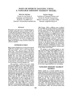
coma. Magnetic resonance imaging (MRI) was then per-
formed and showed a hypointe nse tumor mass on T1-
weigh ted sequences (Figure 4) and hyperintense proper-
ties on T2-weighted spin-echo sequences compared to
surrounding musculature (Figure 5), a signal pattern
characteristic of mo st tumors. The skip lesions of the
talus displayed hyposignal properties on T1- and T2-
weighted sequences. After intravenous gadolinium c he-
late administration, strong contrast enhancement of the
tumor was observed (Figure 6). Skip metastases of the
talus were evidenced as low-signal masses with periph-
eral enhancement (Figure 6).
* Correspondence:
1
Department of Radiology, Ibn Tofail Hospital, Gueliz, Marrakesh, 40000,
Morocco
Full list of author information is available at the end of the article
Jalal et al. Journal of Medical Case Reports 2011, 5:451
/>JOURNAL OF MEDICAL
CASE REPORTS
© 2011 Jalal et al; licensee BioMed Central Ltd. This is an Open Access article distributed u nder the terms of the Creative Comm ons
Attribu tion License ( which permits unrestricted use, distribution, an d reproduction in
any medium, provid ed the original work is properly cited.
A biopsy was performed and histopa thol ogy showed a
malignant small round-cell tumor, identified as Ewing’s
sarcoma at immunohistochemistry study. Chest radio-
graphy and liver ultrason ography excluded the presence
of any distant metastases. Our patient started neoadju-
vant chemotherapy and underwent a below-knee ampu-
tation. Postoperative histology confirmed the diagnosis.
Our p atient remained disease-free for six months after
diagnosis. Based on these findings, a diagnosis of
Ewing’s sarcoma of the calcaneus was made.
Discussion
Ewing’s sarcoma is a rare malignant neoplasm, predomi-
nantly affecting young patients of the ages five to 20
years. It involves the diaphyses of long bones and occurs
Figure 1 Anteropo sterior radiographs of patient’s foot show a
lesion in the calcaneus condensed with aggressive periosteal
reaction and soft-tissue swelling.
Figure 2 Lateral radiographs of the patient’ s foot show a
condensed lesion in the calcaneus with aggressive periosteal
reaction and soft-tissue swelling.
Figure 3 CT image of the patient’s foot, revealing a soft-tissue
mass originating from the calcaneus, permeative destruction
of the entire bone with aggressive spiculated periosteal
reaction and cortical destruction.
Jalal et al. Journal of Medical Case Reports 2011, 5:451
/>Page 2 of 4
less commonly in flat bones [1]. Clinical and laborato ry
features include local pain, soft-tissue swelling and
erythema, occasionally accompanied with fever, anemia,
leukocytosis, and accelerated erythroc yte sedimentation
rate [2]. It rarely affects the feet.
Cook listed 29 cases of Ewing’s sarcoma of the calca-
neus in the literature since 1921 [3]. These rare cases
are usually misdiagnosed, leading to treatment delay,
which is detrimental to the outcome.
According to a retrospective study concerning 235
patients with non-metastatic Ewing’ s sarcoma of the
bone, 15 patients were identified with a skip lesion at
diagnosis. However, the skip lesions were located in
adjacent juxta-articular bone in only two cases [4].
The radiographic features of Ewing’s sarcoma in our
case were those of classic Ewing’ s sarcoma: a permea-
tive, lytic and condensed lesion with cortical destruction,
aggressive periosteal reaction, large extraosseous soft-tis-
sue component and often sclerotic reaction [5,6]. In
spite of clinical and radiological findings, Ewing’ ssar-
coma can be misinterpreted as osteomyelitis, cartilagi-
nous tumor, giant cell lesion, lymphoma or
osteosarcoma, and the distinction often requires exten-
sive evaluation using varied imaging modalities [7].
CT can reveal a soft-tissue mass of the foot, such as
permeative lytic lesions of the bone with aggressive peri-
osteal reaction and cortical destruction, but the distinc-
tion between osseous remnants, reactive change s and
tumor matrix can sometimes be challenging [8]. Bone
scintigraphy of the whole skeleton demonstrates a focus
of incr eased uptake o f technetium-99 m-methylene
diphosphonate [8,9].
T2-weighted MRI cannot adequately distinguish tumor
from necrosis, and lesion boundaries are frequently
Figure 4 MRI of the patient’s foot shows a hypointense tumor
mass on T1-weighted spin-echo sequences compared to
surrounding musculature. The skip lesion of the talus displays a
hyposignal on T1-weighted sequences.
Figure 5 MRI of the patient’s foot shows a hyperintense tumor
mass on T2-weighted spin-echo sequence images compared to
surrounding musculature. The skip lesion of the talus displays a
hyposignal on T2-weighted sequences.
Figure 6 T1-weighted fa t saturation sequence after
intravenous gadolinium chelate administration reveals strong
contrast enhancement of the tumor. We note the skip lesion in
the talus as a low signal mass with peripheral enhancement.
Jalal et al. Journal of Medical Case Reports 2011, 5:451
/>Page 3 of 4
overestimated because of the presence of edema and
hem orr hage [9]. The enhancement pattern after admin-
ist rati on of contrast medium on MRI allows differentia-
tion between a tumor and peritumoral reactive edema.
Furthermore, MRI can often distinguish the large solid
sarcomatous soft-tissue mass around the involved bone
from edema or an osteomyelitic abscess. MRI findings
can narrow the differential diagnosis, but a specific diag-
nosis can rare ly be established. Therefore, a biopsy of
the tumor with histopathological analysis is needed to
confirm the diagnosis. Staging, prior to biopsy, is essen-
tial to document the local and distant spread of the
tumor. In Ewing’s sarcoma, the metastatic pattern may
be pulmonary involvement, bone or bone marrow
spreading, skip metastases, or combined metastatic dis-
ease [7,9].
The imaging features of local spread of Ewing’ ssar-
coma, involving small bon es to adjacent bon es, have not
been described in the recent literature. It wasn’t possible
to determine the exact local extent of the tumor by
means of conventional radiography and CT [5,9].
Due to its superior contrast resolution and multipla-
nar capabilities, MRI is more sensitive than other ima-
ging techniques, especially for the investigation of tumor
spread to bony structures and bone ma rrow. MRI
should always be performed in the analysis of Ewing’s
sarcoma since it allows accurate evaluation of the tumor
extent, which is decisive for treatment [10].
Skip lesions in patients with otherwise non-metastatic
skeletal Ewing’s sarcoma may be of the same importance
as the molecular detection of marrow metastases, and
possibly confer a worse prognosis. Newer imaging mod-
alities like positron emission tomography-computed
tomography and careful staging work-up may indicate
that skip metastases in Ewing’s sarcoma are more com-
mon than previously suspected [8,10].
Conclusions
This case report confirms that the routine radiological
management of Ewing’s sarcoma should include radio-
graphy and MRI of the affected region, together with
wholeskeletonbonescintigraphyandCTofthechest.
MRI is essential in the determination of the true e xtent
of the tumor. It is important to bear in mind that early
recognition of an unusual appearance and location of
Ewing’s sarcoma is necessary for its adequate treatment.
Consent
Written informed consent was obtained from the father
of our patient for publication of this case report and any
accompanying images. A copy of the written consent is
available for review by the Editor-in-Chief of this
journal.
Abbreviations
CT: computed tomography; MRI: magnetic resonance imaging
Acknowledgements
We wish to acknowledge Prof. Arak Abdelfattah for his critical appraisal.
Author details
1
Department of Radiology, Ibn Tofail Hospital, Gueliz, Marrakesh, 40000,
Morocco.
2
Department of Traumatology, Ibn Tofail Hospital, Gueliz,
Marrakesh, 40000, Morocco.
Authors’ contributions
HJ, ZB and HE made, analyzed and interpreted our patient’s imaging
examinations. MM and TF are the traumatologists whom operated on our
patient and made major contributions to the manuscript. The manuscript
was prepared by HJ under the supervision of OE and AO. All authors read
and approved the final manuscript.
Competing interests
The authors declare that they have no competing interests.
Received: 22 February 2011 Accepted: 12 September 2011
Published: 12 September 2011
References
1. Davies AM, Makwana NK, Grimer RJ, Carter SR: Skip metastases in Ewing’s
sarcoma: a report of three cases. Skeletal Radiol 1997, 26:379-384.
2. Pritchard DJ, Dahlin DC, Dauphine RT, Taylor WF, Beabout JW: Ewing’s
sarcoma: A clinicopathological and statistical analysis of patients
surviving five years of longer. J Bone Joint Surg 1975, 57:10-16.
3. Cook MA, Manfredi OL: Ewing’s sarcoma of the hand: a case report. Bull
Hosp Jt Dis 1996, 55:75-77.
4. Jiya TU, Wuisman PI: Long-term follow-up of 15 patients with non-
metastatic Ewing’s sarcoma and a skip lesion. Acta Orthop 2005,
76:899-903.
5. Siddiqui YS, Zahid M, Sabir AB, Asif N, Kumar G, Akhtar M: Calcaneal
Ewing’s sarcoma with skip metastases to the adjacent tarsal bones. J
Clin Diag Res 2011, 5:117-119.
6. Sandrasagra FA: Ewing’s sarcoma of metatarsal bone. Ceylon Med J 1973,
18:58-61.
7. Wilkins RM, Pritchard DJ, Burgert EO, Unni K: Ewing’s sarcoma of bone:
experience with 140 patients. Cancer 1986, 58:2551-2555.
8. Baraga JJ, Amarami KK, Swee RG, Wold L, Unni KK: Radiographic features
of Ewing’s sarcoma of the bones of the hand and feet. Skeletal Radiol
2001, 30:121-126.
9. Zelazny A, Reinus WR, Wilson AJ: Quantitative analysis of the plain
radiographic appearance of Ewing’s sarcoma of bone. Invest Radiol 1997,
32:59-65.
10. Reinus WR, Gilula LA: Radiology of Ewing’s sarcoma: Intergroup Ewing’s
Sarcoma Study (IESS). Radiographics 1984, 4:929-944.
doi:10.1186/1752-1947-5-451
Cite this article as: Jalal et al.: Contribution of magnetic resonance
imaging in the diagnosis of talus skip metastases of Ewing’s sarcoma of
the calcaneus in a child: a case report. Journal of Medical Case Reports
2011 5:451.
Jalal et al. Journal of Medical Case Reports 2011, 5:451
/>Page 4 of 4