báo cáo khoa học: " A large gastrointestinal stromal tumor of the duodenum: a case report" doc
Bạn đang xem bản rút gọn của tài liệu. Xem và tải ngay bản đầy đủ của tài liệu tại đây (1.16 MB, 3 trang )
CAS E REP O R T Open Access
A large gastrointestinal stromal tumor of the
duodenum: a case report
Basem Morcos
*
and Firas Al-Ahmad
Abstract
Introduction: Gastrointestinal stromal tumors of the duodenum are uncommon. They can reach a large size.
Diagnosis can be elusive and managing them can be difficult. Our case report aims to increase awareness and
highlight some issues related to the diagnosis and management of duodenal gastrointestinal stromal tumors.
Case presentation: We present the case of a 38-year-old Middle Eastern woman with a large, slowly-growing
gastrointestinal stromal tumor of the duodenum. Her complaints were minor epigastric discomfort and swelling.
A pancreaticoduodenectomy with complete tumor excision was performed. She was doing very well with no
evidence of disease recurrence when she was last seen 34 months after her operation.
Conclusion: Gastrointestinal stromal tumors of the duodenum should be suspected in any patie nt with a
duodenal wall mass. Extramural growth and central ulceration with or without bleeding should alert the
endoscopist to the possibility of a duodenal gastrointestinal stromal tumor diagnosis. There is more than one
surgical approach available; however, complete surgical excision, with negative margins, is the absolute
requirement. Preoperative imatinib mesylate can be considered in unresectable or borderline resectable cases.
Introduction
The most common sites for gastrointestinal stromal
tumors (GIST) are the stomach and, to a lesser extent, the
small intestine [1]. Small intestinal GIST can occur any-
where along the length of the bowel and can be multiple.
The duodenum is involved in about 10% to 20% of small
intestinal GIST [2]. Although duodenal GIST is similar
pathologically to that involving other organs, they do have
some peculiar features. GISTs in the duodenum pose par-
ticular challenges for diagnosis and management.
We describe the case of a large duodenal GIST including
its presentation, dia gnosis, and the type of surgery per-
formed, as well as a review of issues related to GIST in the
duodenum.
Case presentation
A 38-year-old Middle Eastern woman presented with a
slowly enlarging abdominal mass of 12 years duration.
According t o the patient, a surgeon had attempted to
resect the mass 12 years earlier, but could not do so due
to excessive bleeding from the tumor. She was offered no
further treatment.
At presentation, her main complaint was epigastric dis-
comfort. She also gave a history of so me mild back pain
and occasional abdomina l pain. Her appeti te was good
and she had not lost weight. There was no history of
vomiting, ch ange in bowel habits or melena. She had
been diagnosed with a peptic ulcer many years ago.
On examination she looked healthy with no clinical
jaundice or pallor. Abdominal examination revealed a
large upper abdominal mass with thinned overlying skin.
It had minimal mobility and was not tender. The rest of
the examination was normal. Her hemoglobin level was
10.8 g/dL, with hypochromic microcytic red blood cell
indices. Otherwise, all blood tests were normal. A com-
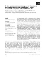
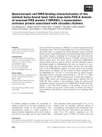
puted tomography (CT) scan of the abdomen revealed a
20 cm retroperitoneal mass in the region of the head of
the pancreas (Figure 1). It appeared to push and stretch
the surrounding structures. There was no e vidence of
metastases to the liver or lung. Upper gastrointestinal
endoscopy was performed , showing a 2.5 cm ulcer in the
second part of the duodenum with a clot at its center.
There was no intraluminal mass. A deep biopsy was taken,
but was not diagnostic.
* Correspondence:
Department of Surgical Oncology, King Hussein Cancer Center, Queen Rania
Al Abdullah Street, P.O.Box 1269 Al-Jubeiha, Amman, 11941, Jordan
Morcos and Al-Ahmad Journal of Medical Case Reports 2011, 5:457
/>JOURNAL OF MEDICAL
CASE REPORTS
© 2011 Morcos and Al-Ahmad; licensee BioMed Central Ltd. This is an Open Access article distributed under the terms o f the Creative
Commons Attribution License ( which permits u nrestricted use, distribution, and
reproduction in any medium, provided the original work is properly cited.
Tumor e mbolization was planned to decrease tumor vas-
cularity before resection. Angiography revealed that the
hepatic artery was the main feeding vessel; however, embo-
lization was not possible be cause the celiac axis was kinked
and the catheter could not be advanced into the feeding
artery. After preparation she was taken to the operating
theater. A midline incision over the previous scar was per-
formed. The tumor was very vascular with large v enous tri-
butaries draining into the portal circulation. It lay posterior
to the pancreatic head and duodenum, pushing them ante-
riorly. A pancreati coduodenectomy (Whipple pro cedure)
was performed with the dissection kept outside the pseu-
docapsule of the tumor, taking care not to rupture the
tumor. The patient tolerated the procedure well and had
an uneventful recovery. Histopathological examination
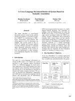
revealed a 22 cm tumor arising from the second part of the
duodenum. The tumor showed m oderate cellularity and
mildly atypical spindle cells arranged in fascicles with a low
mitotic count (1/50 high power field) and no necrosis
(Figure 2). Prominent skeinoid fibers were seen. The tumor
was negative for c-kit, SMA and S100 protein, but positive
for CD34. Although it was c-kit negative, the features were
consistent with the diagnosis of GIST. The tumor was con-
sidered of high malignant potential because of its size. Ima-
tinib mesylate (IM) was considered as an adjuvant
treatment but the patient could no t afford it. She contin-
ued to do well, however, and was f ree of any recurrence
the last time she attended the clinic, 34 mo nths after the
operation.
Discussion
GISTs are the most common mesenchymal tumors of the
gastrointestinal tract [1]. They are most commonly fo und
in the stomach and small bowel. Uncommon sites
include the colon, rectum, esophagus and even the liver
and mesentery. They mainly affect adults and are uncom-
mon in children [3]. The duodenum is an uncommon site
for GIST. It comprises 10%-20% of small-intestinal
GISTs, or only three to five percent of all GIST cases [4].
Most data on duodenal GIST are from single case reports
or from a few small series [4,5]. Duodenal GIST is usually
asymptomatic when small in size and can reach a large
size before causing any symptom. As the tumor enlarges
it causes variable symptomatology. The most common
presentation is gastrointestinal bleeding which may be
chronic and mild or sudden and massive [6] . Although
our patient had a large tumor, she had mild anemia. The
next most common presentations are abdominal discom-
fort, pain and swelling [5].
Diagnosis can be made with upper gastrointestinal endo-
scopy [5]. The tumor is usually exophytic, and appears as a
submucosal swelling. Sometimes it presents only as an
ulcer, as in our case. The biopsy should be deep, but may
not always be diagnostic. Endoscopic ultrasound can help in
delineating the submucosal tumor. A CT scan of the abdo-
men usually shows a retroperitoneal tumor at the site of the
duodenum and head of the pancreas [7]. However, CT
scans a re not always helpful in specifying the origin of the
mass. In a number of c ases reported in the literature, the
mass was misdiagnosed as arising from the head of the pan-
creas [8].
The treatment of choice for duodenal GIST is complete
surgical excision. This can be performed by local or seg-
mental duodenal resection with preservation of the pan-
creas for small tumors [2]. As for larger tumors, a
pancreaticoduodenectomy is required. The surgical
choice depends not only on the size of the tumor but
also on the location in the duodenal wall and the relation
to the ampulla of Vater. It is not clear what the optimal
surgical margin should be, but a negative one is essential
to prevent local recurrence of the tumor. No lymph node
Figure 1 Retropancreatic tumor. A preoperative CT scan showing
the large retropancreatic tumor.
Figure 2 Tumor histopathology. Hematoxylin and eosin (H&E)
slide. Notice the spindle cells with abundance of skeinoid fibers
which are features of gastrointestinal stromal tumors.
Morcos and Al-Ahmad Journal of Medical Case Reports 2011, 5:457
/>Page 2 of 3
dissection is required since they are very unlikely t o be
involved [1].
The outcome depends on the pathologica l features of
the tumor and the completeness of surgical resection.
Large tumors with high mitotic counts behave much
worse than small tumors with low mitotic counts, which
are considered benign [9]. Local recurrence is higher in
tumors not completely removed or with a posit ive
microscopic margin. Most GISTs respond to IM, so
patients with tumors with a high malignant potential
should be offered IM as an adjuvant therapy. Preopera-
tive IM can be given in cases of unresectable or border-
line resectable cases. This might improve resectability.
Conclusion
Duodenal GIST should be suspected in any patient with a
duodenal wall mass. Extramural growth and central
ulceration with or without bleeding should alert the
endoscopist to the possibility of this diagnosis. There is
more than one surgical ap proach available, but the abso-
lute requirement is complete surgical excision. Preopera-
tive IM can be considered in unresectable or borderline
resectable cases.
Consent
Written informed consent was obtained from the patient
for publication of this case report and accompanying
images. A copy of the written consent is available for
review by the Editor-in-Chief of this journal.
Authors’ contributions
BM performed the literature review, collected the photos and wrote the
article. FA collected some papers for review and provided input for the
article. All authors read and approved the final manuscript.
Competing interests
The authors declare that they have no competing interests.
Received: 14 May 2011 Accepted: 14 September 2011
Published: 14 September 2011
References
1. Connolly EM, Gaffney E, Reynolds JV: Gastrointestinal stromal tumors. Br J
Surg 2003, 90:1178-1186.
2. Pidhorecky I, Cheney RT, Kraybill WG, Gibbs JF: Gastrointestinal stromal
tumors: Current diagnosis, biologic behaviour and management. Ann
Surg Oncol 2000, 7:705-712.
3. Hayashi Y, Okazaki T, Yamataka A, Toshihiro Y, Yamashiro Y, Tsurumaru M,
Kajiyama Y, Miyano T: Gastrointestinal stromal tumor in a child and
review of the literature. Pediatr Surg Int 2005, 21:914-917.
4. Miettinen M, Kopczynski J, Makhlouf H, Sarlomo-Rikala M, Gyorffy H,
Burke A, Sobin LH, Lasota J: Gastrointestinal Stromal Tumors, intramural
Leiomyomas, and Leiomyosarcomas in the Duodenum. Am J Surg Path
2003, 27:625-641.
5. Goh B, Chow P, Kesavan S, Yap W, Wong W: Outcome after surgical
treatment of Suspected Gastrointestinal Stromal Tumors Involving the
Duodenum: Is Limited Resection Appropriate? J Surg Oncol 2008,
97:388-391.
6. Winfield RD, Hochwald SN, Vogel SB, Hemming AW, Liu C, Cance WG,
Grobmyer SR: Presentation and management of gastrointestinal stromal
tumors of the duodenum. Am Surg 2006, 72:719-722.
7. King M: The radiology of gastrointestinal stromal tumours (GIST). Cancer
Imaging 2005, 5:150-156.
8. Uchida H, Sasaki A, Iwaki K, Tominaga M, Yada K, Iwashita Y, Shibata K,
Matsumoto T, Ohta M, Kitano S: An extramural gastrointestinal stromal
tumor of the duodenum mimicking a pancreatic head tumor. J
Hepatobiliary Pancreat Surg 2005, 12:324-327.
9. Fletcher CD, Berman JJ, Corless C, Gorstein F, Lasota J, Longley BJ,
Miettinen M, O’Leary TJ, Remotti H, Rubin BP, Shmookler B, Sobin LH,
Weiss SW: Diagnosis of Gastrointestinal stromal tumors: A consensus
approach. Hum Pathol 2002, 33:459-465.
doi:10.1186/1752-1947-5-457
Cite this article as: Morcos and Al-Ahmad: A large gastrointestinal
stromal tumor of the duodenum: a case report. Journal of Medical Case
Reports 2011 5:457.
Submit your next manuscript to BioMed Central
and take full advantage of:
• Convenient online submission
• Thorough peer review
• No space constraints or color figure charges
• Immediate publication on acceptance
• Inclusion in PubMed, CAS, Scopus and Google Scholar
• Research which is freely available for redistribution
Submit your manuscript at
www.biomedcentral.com/submit
Morcos and Al-Ahmad Journal of Medical Case Reports 2011, 5:457
/>Page 3 of 3