báo cáo khoa học: " Pseudoclavibacter-like subcutaneous infection: a case report" ppt
Bạn đang xem bản rút gọn của tài liệu. Xem và tải ngay bản đầy đủ của tài liệu tại đây (291.92 KB, 3 trang )
CAS E REP O R T Open Access
Pseudoclavibacter -like subcutaneous infection: a
case report
François Lemaitre
1
, Andreas Stein
2
, Didier Raoult
1,3
and Michel Drancourt
1,3*
Abstract
Background: Arthrobacter-like organisms, including Pseudoclavibacter organisms, have rarely been documented as
being responsible for infection in humans.
Case presentation: An 81-year-old French man developed a subcutaneous infection despite antibiotic treatment
combining clindamycin and metronidazole for chronic wound infection. A skin biopsy showed numerous
polymorphonuclear cells and no bacteria, but a subcutaneous swab yielded numerous polymorphonuclear cells, a
few Gram-positive cocci, Gram-negative cocci, and Gram-positive rods. The Gram-positive rod sequence exhibited
99% sequence similarity with uncultured Pseudoclavibacter sp. [GenBank:EF419350] and 99% sequence similarity
with uncultured Pseudoclavibacter sp. [GenBank:EF419347]. The genetic data and unique peptide profile of this
Pseudoclavibacter-like isolate, determined by matrix-assisted laser desorption ionization-time of flight mass
spectrometry, underscored its uniqueness.
Conclusions: Pseudoclavibacter-like organisms are identifiable in cutaneous and subcutaneous infections in
humans.
Keywords: Pseudoclavibacter, 16S rRNA gene, MALDI-TOF, identification, skin infection
Background
Pseudoclavibacter is an emerging bacterial genus created
a few years ago to accommodate environmental Brevi-
bacterium organisms [1]. Indeed, Arthrobacter-like bac-
teria have rarely be en isolated in patient s, and a
Pseudoclavibacter organism has been reported to be iso-
lated only once, from an aortic valve of a 74-year-old
man [2].
Case presentation
An 81-year-old Frenc h man was admitted to our hospi-
tal for erysipelas of the right leg. The patient had sud-
denly developed this infection despite antibiotic
treatment combining clindamycin and metronidazole for
a chronic wound infection of the same leg with previous
documentation of clindamycin-susceptible Staphylococ-
cus a ureus, Klebsiella oxytoca, Serratia marcescens,and
Corynebacterium spp., but no anaerobe. His leukocyte
count was 10.32 g/L with 72% polymorph onuclear cells,
18% lymphocytes, and 8% monocytes. Inflammatory syn-
drome was apparent with a C-reactive protein level of
119 mmol/L and a fibrinogen level of 8.6 g/L. Antibo-
dies against streptolysin O and streptococcal DNase
were not detectable in the patient’s serum. Direct micro-
scopic examination of a skin biopsy showed numerous
polymorphonuclear cells and no bacteria. Culture
remained sterile after a five-day inoculation on Colum-
bia agar with 5% sheep blood (bioMérieux, Marcy-
l’ Etoile, France), Chocolate agar with PolyViteX agar
(bioMérieux) and MacConk ey agar (bioMérieux) at 37°C
in 5% CO
2
. Three days later a subcutaneous swab
yielded numerous polymorphonuclear cells, and semi-
quantitative direct examination indicated an average of
15 to 30 organisms per microscopic field composed of
an equal proportion of Gram-positive cocci, Gram-nega-
tive cocci, and Gram-positive rods. Culture under the
same conditions described above yielded small, gray
colonies after 48-hour inoculation on Columbia agar
with 5% sheep blood. The Gram-positive rod was oxi-
dase-negative and catalase-positive. Inoculation of an
API Coryne system identification strip (bioMérieux),
performed twice, yielded no reaction and thus no
* Correspondence:
1
Pôle des Maladies Infectieuses, Fédération de Microbiologie Clinique,
Hôpital de la Timone, rue Saint-Pierre, 13005 Marseille, France
Full list of author information is available at the end of the article
Lemaitre et al. Journal of Medical Case Reports 2011, 5:468
/>JOURNAL OF MEDICAL
CASE REPORTS
© 2011 Lemaitre et al; licensee BioMed Central Ltd. This is an Open Access article distributed under the terms of the Creative
Commons Attribution License (http://creativecomm ons.org/licenses/by/2.0), which permits unrestricted use, distribution, and
reproduction in any medium, provided the original work is properly cited.
identi fying profile. No other organism was isolated from
this specimen. Antibiotic susceptibility assessed on
Mueller-Hinton agar (bioMérieux) using the disc
method (Mast Diagnostics, Amiens, France) and break
points as previously reported [3] yielded susceptibility to
amoxicillin (minimum inhibitory concentration (MIC) ≤
2 mg/L), rifampin (MIC ≤ 4 mg/L), doxycycline (MIC ≤
4 mg/L), and vancomycin (MIC ≤ 4mg/L)andresis-
tance to co-trimoxazole, clindamycin, and metronida-
zole. The latter three antibiotics yielded no growth
inhibition zone. To identify the isolate, we amplified (by
polymerase chain reaction) and sequenced 1431 bases of
the 16S rRNA gene [GenBank:FJ375951] [4]. This
sequence exhibi ted 99% sequence simil arity with uncul-
tured Pseudoclavibacter sp. [GenBank:EF419350] and
99% sequence similarity with uncultured Pseudoclavi-
bacter sp. [GenBank:EF419347]. The third hit exhibited
only 97% sequence similarity with Zimmermannella
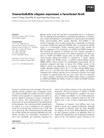
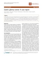
bifida [GenBank:AB012589]. The peptide profile of the
isolate was determined by matrix-assisted laser deso-
rption ionization-time of fli ght (MALDI-TOF) mass
spectrometry as previously described [5] ( Figure 1).
MALDI-TOF-based identification was achieved by com-
paring the isolate profile with the 3438 bacterial profiles
deposited in the MALDI BioTyper database (Bruker
Corp. Bremen, Germany), which includes 56 Arthrobac -
ter,17Brevibacterium,threePseudoclavibacter,andno
Zimmermannel la organism profiles (as of June 2010).
The isolate was not ident ified with any of the species in
the database, with the best identification score being
1.326 with Corynebacterium afermentans.Theisolate
has been deposited in the Collection de Souches de
l’ Unité des Rickettsies, Marseilles, France (CSUR P29).
The clinical evolution was favorable under antibiotic
treatment combining intravenous imipenem and vanco-
mycin. Oral treatment with amoxicillin/clavulanate
replaced intravenous antibiotics on day six.
Identification of the isolate was made possible only
after 16S rRNA gene sequence analysis, as the isolate
was not reactive on identification strips and did not
exhibit any identifying phenotype. T he 16S rRNA gene
sequence comparison indicated that this isolate is repre-
sentative of a previously uncultured organism. This case
illustrates a new paradigm in using 16S rRNA gene
sequence-based identification of organisms in clinical
micro biology laboratories. Indeed, most of the emerging
bacteria have been described previously on the basis of
an original bacterial isolate exhibiting a 16S rRNA gene
sequence with < 98.7% sequence similarity with any
other sequence [4,6,7]. In our case report, however, the
16S rRNA gene sequence was already ava ila ble in Gen-
Bank before we recovered the isolate. Indeed, extensive
genomic and metagenomic explor ations of complex
environmental and mucosa-associated flora yielded a
tremendous amount of the original 16S rRNA gene
sequence from as-yet-uncultured organisms [8,9]. In our
case report, isolation of an organism exhibiting a 16S
rRNA gene sequence identical to that of a previously
uncultured organism underscores the uniqueness of this
isolate.
Figure 1 Matrix-assisted laser desorption ionization-time of flight (MALDI-TOF) mass-spectrometry peptide profile of a
Pseudoclavibacter-like organism. This profile could be used as a reference for rapid identification of this bacterial species.
Lemaitre et al. Journal of Medical Case Reports 2011, 5:468
/>Page 2 of 3
Conclusions
In the case of our patient, the Pseudoclavibacter-like
organism was most probably involved in his clinical
infection, since this Gram-positive rod was observed in
the presence of pus during the direct examination of a
subcutaneous specimen. It grew in pure culture from a
patient who was taking two antibiotics to which the
Pseudoclavibacter-like organism was found to be resis-
tant, thus supporting t he hypothesis that growth of the
Pseudoclavibacter-like organism was indeed selected by
the antibiotic treatment. Also, P seudoclaviba cter sp. and
other Arthrobacter-like organisms have never been
reported as po tential contaminants of culture, and Pseu-
doclavibacter spp. have not been isolated in our labora-
tory, with the exception of this patient. The 16S rRNA
gene sequence of identical Pseudoclavibacter-like organ-
isms was found in the diseased skin of patients with
psoriasis before we obtained the first isolate [10]. This
fact and the data presented i n this case report suggest
that Pseudoclavibacter-like organisms are organisms
involved in skin diseases. Pseudoclavibacter-like organ-
isms are bacterial organisms identifiable in cutaneous
andsubcutaneousinfectionsinhumansonthebasisof
auniquepeptideprofileobtainedbyMALDI-TOFana-
lysis and unique 16S rRNA gene sequencing.
Consent
Written informed consent was obtained from the patient
for publication of this case report and any accompany-
ing images. A copy of the written c onsent is available
for review by the Editor-in-Chief of this journal.
Author details
1
Pôle des Maladies Infectieuses, Fédération de Microbiologie Clinique,
Hôpital de la Timone, rue Saint-Pierre, 13005 Marseille, France.
2
Pôle des
Maladies Infectieuses, Service de Maladies Infectieuses et Tropicales, Hôpital
de la Conception, boulevard Baille, 13005 Marseille, France.
3
Unité de
Recherche sur les Maladies Infectieuses et Tropicales Emergentes, CNRS-IRD
UMR 6236, Faculté de Médecine, IFR 48, Université de la Méditerranée, 27
Boulevard Jean Moulin, F-13385 Marseille cedex 5, France.
Authors’ contributions
FL reviewed the medical and laboratory charts and was involved in drafting
the manuscript. AS took care of the patient. DR and MD drafted the
manuscript. All authors read and approved the final manuscript.
Competing interests
The authors declare that they have no competing interests.
Received: 28 March 2011 Accepted: 20 September 2011
Published: 20 September 2011
References
1. Manaia CM, Nogales B, Weiss N, Nunes OC: Gulosibacter molinativorax gen.
nov., sp. nov., a molinate-degrading bacterium, and classification of
‘Brevibacterium helvolum’ DSM 20419 as Pseudoclavibacter helvolus gen.
nov., sp. nov. Int J Syst Evol Microbiol 2004, 54:783-789.
2. Mages IS, Frodl R, Bernard KA, Funke G: Identities of Arthrobacter spp. and
Arthrobacter-like bacteria encountered in human clinical specimens. J
Clin Microbiol 2008, 46:2980-2986.
3. Martínez-Martínez L, Ortega MC, Suárez AI: Comparison of E-test with
broth microdilution and disk diffusion for susceptibility testing of
coryneform bacteria. J Clin Microbiol 1995, 33:1318-1321.
4. Drancourt M, Berger P, Raoult D: Systematic 16S rRNA gene sequencing
of atypical clinical isolates identified 27 new bacterial species associated
with humans. J Clin Microbiol 2004, 42:2197-2020.
5. Seng P, Drancourt M, Gouriet F, La Scola B, Fournier PE, Rolain JM, Raoult D:
Ongoing revolution in bacteriology: routine identification of bacteria by
matrix-assisted laser desorption ionization time-of-flight mass
spectrometry. Clin Infect Dis 2009, 49:543-551.
6. Stackebrandt E, Ebers J: Taxonomic parameters revisited: tarnished gold
standards. Microbiol Today 2006, 33:152-155.
7. Janda JM, Abbott SL: 16S rRNA gene sequencing for bacterial
identification in the diagnostic laboratory: pluses, perils and pitfalls. J
Clin Microbiol 2007, 45:2761-2764.
8. Medini D, Serruto D, Parkhill J, Relman DA, Donati C, Moxon R, Falkow S,
Rappuoli R: Microbiology in the post-genomic era. Nat Rev Microbiol 2008,
6:419-430.
9. Hugenholtz P, Tyson GW: Metagenomics. Nature 2008, 455:481-483.
10. Gao Z, Tseng CH, Strober BE, Pei Z, Blaser MJ: Substantial alterations of
the cutaneous bacterial biota in psoriatic lesions. PLoS One 2008, 3:e2719.
doi:10.1186/1752-1947-5-468
Cite this article as: Lemaitre et al.: Pseudoclavibacter-like subcutaneous
infection: a case report. Journal of Medical Case Reports 2011 5:468.
Submit your next manuscript to BioMed Central
and take full advantage of:
• Convenient online submission
• Thorough peer review
• No space constraints or color figure charges
• Immediate publication on acceptance
• Inclusion in PubMed, CAS, Scopus and Google Scholar
• Research which is freely available for redistribution
Submit your manuscript at
www.biomedcentral.com/submit
Lemaitre et al. Journal of Medical Case Reports 2011, 5:468
/>Page 3 of 3