báo cáo khoa học: " Hypothyroidism in a five-year-old boy with rhabdomyolysis and recent history of cardiac tamponade: a case report" pptx
Bạn đang xem bản rút gọn của tài liệu. Xem và tải ngay bản đầy đủ của tài liệu tại đây (444.16 KB, 4 trang )
CAS E REP O R T Open Access
Hypothyroidism in a five-year-old boy with
rhabdomyolysis and recent history of cardiac
tamponade: a case report
Juan José Delgado Hurtado
1*
, Waleska Guevara
2
, Evelyn Ramos
2
, Claudia Lorenzana
2
and Susana Soto
2
Abstract
Introduction: Cardiac tamponade is a rare manifestation of hypothyroidism, and a less rare cause of pericardial
effusion. The accumulation of the pericardial fluid is gradual, and often does not compromise cardiac
hemodynamic function. There is a relationship between the severity and chro nicity of the disease with the
presence of pericardial effusion. There are few cases describing associated pericardial tamponade published in the
literature. When a tamponade occurs, a concomitant provocative factor such as a viral pericarditis may be related.
Our patient’s case appears to be the youngest patient described so far.
Case presentation: We report the case of a previously healthy five-year-old Hispanic (non-indigenous) boy who
developed rhabdomyolysis with a history of a recent pericardial effusion and tamponade two months before that
required the placement of a percutaneous pericardial drainage. Pericardial effusion was considered to be viral . Later
on readmission, clinical primary hypothyroidism was diagnosed and thought to be associated with the previous
cardiac tamponade. He developed rhabdomyolysis, which was considered to be autoimmune and was treated with
steroids. The level of creatine phosphate kinase and creatine kinase MB fraction returned to within the reference
rangeone week after our patient was started on steroids and three weeks after he was started on thyroid
hormones.
Conclusions: Physicians should consider hypothyroidism as a differential diagnosis in patients with pericardial
effusion. Pericardial effusion may progress and cause a cardiac tamponade with hemodynamic instability. The fact
that our patient did not have any manifestations of hypothyroidism might have delayed diagnosis.
Introduction
Pericardial tamponade is a rare complication of
hypothyroidism. Previous case series have reported peri-
cardial effusion in 50% to73%ofchildrenwith
hypothyroidism [1-3] sometimes associated with Down’s
syndrome [4], but there have been few describing car-
diac tamponade associated with hypothyroidism [5].
Aside from bradycardia, cardiac pericardial effusion is
the mo st common cardiac manifestation of clinical
hypothyroidism in adults [6]. Effusion in the hypothyr-
oid patient is caused by an increased capillary leak,
which increases the album in rich fluid build-up in the
pericardial spa ce and slow lym phatic drainage [7].
Cardiac tamponade is probably rare because the pericar-
dial effusion accumulation is gradual and allows the
pericardium to stretch, allowing normal maintenance of
cardiac function [8]. Thyroid hormones also have an
important effect on the expression of certain gene pro-
teins, including: a myosin heavy chain, potassium chan-
nels, b1 adrenergic receptor and a calcium ATPase,
which explain the cardiac manifestations. Characteristi-
cally, the protein content of pericardial effusion is
increased when effusion is due to hypothyroidism. The
treatment of pericardial effusion related to hypothyroid-
ism is usually medical. Pericardial effusion generally
improves in the hypothyroid patient two months to a
year after thyroid hormon es are started and the patient
becomes euthyroid [4,6,9]. Our patient’s case required
the placement of a percutaneous drainage due to hemo-
dynamic instability.
* Correspondence:
1
Universidad Francisco Marroquín, 19 Ave. 8-44 Zona 15, Vista Hermosa I,
Guatemala City, Guatemala
Full list of author information is available at the end of the article
Delgado Hurtado et al. Journal of Medical Case Reports 2011, 5:515
/>JOURNAL OF MEDICAL
CASE REPORTS
© 2011 Delgado Hurtado et al; licensee BioMed Central Ltd. This is an Open Access article distributed under the terms of the Creative
Commons Attribution License ( es/by/2.0), which permits unrestricted use, distribution, and
reproduction in any medium, provided the original work is properly cited.
Case presentation
A five-year-old Hispanic (non-indigenous) boy was
admitted to our hospital with a history of intermittent
fever, and was found to have elevated muscle enzymes.
His mother reported that he had been taken to a doc-
tor five days earlier and the apparent diagnosis was an
intestinal infection, for which he was started on tri-
methoprim sulfamethoxazole and acetaminophen. The
fever was associated with productive cough. After no
apparent improvement, she decided to bring her child
to our emergency department.
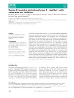
The child had b een healthy four months before. He
had been admitted to our hospital two months prior
due to dyspnea and hypoxia, and was diagnosed with a
pericardial effusion that ca used a tamponade on an
echocardiogram, causing right atrial and ventricular col-
lapse (Figure 1). Emergency percutaneous pericardial
catheter drainage was placed, draining 200 mL of serous
clear fluid. A culture from the pericardial effusion was
reported sterile; cytology reported no microorganisms or
leukocyt es, but an increased level of proteins (3400 mg/
dL). Acid-fast stain results were negative, as were the
results of adenosine deaminase testing. Rheumatoid tests
were also performed: anti-double- stranded DNA results
were negative and serum complement (C3 and C4)
levels were within normal limits. HIV rapid test and
serum antibody (IgM) tests for cytomegalovirus and
Epstein Barr virus were negative as well. Coxsackie ser-
ology was not available. Hypoalbuminemia was also
noted at 2 g/dL (reference range 3.4 to 4.8 g/dL) at
admission, associated with proteinuria. Tests were
inconclusive and the pericardial effusion was considered
to be viral. Because of his overall health improvement,
he was discharged from hospital o ne month later, with
follow-up planned.
Our patient did not have any important family,
traumatic, or surgical history. He had had viral hepa-
titis three years previously. No allergies or medication
taking were reported. He had normal psychomotor
development, and vaccinations had been given on
schedule.
On physical examination, his vital signs were within
normal limits and he was not febrile. His weight was
13.6 kg and his height 98 cm, considered low stature for
his age (Z = -3.9). A thyroid palpation revealed no goi-
ter, nodules or pain. Heart sounds were rhythmic and
no pericardial rub was heard. An anterior mid-thoracic
scar was noted where the percutaneous pericardial
catheter had been placed. He had hepatomegaly that
was later confirmed on an ultrasound. Results of an
anterior chest X-ray were normal.
Hemat ology tests on admission revealed a total leuko-
cyte count of 6.83 K/UL, hemoglobin level of 12.5 g/dL
and a platelet count of 310 K/μL. His blood chemistry
results reveale d elevated muscles enzymes: lactate dehy-
drogenase (LDH) of 1117 (reference range 240 to 480
IU/L), a creatine phosphokinase (CPK) of 2874 U /L
(reference range 38 to 170 U/L) and creatine kinase MB
(CK-MB) of 60 U/L (reference range 0 to 24 U/L). His
renal function tests were within normal limits. Hypertri-
gliceridemia was also noted at 408 mg/dL (reference
range 30 to 200 mg/dL). Urine test results showed 500
mg/dL proteinuria and microscopic analysis showed five
to eight erythrocytes per field. During the time our
patient was hospitalized, his CPK levels were moderately
increased (2435 to 4615 U/L). Our patient did not
report muscle pain or weakness, and results of a neuro-
logical examination were normal. A repeat echocardio-
gram was performed due to his history, showing normal
systolic and diastolic ventricular function with no peri-
cardial effusion.
Considering his recent history of cardiac tamponade,
thyroid tests were ordered. The results were diagnostic
of primary clinical hypothyroidism: free T3: < 1.1 pg/mL
(reference range 1.1 to 3 pg/mL), total T3: 34.9 ng/dL
(reference range 100 to 200 ng/dL), free T4: 0.2 ng/dL
Figure 1 Echocardiogram showing a large anterior and posterior pericardial effusion causing right atrial and ventricular collapse.
Delgado Hurtado et al. Journal of Medical Case Reports 2011, 5:515
/>Page 2 of 4
(reference range 0.7 to 1.85 ng/dL), total T4: 1.04 μg/dL
(reference range 4.5 to 12 μg/dL) and thyrotropin
299.380 μIU/mL (reference range 0.490 to 4.670 μIU/
mL). A thyroid scan with technetium (Figure 1) and a
thyroid ultrasound were performed showing a normally
located and absorbing thyroid gland without nodules
(Figure 2). Anti-p eroxid ase (APO) and anti-thyroglobu-
lin antibody test results were negative. Levothyroxine
was started at a dose of 9 μg/kg per day. Bone age was
also calculated with a radiograph of the left hand and
was reported delayed by two years.
An electromyogram reported a myophatic inflamma-
tory process involving mainly proximal muscles com-
patible with myositis. A muscle biopsy was performed
that revealed colliquative necrosis without inflamma-
tory cell infiltration (Figures 3 and 4). An anti-Jo-1
antibody test result was negative. Rhabdomyolisis was,
however, considered to be autoimmune and steroids
were given. Three weeks after the thyroid hormones
and one week after steroids had been started, muscle
enzyme levels (CPK, CK-MB, and LDH) had returned
to normal levels.
Discussion
The cause of our patient’s hypothyroidism was not diag-
nosed. Case series and case reports have suggested that
there is a relationship between the severity and chroni-
city of hypothyroidism with the presence of pericardial
effusion [2], which can be assessed by the short stature
seen in our patient as well as the delayed bone age
reported.
Our patient was evaluated by a rheumatologist, who
considered that the rhabdomyolisis was prob ably due to
an autoimmune disease. Our patient had moderately ele-
vated muscle enzymes over a period of approximately
one month, and there was a great concern acute renal
failure might develop. He was given steroids and, fortu-
nately, one week later his muscle enzyme levels had
returned to normal. The cause of rhabdomyolysis was
not confirmed. Viruses such as Cocksackie can cause
pericarditis along with myositis. The fact that the mus-
cle biopsy had no inflammatory cell infiltration might
suggest a viral myositis. When a cardiac tamponade
occurs in a hypothyroid patient, a concomitant provoca-
tive factor can be associated (for example, viral pericar-
ditis) [10]. This could be the triggering infection that
Figure 2 Thyroid scan with technetium showing a normal absorbing thyroid.
Figure 3 Muscle biopsy. Eoshinophilic muscle fibers showing
necrosis with no inflammatory cell infiltration.
Figure 4 Muscle fibers with colliquative necrosis.
Delgado Hurtado et al. Journal of Medical Case Reports 2011, 5:515
/>Page 3 of 4
exacerbated the cardiac tamponade in a previous
hypothyroidism-related pericardial effusion in our
patient. Hypothyroidism is a cause of rhabdomyolysis in
adults, but has been described in the pediatric popula-
tion as well [9].
Conclusions
Cardiac tamponade is a rare manifestation of hypothyr-
oidism. The early diagnosis of pericardial effusion due
to hypothyroidism may prevent serious complications
such as the cardiac tamponade seen in our patient. As
has been described in other case reports, treatment with
thyroid hormone replacement reduces the pericardial
effusion. The fact that our patient did not have any
common clinical manifestations of hypothyroidism
might have delaye d the diagnosis. Therefore, hypothyr-
oidism should be considered in every patient who pre-
sents with a pericardial effusion. M ultidisciplinary
involvement is important in the treatment of patients
with a difficult diagnosis.
Consent
Written informed consent was obtained from the
patient’ s legal guardian for publication of this case
report and any accompanying images. A copy of the
written consent is available for review by the Edito r-in-
Chief of this journal.
Acknowledgements
We would like to thank Dr Elizabeth Orellana of the Pathology Department
of Esperanza Hospital, Universidad Francisco Marroquin for providing the
photograph of the biopsy as well as the Cardiovascular Surgery Unit
(UNICAR) for providing the original echocardiogram report along with the
photograph. Matt Hodgkinson helped editing the original manuscript.
Author details
1
Universidad Francisco Marroquín, 19 Ave. 8-44 Zona 15, Vista Hermosa I,
Guatemala City, Guatemala.
2
Department of Pediatrics, Roosevelt Hospital,
5ta Avenida Calzada Roosevelt, Zona 11, Guatemala City, Guatemala.
Authors’ contributions
JJD wrote the case report. WG was the doctor in charge of our patient and
revised the case report. ER revised the case report. CL and SS were
consulting physicians and revised the case report. All authors read and
approved the final manuscript.
Competing interests
The authors declare that they have no competing interests.
Received: 6 July 2011 Accepted: 10 October 2011
Published: 10 October 2011
References
1. Bhupathi R, Kothari SS, Gupta AK, Menon PS: Cardiac function in
hypothyroid children: effect of replacement therapy. Indian Pediatr 1999,
36:779-784.
2. Farooki ZQ, Hoffman WH, Perry BL, Green EW: Myocardial dysfunction in
hypothyroid children. Am J Dis Child 1983, 37:65-68.
3. Hayford JT, Schieken RM, Thompson RG: Cardiac function in primary
hypothyroidism. Am J Dis Child 1980, 34:556-559.
4. Bereket A, Yang TF, Dey S, Blethen SL, Biancaniello TM, Wilson TA: Cardiac
decompensation due to massive pericardial effusion. A manifestation of
hypothyroidism in children with Down’s syndrome. Clin Pediatr (Phila)
1994, 33:749-745.
5. Sanda S, Newfield RS: A child with pericardial effusion and cardiac
tamponade due to previously unrecognized hypothyroidism. J Natl Med
Assoc 2007, 99:1411-1413.
6. Ladenson PW: Recognition and management of cardiovascular disease
related to thyroid dysfunction. Am J Med 1990, 88:638-641.
7. Parving A, Ostri B, Bretlau P, Hansen JM, Parving HH: Mechanisms of
edema formation in myxedema-increased protein extravasation and
relatively slow lymphatic drainage. N Engl J Med 1979, 301:460-465.
8. Sternbach GL: Pericarditis. Ann Emerg Med 1988, 17:214-220.
9. Galli-Tsinopoulou A, Stylianou C, Kokka P, Panagopoulou P, Nousia-
Arvanitakis S: Rhabdomyolysis, renal failure, pericardial effusion, and
acquired von Willebrand disease resulting from hypothyroidism in a 10-
year-old girl. Thyroid 2008, 18:373-375.
10. Manolis A, Varriale P, Ostrowski RM: Hypothyroid cardiac tamponade. Arch
Intern Med 1987, 147:1167-1169.
doi:10.1186/1752-1947-5-515
Cite this article as: Delgado Hurtado et al.: Hypothyroidism in a five-
year-old boy with rhabdomyolysis and recent history of cardiac
tamponade: a case report. Journal of Medical Case Reports 2011 5:515.
Submit your next manuscript to BioMed Central
and take full advantage of:
• Convenient online submission
• Thorough peer review
• No space constraints or color figure charges
• Immediate publication on acceptance
• Inclusion in PubMed, CAS, Scopus and Google Scholar
• Research which is freely available for redistribution
Submit your manuscript at
www.biomedcentral.com/submit
Delgado Hurtado et al. Journal of Medical Case Reports 2011, 5:515
/>Page 4 of 4