báo cáo khoa học: "Aberrant DNA methylation of cancer-related genes in giant breast fibroadenoma: a case report" docx
Bạn đang xem bản rút gọn của tài liệu. Xem và tải ngay bản đầy đủ của tài liệu tại đây (1.42 MB, 4 trang )
CAS E REP O R T Open Access
Aberrant DNA methylation of cancer-related
genes in giant breast fibroadenoma: a case
report
Diego M Marzese
1,2
, Francisco E Gago
2,3
, Javier I Orozco
2,3
, Olga M Tello
3
, María Roqué
1
and
Laura M Vargas-Roig
2,4*
Abstract
Introduction: Giant fibroadenoma is an uncommon variant of benign breast lesions. Aberrant methylation of CpG
islands in promoter regions is known to be involved in the silencing of genes (for example, tumor-suppressor
genes) and appears to be an early event in the etiology of breast carcinogenesis. Only hypermethylation of
p16INK4a has been reported in non-giant breast fibroadenoma. In this particular case, there are no previously
published data on epigenetic alter ations in giant fibroadenomas. Our previous results, based on the analysis of 49
cancer-related CpG islands have confirmed that the aberrant methylation is specific to malignant breast tumors
and that it is completely absent in normal breast tissue and breast fibroadenomas.
Case presentation: A 13-year-old Hispanic girl was referred after she had noted a progressive development of a
mass in her left breast. On physical examination, a 10 × 10 cm lump was detected and axillary lymph nodes were
not enlarged. After surgical removal the lump was diagnosed as a giant fibroadenoma. Because of the high growth
rate of this benign tumor, we decided to analyze the methylation status of 49 CpG islands related to cell growth
control. We have identified the methylation of five cancer-related CpG islands in the giant fibroadenoma tissue:
ESR1, MGMT, WT-1, BRCA2 and CD44.
Conclusion: In this case report we show for the first time the methylation analysis of a giant fibroadenoma. The
detection of methylation of these five cancer-related regions indicates substantial epigenomic differences with
non-giant fibroadenomas. Epigenetic alterations could explain the higher growth rate of this tumor. Our data
contribute to the growing knowledge of aberrant methylation in breast diseases. In this particular case, there exist
no previous data regarding the role of methylation in giant fibroadenomas, considered by definition as a benign
breast lesion.
Introduction
Fibroadenoma represents the most frequent breast
lesion in adolescents and young women with the giant
fibroadenoma (GF) being an uncommon variant. GFs,
which occur mostly in adolescent girls, are characterized
by their large size (more than 5 cm). They are encapsu-
lated masses and generally asymptomatic. Their rapid
growth (between two and five months) is associated
with skin congestion and ocasionally ulceration. It is
thoughtthatincreasedestrogen receptor sensitivity is
responsible for the etiology of GF [1].
Aberrant methylation of CpG islands (CpGIs) in pro-
moter regions is known to be involved in the silencing of
tumor-suppressor genes, steroid receptors, cell adhesion
molecules and cell cycle regulator genes and appears to
be an early event in the etiology of breast carcinogenesis
[2]. The aberrant methylation of cell cycle regulator
genes leads to a higher proliferation rate [3].
Our previous results, based on the analysis of 49 can-
cer-related CpGIs, have confirmed that the aberrant
methylation is specific to malignant breast tumors and
that it is completely absent in normal breast tissue and
breast fibroadenomas [4]. Other authors have reported
* Correspondence:
2
School of Medical Sciences, National University of Cuyo, Parque General San
Martín s/n, CP 5500, Mendoza, Argentina
Full list of author information is available at the end of the article
Marzese et al. Journal of Medical Case Reports 2011, 5:516
/>JOURNAL OF MEDICAL
CASE REPORTS
© 2011 Marzese et al; licensee BioMed Cent ral Ltd. This is an Open Access article distri buted under the terms of the Creative Commons
Attribution License ( .0), which perm its unrestricted use, distribution, and reproduction in
any mediu m, provided the original work is properly cited.
aberrant methylation of p16INK4a not only in malignant
breast lesions but also in fibroadenoma and normal
mammary tissues [5]. There are no previous data of epi-
genetic alterations in giant fibroadenomas. The estab-
lished precursors of breast carcinoma are atypical ductal
hyperplasia, ductal carcinoma in situ , and lobular neo-
plas ia. The malignant transformation of a fibroadenoma
is a rare event, with about 1 00 cases reported in the
world literature. Despite this fact we decided to analyze
the methylation status of a GF which is a rapidly grow-
ing benign breast lesion [ 6], because the methylation of
the analyzed genes is associated with a greater capacity
for cell growth [3].
Case presentation
A 13-year-old Hispanic girl was referred after she had
noted the progressive development of a mass in her left
breast. On physical examination, a 10 × 10 cm lump
was detected. Her axillary lymph nodes were not
enlarged. Surgery was performed and a GF was
removed. At present, with a follow-up of three years,
both breasts are symmetrical, normally developed, and
no signs of recurrence have been detected at clinical
evaluations.
Methylation-specific multiplex ligation-dependent
probe amplification (MS-MLPA) assay was performed
on the DNA obtained from the GF to study the methy-
lation status of the 49 CpGIs (Table 1). We have pre-
viously analyzed these regions in invasive ductal
carcinomas, breast fibroadenomas and normal mammary
tissue [4]. The MS-MLP A Kits ME001 and ME002 were
used accord ing to the manufa cturer’s recommendations
(MRC-Ho lland, Amsterdam, Netherlands) with minimal
modifications [4].
The immunohistochemical procedure was performed
as reported prev iously using the monoclonal antibody
clone ER88 (Biogenex, CA, USA) against estrogen recep-
tor alpha protein [7].
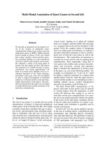
We have detected aberrant meth ylation in five cancer-
related CpGIs, that is estrog en receptor-a [ESR1
(+244bp)], O6-methylguanine-DNA m ethyltransferase
[MGMT (-463bp)], Wilms’ Tumor-1 [WT-1 (-146bp)],
Breast Cancer 2 [BRCA2 (+138bp)] and Hermen Anti-
gen [CD44 (+28bp)] (Figure 1). As a control we have
analyzed six normal breast tissues and three breast
fibroadenomas from 21-, 23- and 29-year-old patients.
None of these samples showed methylation in any of
the 49 CpGIs.
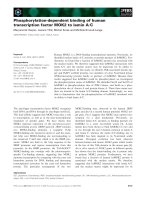
In order to evaluate the effect of the aberrant methyla-
tion on the level of protein expression in the fibroade-
noma, we investigated the expression of ER a protein
observing a moderate intensity in only 15% of the
fibroadenoma epithelial cells (Figure 2).
Discussion
To the best of our knowledge, the only reported ab er-
rant methylation in fibroadenomas is in gene p16INK4a.
Our previous results analyzing a 49-gene regions panel
which does not include the same reported CpGI of
p16INK4a- have not revealed aberrant methylation in
benign breast lesions [4,5].
Our finding of five aberrant methylated regions in the
reported GF suggests that this type of fibroadenoma
presents a different etiology than other benign breast
lesions, at least regarding the methylation profile.
In invasive breast tumors we have detected from two
to 23 aberrantly methylated cancer-related regions,
which indicates that five affected CpGIs is not a high
number for a breast carcinoma (unpublished data). The
surprising novelty, however, is that this finding occurs
in a benign lesion.
These five aberrant methylated genes play diverse
functions in the cell: DNA reparation (MGMT and
BRCA 2), cell cycle control (BRCA2, WT1), proliferation
(WT1, ESR1) and cell adhesion (CD44). The methyla-
tion of three of them (ESR1, MGMT and WT1) has
Table 1 CpG Islands analyzed
Gene Region Gene Region Gene Region Gene Region Gene Region
1 APC -21 bp 11 CDH13 186 bp 21 IGSF4 -56 bp 31 p73 +258 bp 41 RASSF1 +46 bp
2 ATM +309 bp 12 CHFR -103 bp 22 IGSF4 -294 bp 32 p73 +25 bp 42 RB1 -226 bp
3 ATM +138 bp 13 CHFR -96bp 23 MGMT -463 bp 33 PAX5 -120 bp 43 RB1 -449 bp
4 BRCA1 -20bp 14 DAPK1 +527 bp 24 MLH1 +55 bp 34 PAX6 -52 bp 44 STK11 +416 bp
5 BRCA1 +86bp 15 ESR1 +244 bp 25 MLH1 -320 bp 35 PTEN -813 bp 45 THBS1 -791 bp
6 BRCA2 +221 bp 16 FHIT +225 bp 26 p15 +473 bp 36 PTEN -66 bp 46 TIMP3 +1019 bp
7 BRCA2 +138 bp 17 GATA5 +271 bp 27 p16 -817 bp 37 PYCARD +437 bp 47 VHL +115 bp
8 CASP8 +291bp 18 GSTP1 +148 bp 28 p16 +200 bp 38 RARb -357 bp 48 VHL -3 bp
9 CD44 +411 bp 19 GSTP1 +468 bp 29 P27 +307 bp 39 RARb -180 bp 49 WT1 -210 bp
10 CD44 +28 bp 20 HIC1 -6 bp 30 P53 +100 bp 40 RASSF1 -136 bp
The table shows the 49 genomic regions tested during the study. Positive and negative signs are related to the transcription start base pair.
Marzese et al. Journal of Medical Case Reports 2011, 5:516
/>Page 2 of 4
been widely reported in breast tumors [2,4,8]. Methyla-
tion of WT1 has not been found in normal tissue [9].
Previous studies have reported the methylation of
BRCA2 in breast tumor but t o the best of our knowl-
edge, our stud y is the first to find methylated BRCA2 in
benign breast disease [10]. Regarding gene CD44, as far
as we know, its methylation status has not been
reported in mammary tissue before, even though new
evidence suggests its methylation in the breast cancer
cell line MCF7 [11]. Methylation of the ESR1 promoter
and its first exon has been observed to be correlated
with loss of the expression of ERa receptor, even
though some breast cancer specimens maintain its
expression (ER+) [12-14]. Tests based on ERa staining
in fibroadenoma reveal a pronounced heterogeneity
(range between 1% and 85%) showing no age correlation
[15]. Our specimen expresses 15% of ERa protein,
which is considered low. Even though we are not able
to establish the percentage of methylated ESR1 genes in
the GF, given its heterogeneity, this low protein expres-
sion is in accordance with the determined methylated
gene profile. The methylation of these five regions could
be responsible in part for the high growth rate present
in the analyzed GF.
Conclusions
Our data contribute to the growing knowledge of aber-
rant methylation in breast diseases. In this particular
casetherewerenopreviouslypublisheddataregarding
the role of methylation in GFs, considered by definition
to be a benign breast lesion. These findings should be
taken into account to evaluate whether it is associated
Figure 1 DetectionofaberrantDNAmethylationinthegiantfibroadenoma. A: MS-MLPA analysis of DN A isolated from non-giant
fibroadenoma. None of the analyzed regions are methylated. Only the PCR products from control probes are detected. B and C: MS-MLPA
analysis of DNA isolated from the giant fibroadenoma. The methylation specific peaks are marked with an asterisk (*). Panel B shows the
presence of methylation in BRCA2, CD44 and ESR1 genes and panel C shows the methylation of WT1, ESR1 and MGMT genes.
Marzese et al. Journal of Medical Case Reports 2011, 5:516
/>Page 3 of 4
with the different etiology of non-GFs and GFs. Furth er
studies will be necessary to draw more definitive conclu-
sions about the meaning of the methylation de-regula-
tion in this type of disease.
Consent
Written informed consent was obtained from the
patient’s next-of-kin for publication of this case report
and any accompanying images. A copy of the written
consent is available for review by the Editor-in-Chief of
this journal.
The study was approved by the Bioethics Committee
of the School of Medical Sciences, National University
of Cuyo, Mendoza, Argentina.
Abbreviations
BRCA2: Breast Cancer 2; CD44: Hermen Antigen; CpGIs: CpG islands; ERα:
estrogen receptor α protein; ESR1: estrogen receptor-α; GF: giant
fibroadenoma; MGMT: O6-methylguanine-DNA methyltransferase; MS-MLPA:
Methylation-specific multiplex ligation-dependent probe amplification;
p16INK4a: Cyclin-dependent kinase inhibitor 2A; WT-1: Wilms’ Tumor-1
Acknowledgements
Funding for this study was provided by SECTyP, National University of Cuyo
(06-J343) and the School of Medical Sciences, National University of Cuyo,
Mendoza, Argentina.
Author details
1
Cellular and Molecular Laboratory, IHEM-CCT-CONICET, Parque General San
Martín s/n, CP 5500, Mendoza, Argentina.
2
School of Medical Sciences,
National University of Cuyo, Parque General San Martín s/n, CP 5500,
Mendoza, Argentina.
3
Gineco-Mamario Institute, San Lorenzo 536, CP 5500,
Mendoza, Argentina.
4
Tumor Biology Laboratory, IMBECU-CCT-CONICET,
Avda Adrian Ruiz Leal s/n, Parque General San Martín, CP 5500, Mendoza,
Argentina.
Authors’ contributions
DMM performed the methylation study and revised the manuscript critically.
FEG participated in the study design with JO. OT carried out the
pathological studies. MR participated in interpretation of data and revised
the manuscript critically. LMV-R designed the study and wrote the
manuscript. All the authors discussed the results and read and approved the
final manuscript.
Competing interests
The authors declare that they have no competing interests.
Received: 17 March 2011 Accepted: 18 October 2011
Published: 18 October 2011
References
1. Gobbi D, Dall’Igna P, Alaggio R, Nitti D, Cecchetto G: Giant fibroadenoma
of the breast in adolescents: report of 2 cases. J Pediatr Surg 2009, 44:
e39-41.
2. Agrawal A, Murphy RF, Agrawal DK: DNA methylation in breast and
colorectal cancers. Mod Pathol 2007, 20:711-721.
3. Jones PA, Baylin SB: The epigenomics of cancer. Cell 2007, 128:683-692.
4. Marzese DM, Gago FE, Vargas-Roig LM, Roque M: Simultaneous analysis of
the methylation profile of 26 cancer related regions in invasive breast
carcinomas by MS-MLPA and drMS-MLPA. Mol Cell Probes 2010,
24:271-280.
5. Di Vinci A, Perdelli L, Banelli B, Salvi S, Casciano I, Gelvi I, Allemanni G,
Margallo E, Gatteschi B, Romani M: p16(INK4a) promoter methylation and
protein expression in breast fibroadenoma and carcinoma. Int J Cancer
2005, 114:414-421.
6. Chintamani , Khandelwal R, Tandon M, Yashwant K, Kulshresthal P, Aeron T,
Bhatnagar D, Bansal A, Saxena S: Carcinoma developing in a
fibroadenoma in a woman with a family history of breast cancer: a case
report and review of literature. Cases Journal 2009, 2:9348.
7. Vargas-Roig LM, Cuello-Carrión FD, Fernández-Escobar N, Daguerre P,
Leuzzi M, Ibarra J, Gago FE, Nadin SB, Ciocca DR: Prognostic value of Bcl-2
in breast cancer patients treated with neoadjuvant anthracycline based
chemotherapy. Molecular Oncology 2008, 2:102-111.
8. Munot K, Bell SM, Lane S, Horgan K, Hanby AM, Speirs V: Pattern of
expression of genes linked to epigenetic silencing in human breast
cancer. Hum Pathol 2006, 37:989-999.
9. Loeb DM, Evron E, Patel CB, Sharma PM, Niranjan B, Buluwela L,
Weitzman SA, Korz D, Sukumar S: Wilms’ tumor suppressor gene (WT1) is
expressed in primary breast tumors despite tumor-specific promoter
methylation. Cancer Res 2001, 61:921-925.
10. Cucer N, Taheri S, Ok E, Ozkul Y: Methylation status of CpG islands at sites
-59 to +96 in exon 1 of the BRCA2 gene varies in mammary tissue
among women with sporadic breast cancer. J Genet 2008, 87:155-158.
11. Müller I, Wischnewski F, Pantel K, Schwarzenbach H: Promoter- and cell-
specific epigenetic regulation of CD44, Cyclin D2, GLIPR1 and PTEN by
methyl-CpG binding proteins and histone modifications. BMC Cancer
2010, 10:297.
12. Ottaviano YL, Issa JP, Parl FF, Smith HS, Baylin SB, Davidson NE: Methylation
of the estrogen receptor gene CpG island marks loss of estrogen
receptor expression in human breast cancer cells. Cancer Res 1994,
54:2552-2555.
13. Lapidus RG, Ferguson AT, Ottaviano YL, Parl FF, Smith HS, Weitzman SA,
Baylin SB, Issa J-PJ, Davidson NE: Methylation of estrogen and
progesterone receptor gene 5’ CpG islands correlates with lack of
estrogen and progesterone receptor gene expression in breast tumors.
Clin Cancer Res 1996, 2:805-810.
14. Hori M, Iwasaki M, Yoshimi F, Asato Y, Itabashi M: Determination of
estrogen receptor in primary breast cancer using two different
monoclonal antibodies, and correlation with its mRNA expression. Pathol
Int 1999, 49:191-197.
15. Shoker BS, Jarvis C, Clarke RB, Anderson E, Munro C, Davies MPA,
Sibson DR, Sloane JP: Abnormal regulation of the oestrogen receptor in
benign breast lesions. J Clin Pathol 2000, 53:778-783.
doi:10.1186/1752-1947-5-516
Cite this article as: Marzese et al.: Aberrant DNA methylation of cancer-
related genes in giant breast fibroadenoma: a case report. Journal of
Medical Case Reports 2011 5:516.
Figure 2 Immunostaining of ERa protein. The figure shows the
staining in the nuclei of a few epithelial cells of the giant
fibroadenoma (400x).
Marzese et al. Journal of Medical Case Reports 2011, 5:516
/>Page 4 of 4