báo cáo khoa học: " Molecular imaging of potential bone metastasis from differentiated thyroid cancer: a case report" docx
Bạn đang xem bản rút gọn của tài liệu. Xem và tải ngay bản đầy đủ của tài liệu tại đây (1.5 MB, 5 trang )
CASE REP O R T Open Access
Molecular imaging of potential bone metastasis
from differentiated thyroid cancer: a case report
Nora Sandu
1,2
, Gabriele Pöpperl
3
, Marie-Elisabeth Toubert
4
, Belachew Arasho
1,5
, Toma Spiriev
1
, Mikael Orabi
1
and
Bernhard J Schaller
1,5*
Abstract
Introduction: Molecular imaging of the spine is a rarely used diagnostic method for which only a few case reports
exist in the literature. Here, to the best of our knowledge we present the first case of a combination of molecular
imaging by single photon emission computer tomography and positron emission tomography used in post-
operative spinal diagnostic assessment.
Case presentation: We present the case of a 50-year-old Caucasian woman experiencing progressive spinal cord
compression caused by a vertebral metastasis of a less well differentiated thyroid cancer. Following tumor
resection and vertebral stabilization, total thyroidectomy was performed revealing follicular thyroid carcinoma pT2
pNxM1 (lung, bone). During follow-up our patient underwent five radioiodine therapy procedures (5.3 to 5.7 GBq
each) over a two-year period. Post-therapeutic I-131 scans showed decreasing uptake in multiple Pulmonary
metastases. However, following an initial decrease, stimulated thyroglobulin remained at pathologically increased
levels, indicating further neoplastic activity. F18 Fludeoxyglucose positron emission tomography, which was
performed in parallel, showed remaining hypermetabolism in the lungs but no hypermetabolism of the spinal
lesions correlating with the stable neurolo gical examinations. While on single photon emission computer
tomography images Pulmonary hyperfixation of I-131 disappeared (most likely indicating dedifferentiation), there
was persistent spinal hyperfixation at the operated level and even higher fixation at the spinal process of L3. Based
on the negative results of the spinal F18 fludeoxyglucose positron emission tomography, a decision was made not
to operate again on the spine since our patient was completely asymptomatic and the neurological risk seemed to
be too high. During further follow-up our patient remained neurologically stable.
Conclusions: Molecular imaging by F18 fludeoxyglucose positron emission tomography helps to exclude
metabolically active spinal metastases and to spare further risky surgery.
Introduction
Fluorine-18 fludeoxyglucose (FDG) positron emission
tomography (PET) is a well established diagnostic mod-
ality for standard oncological staging, restaging, and
treatment m onitoring evaluations, and has a major
impact on patient management [1-3]. A key issue that is
less well studied is the performance of FDG-PET in
accurately depicting bone metastases that would poten-
tially have a large effect on patient treatment [2,4].
Metastases to the sp ine represent a common problem in
large oncology centers and usually present a problem in
radiological diagnosis. The role of PET is still being
assessed in this context.
However, molecular imaging (MI) with FDG-PET
seems a go od additional state-of-the -art method to
demonstrate the viability of previously treated spinal
tumor metastasis or to differentiate malignant from
benign lesion s in the spine [2,4]. Additionally, PET may
help to find the sites of the most metabolic active
lesions for biopsy [2,5]. In thyroid cancer, PET MI is
useful in patients with metastat ic poorly differentiated
tumors with high thyroglobulin (Tg) levels and negative
131
I whole-body scan results [6,7].
Similar to the situation with other tumor types, it is
currently unclear whether FDG-PET is adequate in the
detection o f bone metastasis of thyroid cancer. We
* Correspondence:
1
Department of Neurological Surgery, Lariboisiere Hospital, Universities of
Paris, Paris, France
Full list of author information is available at the end of the article
Sandu et al. Journal of Medical Case Reports 2011, 5:522
/>JOURNAL OF MEDICAL
CASE REPORTS
© 2011 Sandu et al; licensee BioMed Central Ltd. This is an Open Access article distributed under the terms of the Creative Commons
Attribution License ( which permits unre stricted use, distri bution, and reproduction in
any medium, provided the original work is pro perly cited.
describe one of the very few reported clinical cases with
vertebral metastases of a less well differentiated follicular
thyroid carcinoma followed by FDG-PET and I-131 sin-
gle photon emission computer tomography (SPECT).
The unique feature of this case is that the follow-up was
performed b y FDG-PET and SPECT, and we can there-
fore compare the results of these two MI mod alities. MI
by FDG-PET helped to exclude a metabolically active
spinal metastasis.
Case report
We present the case of a 50-year-old Caucasian woman
with a vertebral metastasis of a less well differentiated
thyroid cancer, who was followed over a three-year per-
iod clinically and by spinal FDG-PET and I-131 SPECT
imaging after initial surgery. Table 1 chronologically
summarizes the treatment modalities, corresponding
laboratory test values (thryotropin (TSH) and T g level)
and MI results (FDG-PET and I-131 SPECT) for differ-
ent time points during follow-up.
Our p atient presented to our facility with progressive
spinal cord compression. An MRI scan revealed a ver-
tebral metastasis at the T11 level with intraspinal exten-
sion compressing the spinal cord. Our patient was
operated on via a bilateral posterolateral approach,
allowing for tumor resection and stabilization of her
vertebral column by Cementoplasty and a posterior
arthrodesis. A histopathological examination concluded
‘ metastasis of a less well differentiated t hyroid carci-
noma’, which was confirmed after total thyroidectomy
(follicular thyroid carcinoma pT2 pNx). Following her
first radioiodine therapy a post-therapeutic scan revealed
multiple lung metastases and further bone metastases at
the L 3 level, os ilium and left femur; therefore the
tumor was staged as M1 (lung, bone). During follow-up
our patient received five radioiodine therapies (5.3 to 5.7
GBq each) in total over a two-year period.
During the follo w-up period our patient was regularly
monitored clinically and by means of a tumor marker
(thyroglobulin), PET-CT ([F-18]-FDG) and post-thera-
peutic SPECT (I-131). Clinically and neurologica lly our
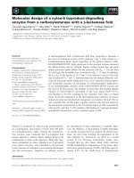
patient was stable over three years of follow-up. Post-
therapeutic radioiodine scans showed decreasing uptake
in most Pulmonary lesions but remaining uptake in spine
lesions (Figure 1). Her stimulated thyroglobulin blood
levels dropped from 2356 μg/L at baseline to 939 μg/L
following the last radioiodine treatment. However, even
after finishing five radioiodine cycles Tg remained on a
pathologically increased level, indica ting some neoplastic
activity. FDG-PET imaging showed slight but remaining
hypermetabolism in the lungs whereas in SPECT imaging
Pulmonary hyperfixation of I-131 disappeared, most
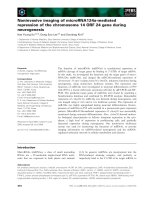
likely indicating dedifferentiation. On the spinal level,
SPECT images showed persistent hyperfixation at the
Table 1 Treatment modalities, corresponding laboratory values (TSH and Tg level) and MI results (FDG-PET and I-131
SPECT) for different time points during follow-up
Parameter Date (MM/YY format) and treatment
08/06 10/06 11/06 05/07 11/07 06/08 11/08 10/09 05/10
Treatment Cementoplasty
and posterior
arthrodesis
Total
thyroidectomy
First RIT
(GBq level
unknown)
Second RIT
(GBq level
unknown)
Third RIT (5.4
GBq)
Fourth RIT (5.5
GBq)
Fifth RIT (5.8
GBq)
Wait and
see
Wait
and
see
TSH (mIU/L) NA NA NA NA 103 119 130 NA < 0.02
Thyroglobulin
(μg/L)
NA NA 2357 805 891 1035 939 NA 606
I-131 SPECT NA NA Positive
uptake:
thyroid bed,
multiple
foci in the
lungs,
osseous
lesions T11,
L3, os ilium,
left femur
No uptake
in the
thyroid bed,
decreasing
uptake in
the lungs,
stable
uptake in
the osseous
lesions
No uptake in
the thyroid
bed, os ilium,
left femur
decreasing
uptake in the
lungs, stable
uptake in the
spine lesions
T11/L3
No uptake in
the thyroid
bed, os ilium,
left femur
decreasing
uptake in the
lungs, stable
uptake in the
spine lesions
T11/L3
No uptake in
the thyroid
bed, os ilium,
left femur
decreasing
uptake in the
lungs, stable
uptake in the
spine lesions
T11/L3
NA NA
FDG-PET NA NA NA NA NA NA Slight uptake
in Pulmonary
metastases, no
uptake in
spinal lesions
T11/L3
Slight
uptake in
Pulmonary
metastases,
no uptake
in spinal
lesions T11/
L3
NA
FDG = fludeoxyglucose; NA = not available; PET = positron emission tomography; RIT = radioiodine treatment; Tg = thyroglobulin; TSH = thryotropin; SPECT =
single photon emission computer tomography.
Sandu et al. Journal of Medical Case Reports 2011, 5:522
/>Page 2 of 5
operated level (T11) and even higher fixation at the
spinal process of L3 (Figure 2) suggestive for remaining,
more differentiated metastases. FDG-PET, however,
showed no hypermetabolism, which correlated with the
stable neurological examination results (Figure 3).
After thorough interdisciplinary discussion, despite the
remaining I-131 uptake it was decided not to operate
again on our patient’ s spine as she was completely
asymptomatic; conventional imaging also remained
stable and the neurological risk seemed to be too high
3
rd RIT 4th RIT 5th RIT
Figure 1 Planar I-131 whole-body scintigraphies after our patient’s third, fourth and fifth radioiodine treatments, demonstrating
decreasing uptake in the pulmonary metastases in the right and left lung parenchyma but stable uptake in the spinal lesions of Th11
and L3.
Figure 2 Single photon emission computer tomography (SPECT) I-131-CT demonstrating a persistent hyperfixation at the operated
level and even higher fixation at the spinal process of L3.
Sandu et al. Journal of Medical Case Reports 2011, 5:522
/>Page 3 of 5
for the thoracic level. During further follow-up our
patient remained neurologically stable.
Discussion
Detection of spinal metastasis by MI is a relatively new,
but clinically important te chnique. Cases such as our
patient’ s, where the different MI modalities can be
directly compared, are important to gain more experi-
ence in the different modalities for spinal MI and to per-
haps find special indications for the one or the other
method. In addition, our case report u nderlines the use-
fulness of FDG-PET in assessing the metabolic activity of
bone metastasis of less well differentiated thyroid cancer.
In our case report, in which different MI techniques
were used for the detection of distant metastases from
thyroid cancer, we were able to demonstrate different
behavior of the pulmonary and osseous lesions. While
the pulmonary nodes presented with decreased radioio-
dine uptake but increased FDG uptake indicating de-dif-
ferentiation, the spinal lesions showed stable radioiodine
uptake without FDG uptake, m ost probably indicating
stable disease. Subsequently, integrated I-131 SPECT/
CT w as found to have an additional value compared to
planar scintigraphy in patients with thyroid cancer for
correct characterization of equivocal tracer uptake seen
on planar imaging, as well as for precise localization of
Figure 3 18F-fludeoxyglucose positron emission tomography/computed tomography (FDG-PET-CT) demonstrating hypometabolism at
the spinal level correlating with the stable neurological examination.
Sandu et al. Journal of Medical Case Reports 2011, 5:522
/>Page 4 of 5
malignant lesions in the skeleton [8,9]. In our patient’ s
case these combined MI findings justified not operating
again on her spine; this turned out to be the right deci-
sion, since our pat ient remained neurologically stable
over further follow-up.
The FDG-PET examinations were performed under
stimulated TSH conditions to increase the diagnostic
sensitivity. It is known that TSH stimulates thyrocyte
metabolism, glucose transport and glycolysis. Since FDG
is a glucose analog, several studies have shown that
recombinant human TSH (rhTSH) stimulation improves
the detection of occult thyroid metastases with FDG-
PET, co mpared with scans performed on TSH suppres-
sion [10]. Beyond I-131 targeting the OPG/RANK/
RANKL axis may offer a nov el therapeutic approach for
malignant osteolytic pathologies [11], but currently
there are no such studies specifically for thyroid cancer
bone metastases.
Conclusions
The presence of bone metastas es alters the prognosi s of
patients with differentiated thyroid carcinoma. Our case
report underlines the fact that FDG-PET can have an
important impact on management in patients with thyr-
oid cancer.
Consent
Written informed consent was obtained from the patient
for publication of this case report and any accompany-
ing images. A copy of the writ ten consent is available
for review by the Editor-in-Chief of this journal.
Author details
1
Department of Neurological Surgery, Lariboisiere Hospital, Universities of
Paris, Paris, France.
2
Department of Neurological Surgery, University of
Lausanne, Lausanne, Switzerland.
3
Department of Nuclear Medicine, Hospital
of Stuttgart, Stuttgart, Germany.
4
Department of Nuclear Medicine, Hospital
of St. Louis, University of Paris, Paris, France.
5
Department of Neurology,
University of Addis Ababa, Addis Ababa, Ethiopia.
Authors’ contributions
NS, GP, MO and BS analyzed and interpreted the data from our patient
regarding the neurosurgical disease and the molecular. MET performed the
histological examination of the kidney, and together with NS, GP, MO, BA, TS
and BS was a major contributor to writing the manuscript. All authors read
and approved the final manuscript.
Competing interests
The authors declare that they have no competing interests.
Received: 4 July 2011 Accepted: 23 October 2011
Published: 23 October 2011
References
1. Hillner BE, Siegel BA, Liu D, Shields AF, Gareen IF, Hanna L, Stine SH,
Coleman RE: Impact of positron emission tomography/computed
tomography and positron emission tomography (PET) alone on
expected management of patients with cancer: initial results from the
National Oncologic PET Registry. J Clin Oncol 2008, 26:2155-2161.
2. Sandu N, Pöpperl G, Toubert ME, Spiriev T, Arasho B, Orabi M, Schaller BJ:
Current molecular imaging of spinal tumors in clinical practice. Mol Med
2011, 17:308-316.
3. Schaller B: Usefulness of positron emission tomography in diagnosis and
treatment follow-up of brain tumors. Neurobiol Dis 2004, 15:437-448.
4. Taira AI V, Herfkens RJ, Gambhir SS, Quon A: Detection of bone
metastases: assessment of integrated FDG PET/CT imaging. Radiology
2007, 243:204-211.
5. Even-Sapir E, Metser U, Mishani E, Lievhitz G, Lerman H, Leibovitch I: The
detection of bone metastases in patients with high-risk prostate cancer:
99 m Tc-MDP planar bone scintigraphy, single- and multi-field-of-view
SPECT, 18F-fluoride PET, and 18F-fluoride PET/CT. J Nucl Med 2006,
47:287-297.
6. Muresan MM, Oliver P, Leclere J, Sirveaux F, Brunaud L, Klein M, Zarnegar R,
Weryha G: Bone metastases from differentitated thyroid carcinoma.
Endocr Relat Cancer 2008, 15:37-49.
7. Al-Nahhas A, Khan S, Gogbashian A, Banti E, Rampin L, Rubello D: Review.
18F-FDG PET in the diagnosis and follow-up of thyroid malignancy. In
Vivo 2008, 22:109-114.
8. Tharp K, Israel O, Hausmann J, Bettman L, Martin WH, Daitzchman M,
Sandler MP, Delbeke D: Impact of 131I-SPECT/CT images obtained with
an integrated system in the follow-up of patients with thyroid
carcinoma. Eur J Nucl Med Mol Imaging 2004, 31:1435-1442.
9. Sandu N, Schaller B, Arasho B, Orabi M: Wallis interspinous implantation to
treat degenerative spinal disease: description of the method and case
series. Exp Rev Neurother 2011, 11:799-807.
10. Chin BB, Patel P, Cohade C, Ewertz M, Wahl R, Ladenson P: Recombinant
human thyrotropin stimulation of fluoro-D-glucose positron emission
tomography uptake in well-differentiated thyroid carcinoma. J Clin
Endocrinol Metab 2004, 89:91-95.
11. Fili S, Karalaki M, Schaller B: Mechanism of bone metastasis: the role of
osteoprotegerin and of the host-tissue microenvironment-related
survival factors. Cancer Lett 2009, 283:10-19.
doi:10.1186/1752-1947-5-522
Cite this article as: Sandu et al.: Molecular imaging of potential bone
metastasis from differentiated thyroid cancer: a case report. Journal of
Medical Case Reports 2011 5:522.
Submit your next manuscript to BioMed Central
and take full advantage of:
• Convenient online submission
• Thorough peer review
• No space constraints or color figure charges
• Immediate publication on acceptance
• Inclusion in PubMed, CAS, Scopus and Google Scholar
• Research which is freely available for redistribution
Submit your manuscript at
www.biomedcentral.com/submit
Sandu et al. Journal of Medical Case Reports 2011, 5:522
/>Page 5 of 5