báo cáo khoa học: " Intestinal adhesion due to previous uterine surgery as a risk factor for delayed diagnosis of uterine rupture: a case report" pot
Bạn đang xem bản rút gọn của tài liệu. Xem và tải ngay bản đầy đủ của tài liệu tại đây (384.33 KB, 3 trang )
CAS E REP O R T Open Access
Intestinal adhesion due to previous uterine
surgery as a risk factor for delayed diagnosis of
uterine rupture: a case report
Tomoyuki Kuwata
1,2*
, Shigeki Matsubara
1,2
, Rie Usui
1,2
, Shin-ichiro Uchida
1
, Naohiro Sata
3
and Mitsuaki Suzuki
1,2
Abstract
Introduction: Uterine rupture is a life-threatening condition both to mothers and fetuses. Its early diagnosis and
treatment may save their lives. Previous myomectomy is a high risk factor for uterine rupture. Intestinal adhesion
due to previous myomectomy may also prevent early diagnosis of uterine rupture.
Case presentation: A 38-year-old primiparous non-laboring Japanese woman with a history of myomectomy was
admitted in her 34
th
week due to lower abdominal pain. Although the pain was slight and her vital signs were
stable, computed tomography revealed massive fluid collection in her abdominal cavity, which led us to perform a
laparotomy. Uterine rupture had occurred at the site of the previous myomectomy; however, the small intestine
was adhered tightly to the rupture, thus masking it. The baby was delivered through a low uterine segment
transverse incision. The ruptured uterine wall was reconstructed.
Conclusion: Intestinal adhesion due to a prior myomectomy occluded a uterine rupture, possibly masking its
symptoms and signs, which may have prevented early diagnosis.
Introduction
Uterine rupture is a life-threatening condition both to
mothers and fetuses [1]. Early diagnosis of uterine rup-
ture and awareness of its risk factors are clinically
important. Previous uterinesurgery,suchasCesarean
section, myomectomy or adenomyomectomy, is a risk
factor [2-4]. Here, we report a prelabor uterine rupture
at a previous myoma enucleation site, in which intest-
inal adhesio n to the ruptured site occluded the rupture,
possibly preventing early diagnosis.
Case presentation
A 38-year-old Japanese primiparous woman with a his-
tory of myomectomy four years previously complained
of lower abdominal pain in her 34
th
week. This was her
second pregnancy with spontaneous conception, with
her first pregnancy resulting in spontaneous abor tion at
six weeks one year earlier. Her past history was unre-
markable except for a h istory of myomectomy, which
was performed for infertility (secondary sterility) for
approximately three years. Myomectomy was pe rformed
under laparotomy, and eight intramural myomas in the
uterine body were enucleated. The largest one (40 × 50
mm) existed in the anterior uterine body, which was
enucleated with vertical incision. The enucleation sites
had been reconstructed using routine two-layered
sutures. Her uterine cavity was not entered. Surgery
took 100 minutes and the total amount of hemorrhage
was 550 mL, requiring no transfusion. She had had an
uneventful postsurgery course without fever.
A physical examination revealed tenderness in the
middle of her lower abdomen without guarding. She
showed no vaginal bleeding. Her blood pressure was
106/64 mmHg, pulse rate 81 beats/min, white blood cell
count 9.2 × 10
9
/L, and hemoglobin 9.4 g/dL. She had
no postural hypotension. Cardi otocography (CTG) indi-
cated a r eassuring pattern with weak ut erine contrac-
tions once per hour. A vaginal and abdomi nal
ultrasound revealed no fluid retention in Pouch of Dou-
glas and no apparent uterine rupture; although no
detailed observation of uterine wall continuity was
* Correspondence:
1
Department of Obstetrics and Gynecology, Jichi Medical University, Tochigi,
Japan
Full list of author information is available at the end of the article
Kuwata et al. Journal of Medical Case Reports 2011, 5:523
/>JOURNAL OF MEDICAL
CASE REPORTS
© 2011 Kuwata et al; licensee BioMed Central Ltd. This is an Open Access article distributed under the terms of the Creative Commons
Attribution License ( which permits unrestricted use, distribution, and reproduction in
any medium, provid ed the original work is properly cited.
made. Slight abdominal pain continued with stable vital
signs and unremarkable laboratory data.
Six h ours later, she complained of upper abdominal pain.
Computed tomograph y (CT) revealed fluid accumulation
around her liver. Surgeons diagnosed this condition as
probable perforated viscus or at least acute abdomen
requiring laparotomy. CTG subsequently indicated recur-
rent late deceleration, re quiring an emergent Cesarean sec-
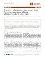
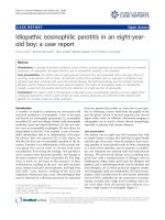
tion. Laparotomy revealed that her small intestine tightly
covered the anterior uterine wall, with bleedin g observed
from the edge of the intestinal covering (Figure 1, arrow).
After separating her small intestine, it became evident that
the anterior uterine wall, corresponding to the previous
myomectomy site, was ruptur ed, with her small intestine
tightly adhering to the ruptured site and thus nearly com-
pletely occluding the rupture (Figure 1). Pouch of Douglas
was not entered due to adhesion. A low segmental trans-
verse incision yielded a 2304-g female baby with Apgar
scores of 2, 4 and 7 at one, five and ten minutes, respec-
tively. Her small intestine was freed from the rupture site.
The 5-cm longitudinal rupture of the anterior uterine wall
was reconstructed. Her total blood loss during the surgery
was 3 750 mL, and s he received a transfusion w ith 2000 mL
hemoperitoneum, 1200 mL allogeneic blood and six units
of fresh frozen plasma. The m other and baby had an
uneventful course without sequelae.
Discussion
In our reported case, intestinal adhesion covered and
occluded a uterine rupture, which may have masked the
symptoms typical to uterine rupture, possibly preventing
early diagnosis. To the best of our knowledge, no pre-
vious report describes this phenomenon. The course of
our patient was considered to be as follows.
The rupture may have occurred around or before the
time of admission; however, the small intestine covering
the ruptured site may have prevented acute massive
bleeding, which may be why vital signs and laboratory
data were stable. Co vering by the small intestine may
have also prevented amniotic rupture or amniotic cavity
protrusion, which may explain the initial absence of a
fetal heart rate pattern indicative of c ord troubles. The
Pouch of Douglas was closed, possibly due to the pre-
vious laparotomy, prohibiting blood retention. The rup-
tured site bled continuously with the blood
accumulating around the liver, causing upper abdominal
pain. The rupture may have increased, causing fetal
heart rate pattern abnormalities.
Kurdoglu et al. [5] reported a uterine rupture case: the
rupture was considered to have occurred due to assisted
fundal pressure at delivery. The diagnosis w as made 32
hours postpartum; postural hypotension was the sign
that attracted the physicians’ attention, leading to the
diagnosis. The present case did not show postural hypo-
tension. Our patient remained lying in bed with little
postural change, which may explain why she showed no
postural hypotension.
Considering that the adhesion was very tight and that
adhesion to the myomectomy site is a frequently observed
phenomenon, the intestinal adhesion to the rupture may
Figure 1 Schematic diagram of the laparotomy findings. The uterine rupture was not initially discernable. Bleeding was observed from the
rupture edge (arrow). Her small intestine tightly adhered the anterior uterine wall. After separating the small intestine, uterine rupture became
evident; her small intestine covered and occluded the uterine rupture. Amniotic membrane beneath the rupture site remained intact.
Kuwata et al. Journal of Medical Case Reports 2011, 5:523
/>Page 2 of 3
have been present well before, and not after, the rupture.
Thus, uterine rupture occurred in the enucleation scar site
on which the small intestine tightly adhered.
A recent article also described uterine rupture
occluded by ‘ fetal legs’. Blihovde et al. [6] described a
prelabor primiparous uterine rupture at the 32
nd
week
of gestation, with the ruptured site being occluded by
the fetal legs. She had abdominal pain but without vagi-
nal bleeding, hemodynamical instability or fetal compro-
mise. The physicians suspected appendicitis; however,
CT revealed the uterine rupture occluded by the pro-
truding fetal legs from the ruptured site, which was con-
firmed by laparotomy. The fetal legs, protruding through
the rupture and occluding it, masked the symptoms and
signs of the rupture, delaying the diagnosis.
The article by Blihovde et al. [6] concluded, ‘clinicians
should consider the diagnosis of uterine rupture when a
patient presents with abdominal pain, e ven without evi-
dence of hypovolemia, vaginal bleeding, contractions, or
fetal compromise’. This statement is supported by the
present case. While intestinal adhesion covered and
delayed the diagnosis of the rupture in our case, fetal legs
had covered, and thus masked, the rupture in their case.
Previous uterine surgery is a well-known risk factor for
uterine rupture even before labor, as previously described
[2-4]. Previous myomectomy, inducing a tight intestinal
adhesion at the site, may mask the symptoms and signs
ofarupture.Wecannotexcludethepossibilitythat
intestinal adhesion might have been a coi ncidental phe-
nomenon. However, two patients were reported in whom
gastric peptic ulcer perforation was covered by the adhe-
sion of the abdominal wall to the perforation sites, which
masked typical symptoms and signs of gastric ulcer per-
foration [7]. We note the similarity between these two
cases a nd the present case. Although i t could not b e
determined whether intestinal adhesion delayed the diag-
nosis of rupture, we must consider this possibility in
pregnant women after myomectomy. Moreover, intest-
inal adhesion occurs not only after myomectomy but also
after any other abdominal surgeries, and thus we must be
cautious about this possibility in dealing with pregnant
women after abdominal surgery.
Conclusions
Myomectomy may be a risk factor for uterine rupture,
not only causing the rupture but also masking it and
thus preventing its early diagnosis.
Consent
Written informed consent was obtained from the patient
for publicatio n of this case report and any accompany-
ing images. A copy of the written consent is available
for review by the Editor-in-Chief of this journal.
Abbreviations
CT: computed tomography; CTG: cardiotocography.
Author details
1
Department of Obstetrics and Gynecology, Jichi Medical University, Tochigi,
Japan.
2
Jichi Perinatal Education Center, Jichi Medical University, Tochigi,
Japan.
3
Department of Surgery, Jichi Medical University, Tochigi, Japan.
Authors’ contributions
TK, SM, SU and RU diagnosed, investigated, followed-up and managed the
patient, and determined the medical significance. SM and TK wrote the
manuscript. TK and NS revised the manuscript. NS and MS provided
important suggestions regarding medical content. All authors read and
approved the final manuscript.
Competing interests
The authors declare that they have no competing interests.
Received: 1 June 2011 Accepted: 23 October 2011
Published: 23 October 2011
References
1. Gupta A, Nanda S: Uterine rupture in pregnancy: a five-year study. Arch
Gynecol Obstet 2011, 283(3):437-441.
2. Morimatsu Y, Matsubara S, Higashiyama N, Kuwata T, Ohkuchi A, Izumi A,
Shibhara H, Suzuki M: Spontaneous uterine rupture during pregnancy
soon after a laparoscopic adenomyomectomy. Reprod Med Biol 2007,
6(3):175-177.
3. Dubuisson JB, Fauconnier A, Deffarges JV, Norgaard C, Kreiker G,
Chapron G: Pregnancy outcome and deliveries following laparoscopic
myomectomy. Hum Reprod 2000, 15(4):869-873.
4. Dow M, Wax JR, Pinette MG, Blackstone J, Cartin A: Third-trimester uterine
rupture without previous cesarean: a case series and review of the
literature. Am J Perinatol 2009, 26(10):739-744.
5. Kurdoglu M, Kolusari A, Yildizhan R, Adali E, Sahin HG: Delayed diagnosis
of an atypical rupture of an unscarred uterus due to assisted fundal
pressure: a case report. Cases J 2009, 2:7966.
6. Blihovde L, Tawfik J, Hill DA: Prelabor third-trimester uterine rupture in an
unscarred uterus with occlusion by fetal small parts: a case report. J
Reprod Med 2010, 55(9-10):437-440.
7. Coulier B, Maldague P, Broze B: Gastric ulcer penetrating the anterior
abdominal wall: ultrasound diagnosis. Abdom Imaging 2003, 28:
(2):248-251.
doi:10.1186/1752-1947-5-523
Cite this article as: Kuwata et al.: Intestinal adhesion due to previous
uterine surgery as a risk factor for delayed diagnosis of uterine rupture:
a case report. Journal of Medical Case Reports 2011 5:523.
Submit your next manuscript to BioMed Central
and take full advantage of:
• Convenient online submission
• Thorough peer review
• No space constraints or color figure charges
• Immediate publication on acceptance
• Inclusion in PubMed, CAS, Scopus and Google Scholar
• Research which is freely available for redistribution
Submit your manuscript at
www.biomedcentral.com/submit
Kuwata et al. Journal of Medical Case Reports 2011, 5:523
/>Page 3 of 3