báo cáo khoa học: "Acute left ventricular dysfunction secondary to right ventricular septal pacing in a woman with initial preserved contractility: a case report" potx
Bạn đang xem bản rút gọn của tài liệu. Xem và tải ngay bản đầy đủ của tài liệu tại đây (1.12 MB, 5 trang )
CAS E REP O R T Open Access
Acute left ventricular dysfunction secondary to
right ventricular septal pacing in a woman with
initial preserved contractility: a case report
Sana Ouali
*
, Soufiene Azzez, Slim Kacem, Afef Lagren, Elyes Neffeti, Rim Gribaa, Fahmi Remedi and
Essia Boughzela
Abstract
Introduction: Right ventricular apical pacing-related heart failure is reported in some patients after long-term
pacing. The exact mechanism is not yet clear but may be related to left ventricular dyssynchrony induced by right
ventricular apical pacing. Right ventricular septal pacing is thought to deteriorate left ventricular function less
frequently because of a more normal left ventricular activation pattern.
Case presentation: We report the case of a 55-year-old Tunisian woman with preserved ventricular function,
implanted with a dual-chamber pacemaker for complete atrioventricular block. Right ventricular septal pacing
induced a major ventricular dyssynchrony, severe left ventricular ejection fraction dete rioration and symptoms of
congestive heart failure. Upgrading to a biventricular device was associated with a decrease in the symptoms and
the ventricular dyssynch rony, and an increase of left ventricular ejection fraction.
Conclusion: Right ventricular septal pacing can induce reversible left ventricular dysfunction and heart failure
secondary to left ventricular dyssynchrony. This complication remains an unpredictable complication of right
ventricular septal pacing.
Introduction
Right ventricular apical (RVA) pacing results in abnormal
left ventricular (LV) el ectrical and mechanical activation
and is associated with an increased risk of developing
heart failure [1-3]. Right ventricular septal (RVS) pacing
has been introduced to avoid this apparent and unpredict-
able complication of RVA pacing, because this pacing site
appears to deliver a more physiological electrical activation
of both ventricl es, visible with a shorter paced QRS com-
plex, than with RVA pacing [4,5].
We report the case of a 55-year-old Tunisian woman
with preserved ventricular function, implanted with a
dual-chamber pacemaker for comple te atrioventricular
block. RVS pacing induced a major ventricular dyssyn-
chrony, severe left ventricular ejection fraction deteriora-
tion and symptoms of congestive heart failure. Upgrading
to a biventricular device was associated with a decrease
in the symptoms and ventricular dyssynchrony, and
increased left ventricular ejection fraction (LVEF).
Case presentation
A 55-year-old Tunisian woman presented with syncope.
An electrocardiogram (ECG) upon admission showed
complete heart block with a narrow QRS complex (<120
ms) and an escape ventricular rate of 45 bpm. Our
patient’s medical history included arterial hypertension.
She did not have diabetes mellitus, and had no family his-
tory of coronary artery disease. A two-dimensional echo-
cardiography showed normal LV function with a 60% EF,
the absence of significant valvulopathy and no regional
wall motion abnormalities or pulmonary artery hyperten-
sion. A conventional dual chamber pacemaker (Medtronic;
Sensia SEDR01, US) was implanted with the right ventri-
cular (RV) lead positioned to her RV septum. The septal
position was confirmed by fluoroscopic images; defined as
a leftward orientation of the lead confirmed by 40° left
anterior oblique projection [6]. The electrocardiograp hic
criteria were defined as a negative deflection of lead I and
* Correspondence:
Department of Cardiology. Sahloul Hospital, Sousse, Tunisia
Ouali et al. Journal of Medical Case Reports 2011, 5:524
/>JOURNAL OF MEDICAL
CASE REPORTS
© 2011 Ouali et al; licensee BioMed Central Ltd. This is an Open Access article distributed under the terms of the Crea tive Commons
Attribution License (http://creativecommons.o rg/licenses/by/2.0), which permits unrestricted use, distribution, and reproduction in
any medium, provided the original work is properly cited.
positive initial R-waves of the paced ventricular complex
in lead ventricular fibrillation (VF) [7]. The pacemaker was
programmed in a DDD mode with lower rate of 50 bpm
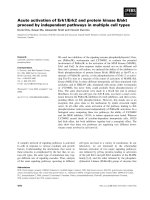
and upper tracking rate of 120 bpm. An ECG before dis-
charge showed atrial synchronized ventricular pacing with
a rate of 80 bpm and QRS duration of 160 ms (Fig ure 1).
Echocardiographic examination two days after pacemaker
implantation demonstrated a normal LV function (55%), a
LV end-diastolic volume (LVEDV) of 84 mL, the absence
of significant valvulopathy and an aortic pre-ejection per-
iod (PEP) of 160 ms. A ventricular dyssynchrony (80 ms
between septal and lateral electromec hanical delays) was
also measured with tissue Doppler imaging (TDI). The
ratio of E (peak transmitral flow velocity in early diastole)
to Ea (peak early diastolic myocardial velocity) velocity (E/
Ea) was estimated at 5.25. Our patient was readmitt ed
seven months later with six days of progre ssive dyspnea
(New York Heart Association (NYHA) class IV). Echocar-
diography showed severe LV akinesis, a depressed LVEF
(28%),aLVEDVof153mL,thepresenceofsignificant
mitral and tricuspid regurgitations (grade II-III), an aortic
PEP of 170 ms, pulmonary artery hypertension (50
mmHg) and an E/Ea ratio of 6. Her troponin level was not
raised. Coronary angiography revealed the absence of
significant obstructive epicardial coronary artery disease
(Figure 2) and left ventriculography demonstrated
depressed LVEF (25%).
Despite instauration of optimal medical therapy, our
patient remained at NYHA functional class III. She was
upgraded to a cardiac resynchronization therapy (CRT)-
device with implantation of a lateral left ventricular lead
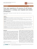
(Figure 3). After one month of CRT, symptoms and exer-
cise tolerance improved markedly from NYHA class III to
class II. A twelve-lead ECG showed QRS shortening after
CRT (Figure 4).
After one month, an echocardiography showed a
decrease in the aortic PEP (130 ms), LV reverse remodel-
ing, with a reduction of the LVEDV from 153 mL to
135 mL, and significant improvement in LVEF to 40%.
She had no symptoms of heart failure or syncope after-
wards and device int errogatio n showed that her cumula-
tive biventricular pacing was 100%.
Discussion
Pacing from RVS sites has been suggested as an alternative
to RVA pacing in an attempt to avoid long-term adverse
consequences on LV function [4]. This case illustrated the
rare phenomenon of rapid development of heart failure
and dramatic decrease of LVEF a fter short-term RVS
pacing for a complete atrioventricular block in a woman
with initially preserved LVEF. This case also sho wed the
reversible nature of RVS pacing-induced heart failure, and
that it may be related to the reversible LV dyssynchrony
induced by RVS pacing, as demonstrated by TDI and an
aortic PEP of 160 ms. There seems to be no other cause to
account for the heart failure in this woman except for RVS
pacing.
There is an increasing body of literature in which the
authors investigate the acute and chronic effects o f
RVS pacing on electrical and mechanical synchrony,
systolic and diastolic ventricular function and cardio-
vascular morbidity and mortality. Alternative RV
pacing sites appear advantageous when compared to
RVA pacing but their superiority has not been uni-
formly proven.
Ten Cate et al. [8] have demonstrated that acute abnor-
mal LV activation e ither forms RVA or RV outflow tract
(RVOT) pacing, resulting in an acute diminished LV func-
tion as assessed with echocardiographic wall motion score,
traced LVEF, electromechanical delay and regional longi-
tudinal LV strain. The authors have suggested that any RV
pacing sites can negatively affect LV function and that
readily available and non-invasive echocardiographic tech-
niques are not helpful to guide the selection of the indivi-
dual optimum pacing site during implantation. In the
same way, Ng et al. [9] demonstrated that standard fluoro-
scopic and electrocardiographic implantation techniques
for RVS pacing resulted in a heterogenous group of
Figure 1 Twelve lead ECG after DDD pacemaker implantation. Note the QRS morp hology with nega tive deflection of lead I and positive
initial R-waves of the paced ventricular complex in lead aVF.
Ouali et al. Journal of Medical Case Reports 2011, 5:524
/>Page 2 of 5
different pacing sites. They found that the patients with
RVS pacing had a lower LVEF, lower circumferential
strain and greater circumferential dyssynchrony than
those patients with RVA pacing, despite achieving a
narrower QRS complex. They concluded that these detri-
mental effects associated with RVS pacing might have
resulted from the heterogeneity of the real pacing sites
included under the umbrella of the RVS pacing concept.
Figure 2 Coronary angiogram of the left coronary artery. Fluoroscopic images at an anteroposterior (AP, Panel A), left anterior oblique (LAO,
Panel B), right anterior oblique (RAO, Panel C) and cranial (Cranial, Panel D) projection, showing the position of the active ventricular pacing lead
at the RV septal region (arrow). Note the proximity of the septal lead tip to the left anterior descending artery.
Figure 3 Anteroposterior (AP, Panel A) and left anterior oblique (LAO, Panel B) fluoroscopic projections showing leads position after
CRT.
Ouali et al. Journal of Medical Case Reports 2011, 5:524
/>Page 3 of 5
In patients with standard indications for pacing, the
prediction of heart failure is difficult a nd the exact
mechanism of RV pacing-related heart failure is not
clear but may be related to LV dyssynchrony induced by
RV pacing [10]. The best treatment option for these
patients remains to be determined. CRT seems to be
superior to RV pacing in patients with either impaired
[11] or preserved LV systolic function [12] and standard
pacing indication.
ThePacingtoAvoidCardiacEnlargementstudy[12]
showed that the mean LVEF declined by almost seven
percentage points (from 61.5 ± 6.6% to 54.8 ± 9.1%) in
the first year of RVA pacing in patients with a normal
ejection fraction. Among nine patients in whom the
LVEF decreased to less than 45% at 12 months, eight
(89%) were in the RV pacing group. The authors suggest
that the ejection fra ction could decrease rapidly in vul-
nerable patients and that these patients might benefit
even more from biventricular pacing [12].
Conclusion
RVS pacing can induce reversible LV dysfunction and
heart failure secondary to LV dyssynchrony. This remains
an unpredictable complication of RV pacing. It should be
highlighted that not all patients develop LV dyssynchrony
and new onset heart failure after RV pacing. Therefore,
early predictive factors [13-15], such as dyssynchrony at
the time of implantation, paced QRS width, age, presence
of atrial fibrillation, concomitant coronary artery disease,
compromised LVEF or antibody status, should be further
evaluated. These factors may reveal the pat ients who are
more prone to LV function deterioration and who are
consequently better candidates for biventricular pacing.
Consent
Written informed consent was obtained from the patient
for publicatio n of this case report and any accompany-
ing images. A copy of the written consent is available
for review by the Editor-in-Chief of this journal.
Authors’ contributions
SO was the major contributor in writing the manuscript. All authors read
and approved the final manuscript.
Competing interests
The authors declare that they have no competing interests.
Received: 20 April 2011 Accepted: 25 October 2011
Published: 25 October 2011
References
1. Leclercq C, Gras D, Le Helloco A, Nicol L, Mabo P, Daubert C:
Hemodynamic importance of preserving the normal sequence of
ventricular activation in permanent cardiac pacing. Am Heart J 1995,
129:1133-1141.
2. Wilkoff BL, Cook JR, Epstein AE, Dual Chamber and VVI Implantable
Defibrillator Trial Investigators: Dual chamber pacing or ventricular backup
pacing in patients with an implantable defibrillator: the Dual Chamber
and VVI Implantable Defibrillator (DAVID) Trial. JAMA 2002, 288:3115-3123.
3. Tops LF, Suffoletto MS, Bleeker GB, Boersma E, van der Wall EE, Gorcsan J III,
Schalij MJ, Bax JJ: Speckle-tracking radial strain reveals left ventricular
dyssynchrony in patients with permanent right ventricular pacing. JAm
Coll Cardiol 2007, 50(12):1180-1188.
4. Mond HG, Vlay SC: Pacing the right ventricular septum: time to abandon
apical pacing. Pacing Clin Electrophysiol 2010, 33(11):1293-1297.
Figure 4 Twelve lead ECG after CRT.
Ouali et al. Journal of Medical Case Reports 2011, 5:524
/>Page 4 of 5
5. Tse HF, Wong KK, Siu CW, Zhang XH, Ho WY, Lau CP: Upgrading
pacemaker patients with right ventricular apical pacing to right
ventricular septal pacing improves left ventricular performance and
functional capacity. J Cardiovasc Electrophysiol 2009, 20(8):901-905.
6. McGavigan AD, Roberts-Thomson KC, Hillock RJ, Stevenson IH, Mond HG:
Right ventricular outflow tract pacing: radiographic and
electrocardiographic correlates of lead position. Pacing Clin Electrophysiol
2006, 29(10):1063-1068.
7. Lieberman R, Grenz D, Mond HG, Gammage MD: Selective site pacing:
defining and reaching the s elected site. Pacing Clin Electrophysiol 2004,
27(6 Pt 2):883-886.
8. ten Cate TJ, Scheffer MG, Sutherland GR, Verzijlbergen JF, van Hemel NM:
Right ventricular outflow and apical pacing comparably worsen the
echocardioghraphic normal left ventricle. Eur J of Echocardiogr 2008,
9(5):672-677.
9. Ng AC, Allman C, Vidaic J, Tie H, Hopkins AP, Leung DY: Long-term impact
of right ventricular septal versus apical pacing on left ventricular
synchrony and function in patients with second- or third-degree heart
block. Am J Cardiol 2009, 103(8):1096-1101.
10. Fung JW, Zhang Q, Yip GW, Yu CM: Reversible left ventricular
dyssynchrony and heart failure induced by right ventricular pacing. Int J
Cardiol 2009, 134(1):117-119.
11. Höijer CJ, Meurling C, Brandt J: Upgrade to biventricular pacing in
patients with conventional pacemakers and heart failure: a double-blind,
randomized crossover study. Europace 2006, 8(1):51-55.
12. Yu CM, Chan JY, Zhang Q, Omar R, Yip GW, Hussin A, Fang F, Lam KH,
Chan HC, Fung JW: Biventricular pacing in patients with bradycardia and
normal ejection fraction. N Engl J Med 2009, 361(22):2123-2134.
13. Zhang XH, Chen H, Siu CW, Yiu KH, Chan WS, Lee KL, Chan HW, Lee SW,
Fu GS, Lau CP, Tse HF: New-onset heart failure after permanent right
ventricular apical pacing in patients with acquired high-grade
atrioventricular block and normal left ventricular function. J Cardiovasc
Electrophysiol 2008, 19(2):136-141.
14. Siu CW, Wang M, Zhang XH, Lau CP, Tse HF: Analysis of ventricular
performance as a function of pacing site and mode. Prog Cardiovasc Dis
2008, 51(2):171-182.
15. Sagar S, Shen WK, Asirvatham SJ, Cha YM, Espinosa RE, Friedman PA,
Hodge DO, Munger TM, Porter CB, Rea RF, Hayes DL, Jahangir A: Effect of
long-term right ventricular pacing in young adults with structurally
normal heart. Circulation 2010, 121(15):1698-1705.
doi:10.1186/1752-1947-5-524
Cite this article as: Ouali et al.: Acute left ventricular dysfunction
secondary to right ventricular septal pacing in a woman with initial
preserved contractility: a case report. Journal of Medical Case Reports
2011 5:524.
Submit your next manuscript to BioMed Central
and take full advantage of:
• Convenient online submission
• Thorough peer review
• No space constraints or color figure charges
• Immediate publication on acceptance
• Inclusion in PubMed, CAS, Scopus and Google Scholar
• Research which is freely available for redistribution
Submit your manuscript at
www.biomedcentral.com/submit
Ouali et al. Journal of Medical Case Reports 2011, 5:524
/>Page 5 of 5