Báo cáo y học: "Half-dose verteporfin photodynamic therapy for bullous variant of central serous chorioretinopathy: a case report" doc
Bạn đang xem bản rút gọn của tài liệu. Xem và tải ngay bản đầy đủ của tài liệu tại đây (627.23 KB, 3 trang )
CAS E REP O R T Open Access
Half-dose verteporfin photodynamic therapy for
bullous variant of central serous
chorioretinopathy: a case report
Winnie WK Ng, Zenith HY Wu and Timothy YY Lai
*
Abstract
Introduction: Central serous chorioretinopathy is characterized by serous neurosensory detachment of the macula
and it usually resolves spontaneously with good visual prognosis. In some patients, however, the serous retinal
detachment might be very extensive and can result in bullous exudative retinal detachment. We evaluated the use
of half-dose verteporfin photodynamic therapy for the treatment of bullous retinal detachment in idiopathic central
serous chorioretinopathy.
Case presentation: We report the case of a 51-year-old Chinese man who presented with blurred vision in his
right eye and superior visual field defect due to bullous variant of centr al serous chorioretinopathy . No
improvement in vision and retinal detachment was noted after three months of observation and a short course of
oral acet azolamide. He was then treated with half-dose verteporfin photodynamic therapy and his visual acuity
improved from 20/70 to 20/25 within one month of treatment. Three months after photodynamic therapy, there
was compl ete resolution of sub-retinal fluid and bullous retinal detachment. No recurrence of central serous
chorioretinopathy was noted in three years of follow-up.
Conclusion: We report the beneficial effect of photodynamic therapy with half-dose verteporfin as a treatment
option for bullous retinal detachment caused by central serous chorioretinopathy.
Introduction
Central serous chorioretinopathy (CSC ) is characterized
by serous neurosensory detachment of the macula and
it usually resolves spontaneously with good visual prog-
nosis. In some patients, however, the serous retinal
detachment might be very ext ensive and a large amount
of sub-retinal fluid can result in bullous exudative ret-
inal detachme nt [1]. Photodynamic therapy (PDT) with
verteporfin has been shown to be effective in the treat-
ment of CSC but the use of conventional dosage of ver-
teporfin (6 mg/m
2
) might be associated with
complications such as iatrogenic choroidal neovasculari-
zation, diffuse retinal epithelial atrophy and more severe
retinal thinning [2-4]. We report the use of half-dose (3
mg/m
2
) verteporfin PDT for treating a patient with the
severe bullous form of CSC.
Case Presentation
A 51-year-old Chinese man with good past health pre-
sented with a 10 day history of reduced vision and a
superior visual field defect of the right eye. He denied a
history of recent steroid use via any route. His best-cor-
rected visual acuity was 0.5 OD and 1.0 OS. An exami-
nation of the fundus showed inferior b ullous retinal
detachment in his right eye with yellowish fibrinous
exudates and retinal pigment epithelial (RPE) changes
without retinal break (Figure 1A). An examination of
the anterior segment and vitreous c avity showed an
absence of any inflammatory reaction suggestive of an
inflammatory cause such as Vogt-Koyanagi-Harada dis-
ease. A B-scan ultrasound confirmed inferior retinal
detachment (Figure 1B) and optical coherence tomogra -
phy (OCT) showed foveal involvement (Figure 1C).
Fluorescein angiography (FA) revealed early hyper-fluor-
escence with diffuse late leakage at the macula (Figure
1D) and indocyanine green angiography (ICGA) showed
dilated choroidal vessels with choroidal hyper-perfusion
* Correspondence:
Department of Ophthalmology & Visual Sciences, Hong Kong Eye Hospital,
The Chinese University of Hong Kong, Hong Kong
Ng et al. Journal of Medical Case Reports 2011, 5:208
/>JOURNAL OF MEDICAL
CASE REPORTS
© 2011 Ng et al; licensee BioMed Central Ltd. This is an Open Access article distributed under the terms of the Creative Commons
Attribution License ( which permits unre stricted use, distri bution, and reproduction in
any medium, provided the original work is properly cited.
consistent with CSC (Figure 1E). An examination of the
fundus of his left eye showed mild RPE changes at the
superior macula, with a mild RPE window defect on FA
and mildly dilated choroidal vessel on ICGA. The find-
ings in his left eye were consistent with resolved CSC.
A diagnosis of bullou s CSC was made and despite a
two-week course of oral acetazolamide (250 mg qid)
and observation for three months, the exudative retinal
detachment persisted and his right eye vision deterio-
ratedto0.3.Hesubsequentlyunderwenthalf-dose(3
mg/m
2
) verteporfin PDT with a spot size of 4500 μmto
cover the area of dilated choroidal vessels in ICGA. One
month after the PDT, his vision improved to 0.8 OD
with a reduction in inferior retinal detachment. After
three months, there was complete absence of sub-retinal
fluid (Figure 2A). A B-scan ultrasound and OCT con-
firmed the resolutio n of exudative retinal detachment
(Figures 2B and 2C). FA and ICGA showed reduced
leakage and choroidal hyperpermeability (Figures 2D
and 2E). He was followed for 38 months, during which
there was no recurrence and his final vision was 1.0 OD.
Discussion
BullousCSCisanuncommonformofCSCassociated
with a lar ge amount of sub-reti nal fluid. The visual prog-
nosis of bullous CSC is generally poorer than those lim-
ited to the macula, as recurrence is common even after
initial complete regression [1]. Traditional management
options of bullous CSC include observation or thermal
laser photocoagulation. However, previous studies have
shown that the outcome of thermal laser photocoagula-
tion is similar with the natural course of the disease in
terms of disease duration and final visual acuity [1].
In recent years, PDT with verteporfin has been uti-
lized for treating patients with CSC. However, full-dose
PDT for treating CSC is not without complications, as
retinal pigment epithelium atrophy, retinal thinning and
choroidal neovascular ization have been r eported after
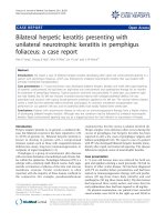
Figure 1 (A) Fundus photo of his right eye at presentation
showing exudative retinal detachment at the macula and
inferior retina. Fibrinous exudate can be seen at the inferior
arcade. (B) B-scan ultrasound axial scan showing inferior exudative
retinal detachment. (C) OCT imaging of the right macula showing
the presence of sub-retinal fluid involving the fovea. (D) Mid-phase
FA showing diffuse fluorescein leakage at the macula with RPE track
inferiorly. (E) Mid-phase ICGA showing dilated choroidal vasculature
with hyperdynamic circulation consistent with central serous
chorioretinopathy. Laser for PDT was applied to the area of
choroidal hyperpermeability as guided by ICGA (red circle).
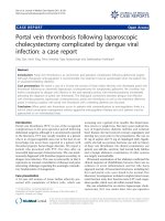
Figure 2 (A) Fundus photo of his right eye three months after
half-dose verteporfin PDT showing complete resolution of the
macular and inferior retinal detachments. (B) B-scan ultrasound
axial scan showing resolution of the inferior exudative retinal
detachment. (C) OCT imaging of the right macula showing absence
of sub-retinal fluid at the macula with thinning of the neurosensory
retina due to CSC. (D) Mid-phase FA showed diffuse RPE window
defect with staining due to sub-retinal fibrosis. (E) Mid-phase ICGA
showing absence of dilated choroidal vasculature.
Ng et al. Journal of Medical Case Reports 2011, 5:208
/>Page 2 of 3
PDT for chronic CSC [2-4]. In order to decrease t he
extent of collateral damage and the risk of adverse
events, a reduced dose of verteporfin has been used to
treat CSC, with the efficacy of PDT remained high
[3,5,6].
To the best of ou r knowledge, the long-term outcome
of half-dose PDT for bullous variant of CSC has not
previously been reported. Our case demonstrated that
this treatment is effective in reducing the bullous exuda-
tive retinal detachment and improving the patient’ s
vision rapidly. Moreover, there was no evidence of any
disease recurrence or c omplications after more than
three years of follow-up. The reasons for the successful
treatment outcome might be related to the idiopathic
cause o f the disease a nd intuition of early treatment in
our patient. Although the longer term complications
and recurrence rate remains unknown, half-dose verte-
porfin PDT for bullous CSC appeared to be an effective
treatment option and m ight be considered as a manage-
ment option for these patients.
Conclusion
Bullous exudative retinal detachment is an uncommon
manifestation of CSC and can result in significant visual
loss. Our patient demonstrated that half-dose vertepor-
fin PDT is an effective treatment option for bullous
CSC, resulting in rapid resolution of exudative retinal
detachment and improvement in vision.
Consent
Written informed consent was obtained from the patient
for publication of this manuscript and any accompany-
ing images. A copy of the written consent is availabl e
for review by the Editor-in-Chief of this journal.
Authors’ contributions
WWKN interpreted the patient data and wrote the first draft of the
manuscript. ZHYW had a role in writing a final draft of the manuscript and
in the preparation of clinical images. TYYL performed the treatment and
follow-up on the patient, and was a major contributor to the writing of the
manuscript. All authors read and approved the final manuscript.
Competing interests
The authors declare that TYYL has received honorium for lecture fees and
serving in the advisory board of Novartis Pharmapeutical Inc. The other
authors (WWKN, ZHYW) have no competing interests.
Received: 20 October 2010 Accepted: 26 May 2011
Published: 26 May 2011
References
1. Otsuka S, Ohba N, Nakao K: A long-term follow-up study of severe variant
of central serous chorioretinopathy. Retina 2002, 22(1):25-32.
2. Yannuzzi LA, Slakter JS, Gross NE, Spaide RF, Costa DL, Huang SJ,
Klancnik JM Jr, Aisman A: Indocyanine green angiography-guided
photodynamic therapy for treatment of chronic central serous
chorioretinopathy: a pilot study. Retina 2003, 23(3):288-298.
3. Lai TY, Chan WM, Li H, Lai RY, Liu DT, Lam DS: Safety enhanced
photodynamic therapy with half-dose verteporfin for chronic central
serous chorioretinopathy: a short term pilot study. Br J Ophthalmol 2006,
90(7):869-874.
4. Shin JY, Woo SJ, Yu HG, Park KH: Comparison of efficacy and safety
between half-fluence and full-fluence photodynamic therapy for chronic
central serous chorioretinopathy. Retina 2011, 31(1):119-126.
5. Chan WM, Lai TY, Lai RY, Liu DT, Lam DS: Half-dose verteporfin
photodynamic therapy for acute central serous chorioretinopathy: one-
year results of a randomized controlled trial. Ophthalmology 2008,
115(10):1756-1765.
6. Zhao MW, Zhou P, Xiao HX, Lv YS, Li CA, Liu GD, Li XX: Photodynamic
therapy for acute central serous chorioretinopathy: the safe effective
lowest dose of verteporfin. Retina 2009, 29(8):1155-1161.
doi:10.1186/1752-1947-5-208
Cite this article as: Ng et al.: Half-dose verteporfin photodynamic
therapy for bullous variant of central serous chorioretinopathy: a case
report. Journal of Medical Case Reports 2011 5:208.
Submit your next manuscript to BioMed Central
and take full advantage of:
• Convenient online submission
• Thorough peer review
• No space constraints or color figure charges
• Immediate publication on acceptance
• Inclusion in PubMed, CAS, Scopus and Google Scholar
• Research which is freely available for redistribution
Submit your manuscript at
www.biomedcentral.com/submit
Ng et al. Journal of Medical Case Reports 2011, 5:208
/>Page 3 of 3