Báo cáo y học: " Pitfalls in the diagnosis of a tumefactive demyelinating lesion: A case report" potx
Bạn đang xem bản rút gọn của tài liệu. Xem và tải ngay bản đầy đủ của tài liệu tại đây (936.56 KB, 4 trang )
CAS E REP O R T Open Access
Pitfalls in the diagnosis of a tumefactive
demyelinating lesion: A case report
Maria Gavra
1*
, Efstathios Boviatsis
2
, Lampis C Stavrinou
2
and Damianos Sakas
2
Abstract
Introduction: In rare instances, demyelinating disorders manifest as tumefactive lesions that simulate brain tumors.
We report a patient with a space-occupying lesion in the parietal lobe, which presented a serious diagnostic
dilemma, between a rare tumefactive demyelinating disease, such as Balo concentric sclerosis and a glioma. This
case report highlights important diagnostic clues in the differential diagnosis of Balo concentric sclerosis.
Case presentation: A 20-year-old Caucasian woman with acute onset of left-sided weakness and numbness was
admitted to hospital with neurologic signs of left-sided hemiparesis and hypoesthesia. Brain magnetic resonance
imaging showed a mass lesion of abnormal signal intensity with concentric enhancing rings in the right parietal
lobe, without perifocal edema. The characteristic concentric pattern detected on the magnetic resonance images
was highly suggestive of Balo disease, and corticosteroids were administered. Evoked potentials, cerebrospinal fluid
analysis, and magnetic spectroscopy findings were not specific, and glioma was also included in the differential
diagnosis. A stereotactic biopsy was not diagnostic.
After one month the patient showed moderate clinical improvement, and during 12 months follow-up, no further
relapses occurred. In the follow-up magnetic resonance imaging, the concentric pattern had completely
disappeared, and only a low-signal, gliotic lesion remained.
Conclusion: We hope this case prese ntation will advance our understanding of clinical and radiologic appearance
of Balo concentric sclerosis, which is a rare demyelinating disease. Although this is a specific entity, it has a broader
clinical impact across medicine, because it must be differentiated from other space-occupying lesions in the central
nervous system.
Introduction
Tumefactive demyelinating brain lesions present a diag-
nostic challenge, because their clinical, radiologic, and
even histologic fe atures may complicate the identifica-
tion of their t rue nature. This often leads to invasive
and costly procedures, which frequently yield non-diag-
nostic results. We report a patient with a right parietal
white matter lesion, who presented a serious diagnostic
dilemma, as the lesion was difficult to differentiate
between a rare demyelinating disease such as Balo con-
centric sclerosis (BCS) and a glioma. The characteristic
magnetic resonance findings of the case, its acute onset,
and its clinical improvement after corticosteroid therapy
finally set the diagnosis of BCS. The risks of the
stereotactic procedures that led to the misdiagnosis of
BCS are discussed.
Case presentation
A 20-year-old Caucasian woman, with no p ast medical
history, presented to the emergency room of a general
hospital, with numbness and weakness of her left-sided
limbs. Neurologic examination revealed no cranial nerve
deficit and 4/5 left-sided hemiparesis. No cerebellar
impairment was noted. She was unable to localize tactile
stimuli or to judge objects’ size and shape. She had no
pain, pressure, or temperature loss. Brain computed
tomography (CT) demonstrated a large (2.1 cm), well-
demarcated hypodense lesion in the right parietal lobe,
without perifocal edema. Magnetic resonance imaging
(MRI) without contrast showed a h ypoisointense con-
centric mass on T
1
-andhyperintenseonT
2
-weighted
images (Figure 1a, b). After contrast, the lesion appeared
* Correspondence:
1
Department of CT and MRI, Children’s Hospital, “Agia Sophia’’, Thivon and
Papadiamantopoulou Street, Athens, Greece
Full list of author information is available at the end of the article
Gavra et al. Journal of Medical Case Reports 2011, 5:217
/>JOURNAL OF MEDICAL
CASE REPORTS
© 2011 Gavra et al; licensee BioMed Central L td. This is an Open Access article distributed under the terms of the Creative Commons
Attribution License ( which permits unrestricted use, distribution, and reproduction in
any medium, provided the original work is properly cited.
to enhance inhomogeneously, in a pattern resembling
separate, alternating enhancing rings (Figure 1c). These
MRI findings were highly suggestive of the concentric
pattern of demyelination (BCS).
Somatosensory evoked potentials ( SSEPs), serum, and
cerebros pinal fluid analysis were normal. Human immu-
nodeficiency virus (HIV) and antinuclear antibody
(ANA) tests were negative. The chest radiograph (CXR)
was normal. Under the presumptive diagnosis of BCS,
the patient received high-dose intravenously adminis-
tered methylprednisolone (500 mg/day for ten days).
The subsequent proton MR spectroscopy (MRS)
revealed reduction in N-acetylaspartate and an increase
in choline, lipids, and lactate. The findings were not
specific and were consistent either with an acute demye-
linating lesion or with a low-grade glioma. Ten days
later, the patient showed moderate clinical improvement
and continued with oral steroid treatment.
A brain CT-guided stereotactic biopsy was scheduled
to establish the diagnosis, as MRS and laboratory find-
ings were not specific. Four specimens within and from
the periphery of the lesion were taken. Histologic exami-
nation failed to show the presence of a significant num-
ber of histiocytes, foamy macrophages, or myelin loss
that would otherwise be exp ected in Balo sclerosis. It
showed, however, mild to moderate nuclear atypia,
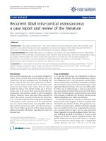
Figure 1 Brain MR image s in a 20-year-old woman. (a) Axial T
2
-weighted image shows a hyperintense mass with concentric pattern in the
right centrum semiovale. (b) Axial T
1
-weighted image reveals hypoisointense concentric rings in the white matter of the right parietal lobe. (c)
Axial T
1
-weighted image after contrast shows concentric enhancing rings. (d) Axial T
2
-weighted image, 1 month after therapy, shows differences
with decrease of the signal intensity at the center of the lesion. (e, f) Coronal T
1
-weighted image after administration of gadolinium
demonstrates a low-signal, non-enhancing lesion.
Gavra et al. Journal of Medical Case Reports 2011, 5:217
/>Page 2 of 4
whereas Ki-67 immunostaining was positive in 1% to 2%
of the nuclei. The pathologist commented that the find-
ings were suggestive of a grade II astrocytoma.
In the face of this diagnostic dilemma, a conservative
approach was adopted. Oral steroid treatment was
continued, and the patient was scheduled for a new 1H-
MRS and MRI scan one month later. The spectroscopic
findings were identical to t he previous ones. However,
the new conventional MRI images showed significant
differences, i n that the signal intensity was lower in the
center of the lesion in T
2
images (Figure 1d), whereas
the enhancin g rings appeared to fade away centrifugally
(Figure 1e, f). The dimensions of the lesion were
unchanged. These findings were considered sufficient
enough to establish the diagnosis of BCS.
No relapse in the symptoms occurr ed during the next
12 months of follow-up. A serial MRI showed a low-
signal, non-enhancing lesion.
Discussion
Diagnosis of tumefactive brain lesions is challenging to
both clinicians and radiologists. Clinical differential
diagnosis includes demyelinating diseases, neoplasms,
and infections such as abscesses. Such lesions with
mass-like characteristics may be the presenting feature
of multiple sclerosis ( MS) , acute disseminated encepha-
lomyelitis, or other rare demyelinating diseases, such as
BCS and Marburg type. BCS is a rare demyelinating dis-
ease considered to be an acute variant of MS, appearing
in young adults and typically follo wing a fulminant
course [1]. It shows a monophasic, rapidly progressive
course, sometimes fatal. Histologically, it is characterized
by a large lesion consisting of rings of demyelination
alternating with rings of intact myelin. MRI is the
method of choice for imaging demyelination lesions,
tumefactive or not. Although the usual appearance of
MS is that of multiple, small, demyelinating plaques, in
some cases, it can simulate a mass lesion, which it
would be hard to d istinguish from a brain tumor [2].
MRI characteristics, such as open-ring enhancement,
peripheral restriction on diffusion-weighted imaging, or
venular enha ncement, may be rewarding in differ entiat-
ing tumefactive MS lesions from neoplastic ones [2].
BCS is also considered within the spectrum of MS. It
shares an apparent basic pathologic similarity to MS,
with the exception of a lamellar pattern. The striking
concentric pattern of demyelination distinguishes this
disorder from other demyelinating diseases. BCS has
characteristic MRI features such as the hypoisointense
conc entric rings on T
1
-wei ghted, the whirlpool hyperin-
tense concentric rings on T
2
-weighted, and the separate
rings of enhancement in a concentric pattern [3]. This
type of concentric pattern has not been described in
association with any other demyelinating/inflammatory
diseases except BCS, and therefore, acute disseminated
encephalomyelitis and Marburg MS were excluded in
our case.
Advanced neuroimaging can provide important in vivo
markers of disease progression. MRS in BCS may show
reduction of N-acetylaspartate and increase in choline
and lipids, reflecting axonal destruction and an elevation
of lactate resonance due to local ischemia from the
ongoing inflammatory process [4]. These resonance
spectra [2,4] are not specific for BCS, and they may
resemble those of brain tumors and acute MS plaques
[2,4,5]. The chronic demyelinating plaque, however,
shows a completely different pattern [4].
A stereotactic biopsy and histologic examination of
the lesion is the final diagnostic approach in equivocal
cases [6]. It is safe and reliable, especially if specimens
from multiple sites within the lesion are targeted. It has
a diagnostic accuracy of 82% to 99% [7]. Acute demyeli-
nating plaque is hypercellular, and on frozen s ections,
this hypercellularity may be mistaken to be indicative of
glioma. The diffuse infiltration of inflammatory cells,
mainly reactive astrocytes and lipid-laden macrophages,
and perivascular cuffing by T-lymphocytes favors the
diagnosis of demyelinating plaque. The presence of
alternating rings of myelin preservation or remyelination
and myelin loss, consistent with demyelination, corre-
sponds to the concentric type of demyelination, or BCS.
Although the presence of reactive astrocytes can raise
the diagnosis of an astrocytoma, in the non-neoplastic
demyelinating plaque, these astrocytes are not significant
in number to establish such a diagnosis safely, nor are
there areas of vascular proliferation, indicative of a
neoplastic process. Staining for myelin and axons and
applying special immunohistochemical stains for macro-
phage markers should help overcome this diagnostic
pitfall [8].
Occasionally, the biopsy is non-diagnostic or dispensa-
ble [5]. In our case, the risk lay in the fact that in BCS,
areas of demyelination alternate with areas of active
gliosis in a dynamic and concentric fashion. Even with
the use of stereotactic procedures, tissue specimens may
be yielded from area of reactive gliosis not just outside,
but also from within the lesion itself, thus giving ambig-
uous or false results. Targeting multiple areas within the
lesion may help overcome this problem.
In our case, histopathologic findings from the biopsy
were misleading.
However, the characteristic concentric rings of demye-
lination alternating with myelination on MRI, the
patient’s considerable clinical improveme nt after steroid
therapy, and the signal differences in follow-up MRI
scans established the diagnosis of BCS.
In serial MRI scans, the concentric ring enhancement
of BCS is expected t o fade away centrifugally, until it
Gavra et al. Journal of Medical Case Reports 2011, 5:217
/>Page 3 of 4
appears as a low-signal, non-enhancing lesion, typical of
a c hronic demyelinating plaque [9], as in our case.
Regarding MRS, it seems that it is the serial changes of
the metabolites’ resonance intensities rather than the
individual values that provide more information about
the nature of the lesion [9].
Conclusion
Demyelinatin g diseases can mimic b rain neoplasms
clinically, radiologically, and histopathologically. BCS is
a rare demyelinating disease, which can manifest as a
mass lesio n. The typical concentric pattern on MR
images, along with clinical features, can lead to accurate
diagnosis and treatment. For suspected cases, it is advi-
sable to use steroid therapy or undergo serial MRI
examinations. However, in borderline cases, pathologic
evidence is beneficial to a final diagnosis.
Consent
Written informed consent was obtained from the patient
for publication of this case report and any accompany-
ing images. A copy of the written consent is available
for review by the Editor-in-Chief of this journal.
Abbreviations
BCS: Balo concentric sclerosis; CT: computed tomography; MRI: magnetic
resonance imaging; MRS: proton magnetic spectroscopy; MS: multiple
sclerosis.
Author details
1
Department of CT and MRI, Children’s Hospital, “Agia Sophia’’, Thivon and
Papadiamantopoulou Street, Athens, Greece.
2
Department of Neurosurgery,
University of Athens Medical School, “Evangelismos” General Hospital, 45-47
Ipsilantou Street 10676, Athens, Greece.
Authors’ contributions
MG collected and analyzed all patient data, conducted a literature review,
and was a major contributor in writing the manuscript. LS collected and
analyzed data related to the patient’s stay in the neurosurgery department
and collected the follow-up information. EB and DS provided clinical details
and technical input, revised the manuscript, and performed changes
throughout the manuscript. All authors read and approved the final
manuscript.
Competing interests
The authors declare that they have no competing interests.
Received: 16 January 2010 Accepted: 7 June 2011
Published: 7 June 2011
References
1. Airas L, Kurki T, Erjanti H, Marttila RJ: Successful pregnancy of a patient
with Balo’s concentric sclerosis. Mult Scler 2005, 11:346-348.
2. Malhotra H, Jain K, Agarwal A, Singh M, Gupta R: Characterization of
tumefactive demyelinating lesions using MR imaging and in vivo proton
MR spectroscopy. Mult Scler 2009, 15:193-203.
3. Li Y, Xie P, Fan X, Tang HB: Balo’s concentric sclerosis presenting with
benign clinical course and multiple sclerosis-like lesions on magnetic
resonance images. Neurol India 2009, 57:66-68.
4. Butteriss DJ, Ismail A, Ellison DW: Use of serial proton magnetic resonance
spectroscopy to differentiate low grade glioma from tumefactive plaque
in a patient with multiple sclerosis. Br J Radiol 2003, 76:662.
5. Enzinger C, Strasser-Fuchs S, Ropele S, Fazekas F: Tumefactive
demyelinating lesions: conventional and advanced magnetic resonance
imaging. Mult Scler 2005, 11:135-139.
6. Xia L, Lin S, Wang ZC, Li SW, Gao CC: Tumefactive demyelinatig lesions:
nine cases and a review of the literature. Neurosurg Rev 2009, 2:171-179.
7. Boviatsis EJ, Kouyialis AT, Stranjalis G: CT-guided stereotactic biopsies of
brain stem lesions: personal experience and literature review. Neurol Sci
2003, 24:97-102.
8. Sugita Y, Terasaki M, Shigemori M: Acute focal demyelinating disease
simulating brain tumors: histopathologic guidelines for an accurate
diagnosis. Neuropathology 2001, 21:25-31.
9. Karaarslan E, Altintas A, Senol U, Siva A: Balo’s concentric sclerosis: clinical
and radiologic features of five cases. Am J Neuroradiol 2001, 22:1362-1367.
doi:10.1186/1752-1947-5-217
Cite this article as: Gavra et al.: Pitfalls in the diagnosis of a tumefactive
demyelinating lesion: A case report. Journal of Medical Case Reports 2011
5:217.
Submit your next manuscript to BioMed Central
and take full advantage of:
• Convenient online submission
• Thorough peer review
• No space constraints or color figure charges
• Immediate publication on acceptance
• Inclusion in PubMed, CAS, Scopus and Google Scholar
• Research which is freely available for redistribution
Submit your manuscript at
www.biomedcentral.com/submit
Gavra et al. Journal of Medical Case Reports 2011, 5:217
/>Page 4 of 4