Báo cáo khoa học: " Upper cervical intramedullary spinal metastasis of ovarian carcinoma: a case report and review of the literature" docx
Bạn đang xem bản rút gọn của tài liệu. Xem và tải ngay bản đầy đủ của tài liệu tại đây (1.05 MB, 4 trang )
CASE REP O R T Open Access
Upper cervical intramedullary spinal metastasis of
ovarian carcinoma: a case report and review of
the literature
Amrendra S Miranpuri
1
, Sharad Rajpal
1
, M Shahriar Salamat
2
and John S Kuo
1*
Abstract
Introduction: Currently there is no generalized approach to treating patients with intra-medullary spinal
metastasis. High cervical spinal cord lesions can be particularly challenging cases, and may even be considered
inoperable by some.
Case report: We present what is, to the best of our knowledge, the first reported case of ovarian carcin oma
(managed primarily with surgery) in a 65-year-old Caucasian woman metastasizing to the upper cervical spinal
cord; we also review the relevant literature and discuss management strategies.
Conclusions: Due to improving systemic cancer therapies, patients with cancer now often survive longer and are
more likely to develop central nervous system metastases. Therefore, neurosurgical oncologists are often
challenged with difficult decisions about how to surgically manage these patients. We recommend individualized
multidisciplinary management based on patient functional status, the need for definitive diagnosis for possible
additional adjuvant therapies, and consideration of extent of systemic disease impacting on desirable quality and
length of survival.
Introduction
Whereas lung and breast cancer represent the most fre-
quently occurring spinal intra-medullary metastatic neo-
plasms, other solid tumors such as ovarian ca rcinoma
can also rarely metastasize to the spinal cord. On ima-
ging studies, the differentialdiagnosesforintra-medul-
lary spinal lesions can include gliomas and vascular
malformations but rare spinal infections such as tuber-
culosis can still be seen in some parts of the world [1,2].
The clinical presentation can range from minor neurolo-
gical symptoms to major symptoms that significantly
alter a patient ’s daily activities.
Surgical resection of intramedullary sp inal metastases
can be assoc iated with signifi cant morbidity. Man age-
ment must therefore be individualized based on patient
functional status, need for definitive diagnosis to guide
additional therapies, and extent of systemic dise ase
impacting on quality and length of survival. Previous
reports have described management strategies for ovar-
ian metastases to the spinal cord. However, we describe
the first ever report of a high cervical ovarian metastasis
managed primarily with surgery. Such an operation has
potential airway and brainstem complications. As
patients with cancer are surviving their primary disease
lon ger, neurosurgical oncologists may be faced with the
challenge of treating what were traditionally believed to
be inoperable lesions. A careful discussion with the
patient a nd their family, comb ined with multidisciplin-
ary input from colleagues from medical and radiation
oncology are important.
Case presentation
A 65-year-old Caucasian woman underwent surgery for
papillary serous ovari an adenocarcinoma involving both
ovaries and with extensive metastases (stage IIIC). An
exploratory laparotomy with total abdominal hysterect-
omy, bilateral salpingo-oophorectomy, and omentectomy
with cancer staging was performed. She also underwent
chemotherapy including carboplatin, paclitaxel, and cis-
platin. Her CA-125 level was normal and there was no
* Correspondence:
1
Department of Neurological Surgery, School of Medicine and Public Health,
University of Wisconsin, Madison, WI, USA
Full list of author information is available at the end of the article
Miranpuri et al. Journal of Medical Case Reports 2011, 5:311
/>JOURNAL OF MEDICAL
CASE REPORTS
© 2011 Miranpuri et al; licensee BioM ed Central Ltd. This is an Open Access article distributed under the terms of the Creative
Commons Attribution License ( which permits unrestricted use, distribution, and
reproduction in any medium, provided the original work is properly cited.
evidence of disease progression at her last clinic visit at
our center. Then, two years later, she re-presented with
progressive neurological symptoms starting initially with
limb dysesthesias and numbness and progressing to
quadriparesis with urinary retention.
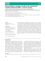
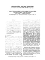
Imaging studies of her spine revealed an enhancing
heterogeneous C2-C5 intramedullary lesion with cord
expansion and edema extending rostrally into the
medulla and caudally to the thoracic spinal cord (Figure
1a). Serum CA-125 was normal at presentation and a
computed tomography (CT) scan of the chest, abdomen,
and pelvis were negative for other lesions. An investiga-
tion for possible sources of infection was negative.
Informed consent was obtained from our patient for
open surgical biopsy and possible debulking. C2-C5
laminectomies were performed for planned ultrasound-
guided dorsal midline biopsy and debulking of the intra-
medullary mass. The tumor was debulked and the rem-
nants of the tumor capsule dissected along the rostral
and caudal margins with care taken not to injure the
surrounding spinal cord. Somatosensory and motor
evoked potentials did not change during surgery. A
post-operative MRI scan showed the expected near total
resection and expected post-l aminectomy changes with-
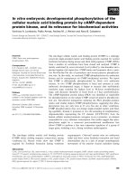
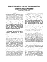
out any associated hematoma (Figure 1b,c). Pathologic
analysis revealed histological and cytological features
consiste nt with papillary serous ovarian adenocarcinoma
(Figure 2), similar to the pathological specimen from
her prior surgery. She made functional improvements
after surgery and was transferred to the rehabilitation
service. She gained the ability to s tand with assistance
using a walker, had antigravity strength in her lower
extremities and 4/5 strength in her upper extremities.
Fractionated radiotherapy was initiated immediately in
the post-operative period during her rehabilitation.
Our patient had improv ement in strength post-opera-
tively but required an emergency re-operation three
weeks later due to sudden paraplegia secondary to spinal
epidural hematoma, after therapy with prophylactic sub-
cutaneous heparin administration. On discharge a week
after epidural hematoma evacuation, she experienced
numbness below the umbilicus and slightly improved to
left toe movement. Unfortunately, our patient di ed five
months after discovery of her spinal metastasis, presum-
ably from a pulmonary embolism.
Discussion
Review of the English language literature via PubMed
database searches revealed five previous case reports of
spinal cord ovarian cancer metastases, of which only
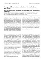
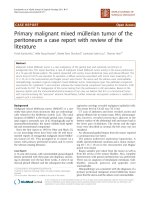
three were tissue confirmed. Data from our report and
the literature are summarized in Table 1.
Our patient’scaseisonlythethirdreportedtissue-
proven case of ovarian carcinoma metastasizing to the
spinal cord and the first reported case of metastasis to
the high cervical spinal cord. Historically, there has
been a role for surgery in resecting a solitary metastatic
lesion to the spinal cord. The limitations to surgical
resection are guided by the risk of morbidity to the
patient, especially with regards to neurological function.
Sundaresan et al. [3] retrospectively reviewed 80
patients with solitary spinal metastasis from a ll cancer
histologies. Overall median survival in that series follow-
ing surgery was 30 months. Survival was superior in the
group with breast and kidney cancers. Morbidity and
recurrence, however, were hi gher in patients receiving
prior radiation therapy. Indications for surgery include
pathological diagnosis, restoration of neurological func-
tion via decompression of mass effect and spinal stabili-
zation [3].
Figure 1 Sagittal cervical spine MRI. (a) Pre-surgical resection, T1 post-contrast demonstrating a 1.3 × 4.4 cm intramedullary enhancing mass
(left panel). (b) Post-surgical resection, T1 pre-contrast (middle panel). (c) Post-surgical resection, T1 post-contrast showing small amount of
residual tumor at caudal margin of tumor (right panel).
Miranpuri et al. Journal of Medical Case Reports 2011, 5:311
/>Page 2 of 4
The degree of tumor resection must be ind ividualized.
Rastelli et al. [4] reported gross total resection in a T11
metastatic ovarian cancer. This patient had near-com-
plete strength improvement and MRI showed no spinal
recurrence 16 months later. Even in the two cases of
subtotal resection reviewed, tissue diagnosis is achieved
while also achieving a less morbid operation as deemed
appropriate by the involved surgeon. Isoyo et al. [5] per-
formed a subtotal resection of a T10 metastatic ovarian
lesion. This patient had no improvem ent in neurological
status but remained alive two years after surgery.
Steroids are beneficial because it provides sympto-
matic relief and reduces peri-tumoral edema with a low
side effec t profile. The other case reports reviewed also
described a 30Gy radiati on dose as a preferred prescrip-
tion for ovarian cancer spinal cord metastases. In select
cases, simultaneous steroids and radiotherapy adminis-
tration without a tissue diagnosis can be considered for
Figure 2 Hematoxylin and eo sin stained section of cervical intramedul lary tumor. This m etastatic neoplasm was compared with prior
hysterectomy and salpingo-oophorectomy of our patient and reveals similar histologic and cytologic features to the ovarian papillary serous
adenocarcinoma.
Table 1 Summary of case reports published for intramedullary ovarian spinal tumors
Reference Lesion level
(enhancing
portion)
Time from primary
diagnosis to spinal
metastasis diagnosis*
Surgical
intervention
Adjuvant therapy Outcome
Current
report
C2-C5 two years Subtotal
resection
30Gy and steroids Strength improved; three weeks post-
operative spinal epidural hematoma; died
five months later
Thomas et
al. [6]
C6-T1 Four and a half years None 30Gy and steroids Strength improved; died six months later
Cormio et
al. [8]
C5-C6 One and a half years None Steroids, chemotherapy,
30Gy
Strength improved; died 10 months later
Isoya et al.
[5]
T10 four years Subtotal
resection
Radiotherapy (dose not
given)
No neurological improvement; alive two
years after surgery
Rastelli et
al. [4]
T11 two years Gross total
resection
30Gy (10 fractions) Near-complete strength improvement; MRI
shows no spinal recurrence 16 months on
Bakshi et
al. [7]
Conus
medullaris and
cauda equina
two years None Steroids, radiotherapy
(dose not given),
chemotherapy
Symptomatic improvement; three-year
complete remission
*Approximate time in some cases based on estimates provided in reference.
Miranpuri et al. Journal of Medical Case Reports 2011, 5:311
/>Page 3 of 4
patients at high surgical risk with poor Karnovsky scores
[6,7]. Cormio et al. [8] demonstrated complete resolu-
tion of neurol ogical symptoms with early steroids and
carboplatin. Prior to the fourth cycle of carboplatin, an
MRI scan of the brain showed diffuse metastatic disease
for which the patient received 30Gy radiotherap y to the
brain and cervical spine. In the above case report, how-
ever, no tissue diagnosis confirmation was obtained. The
MRI scan perfor med after radiot herapy demonstrated
almost complete resolution of the cervical lesion. Thus,
steroids combined with chemot herapy and radiotherapy
can be a viable empiric, alternative treatment regimen in
high-risk surgical patients. Symptomatic and imaging
responses in such cases, however, do not establish the
diagnosis of ovarian spinal metastasis.
Conclusions
There is no current consensus on management of
patients presenting with neurological symptoms and a
potential diagnosis of spinal intramedullary metastasis.
In cases of central nervous system spinal cord metas-
tasesinpatientsexperiencing progressive neurological
symptoms whose medical condition permit surgery, we
advocate open surgical biopsy with resection to confirm
tissue diagnosis, to reduce tumor burden for adjuvant
therapies while mi nimizing surgical mo rbidity, and to
accurately diagnose and treat non-metastatic diseases
that may masquerade as intramedullary spinal metas-
tases. The risks and benefits of such interventions, how-
ever, must be carefully weighed in discussions with
individual patients and their families. As patients with
cancer are surviving their primary disease long er, it will
be critical for neurosurgical oncologists to work closely
with radiation oncologists and medical oncologists to
formulate individualized treatment plans for patients
with central nervous system metastases, based on risk/
benefit analysis while also considering a patient’sdesire
for quality of life and potential extent of survival.
Consent
Written informed consent was obtained from the
patient’s next-of-kin for publication of this case report
and any accompanying images. A copy of the written
consent is available for review by the Editor-in-Chief of
this journal.
Author details
1
Department of Neurological Surgery, School of Medicine and Public Health,
University of Wisconsin, Madison, WI, USA.
2
Department of Pathology and
Laboratory Medicine, School of Medicine and Public Health, University of
Wisconsin, Madison, WI, USA.
Authors’ contributions
ASM, SR and JSK analyzed and interpreted our patient data regarding the
clinical course, surgery and outcome. MSS performed the histological
examination of the tumor. ASM was a major contributor in writing the
manuscript. All authors read and approved the final manuscript.
Competing interests
The authors declare that they have no competing interests.
Received: 11 August 2010 Accepted: 14 July 2011
Published: 14 July 2011
References
1. Waldron JS, Cha S: Radiographic features of intramedullary spinal cord
tumors. Neurosurg Clin N Am 2006, 17:13-19.
2. Ramdurg SR, Gupta DK, Suri A, Sharma BS, Mahapatra AK: Spinal
intramedullary tuberculosis: a series of 15 cases. Clin Neurol Neurosurg
2009, 111:115-118.
3. Sundaresan N, Rothman A, Manhart K, Kelliher K: Surgery for solitary
metastases of the spine: rationale and results of treatment. Spine 2002,
27:1802-1806.
4. Rastelli F, Benedetti G, Di Tommaso L, Mazzoli M, Calbucci F, Crinò L:
Intramedullary spinal metastasis from ovarian cancer. Lancet Oncol 2005,
6:123-125.
5. Isoya E, Saruhash Y, Katsuura A, Takahashi S, Matsusue Y, Hukuda S:
Intramedullary spinal cord metastasis of ovarian tumor. Spinal Cord 2004,
42:485-487.
6. Thomas AW, Simon SR, Evans C: Intramedullary spinal cord metastases
from epithelial ovarian carcinoma. Gynecol Oncol 1992, 44:195-197.
7. Bakshi A, Biswas G, Deshmukh C, Prasad N, Nair R, Parikh PM: Successful
complete regression of isolated intramedullary spinal cord metastases
from epithelial ovarian carcinoma with chemotherapy and radiotherapy.
Indian J Cancer 2006, 43:136-138.
8. Cormio G, Di Vagno G, Di Fazio F, Loverro G, Selvaggi L: Intramedullary
spinal cord metastasis from ovarian carcinoma. Gynecol Oncol 2001,
81:506-508.
doi:10.1186/1752-1947-5-311
Cite this article as: Miranpuri et al.: Upper cervical intramedullary spinal
metastasis of ovarian carcinoma: a case report and review of the
literature. Journal of Medical Case Reports 2011 5:311.
Submit your next manuscript to BioMed Central
and take full advantage of:
• Convenient online submission
• Thorough peer review
• No space constraints or color figure charges
• Immediate publication on acceptance
• Inclusion in PubMed, CAS, Scopus and Google Scholar
• Research which is freely available for redistribution
Submit your manuscript at
www.biomedcentral.com/submit
Miranpuri et al. Journal of Medical Case Reports 2011, 5:311
/>Page 4 of 4