Báo cáo y học: "Fatal invasive cervical cancer secondary to untreated cervical dysplasia: a case report" potx
Bạn đang xem bản rút gọn của tài liệu. Xem và tải ngay bản đầy đủ của tài liệu tại đây (1.39 MB, 5 trang )
CAS E REP O R T Open Access
Fatal invasive cervical cancer secondary to
untreated cervical dysplasia: a case report
Stephan Braun
1
, Daniel Reimer
1
, Isolde Strobl
1
, Ulrike Wieland
2
, Petra Wiesbauer
1
, Elisabeth Müller-Holzner
1
,
Siegfried Fessler
1
, Arthur Scherer
3
, Christian Marth
1
and Alain G Zeimet
1*
Abstract
Introduction: Well-documented cases of untreated cervical intra-epithelial dysplasia resulting in fatal progression of
invasive cervical cancer are scarce because of a long pre-invasive state, the availability of cervical cytology
screening programs, and the efficacy of the treatment of both pre-invasive and early-stage invasive lesions.
Case presentation: We present a well -documented case of a 29-year-old Caucasian woman who was found,
through routine conventional cervical cytology screening, to have pathologic Papanicolaou (Pap) grade III D lesions
(squamous cell abnormalities). She subsequently died as a result of human papillomavirus type 18-associated
cervical cancer after she refused all recommended curative therapeutic procedures over a period of 13 years.
Conclusion: This case clearly demonstrates a caveat against the promotion and use of complementary alternative
medicine as pseudo-immunologic approaches outside evidence-based medicine paths. It also demonstrates the
impact of the individualized demands in diagnosis, treatment and palliative care of patients with advanced cancer
express their will to refuse evidence-based treatment recommendations.
Introduction
Cases of intra-epithelial disease of the cervix are almost
entirely attributable to human papillomavirus (HPV)
infection. A minority of women exposed to HPV develop
a persistent infection that affects the squamocolumnar
junction where the ectocervix and endocervix meet.
Within that junction, dynamic changes of the epitheli um
occur due to puberty, pregnancy, menopause and hormo-
nal stimulation. The epithelium is vulnerable to noxae
associated with smoking, contraceptive use and infection
with other sexually transmitted diseases. Alterations of
the epithelium a re assessed by conventional cervical
cytology screening and are scored according to either the
Bet hesd a or the Papanicolaou system. The occurrenc e of
reactive changes and/or cell abnormalities triggers either
repetitions of the cytology screening to exclude tempor-
ary alterations or a cervical biopsy for histological diag-
nosis of cervical intra-epithelial neoplasia and cervical
cancer. With the advent of HPV vaccination [1] and HPV
screening [2] to identify women at risk of lesions with
atypical or malignant cells prior to clinical manifestation,
in current clinical practice a patient’sHPVstatusshould
play a central role in the prevention of HPV-associated
diseases [3].
Invasive cervical cancer has a long pre-invasive state,
and cervical cytology screening programs are available.
Moreover, HPV vaccination has been shown to be a
successful tool of primary prevention [1], and t reatment
of pre-invasive lesions is effective. Invasive cancer is
considered a preventable cancer in the so-called highly
developed Western countries [3]. Consequently, invasive
cancer of the cervix has become increasingly infrequent
in this part of the world, but it remains a significant
health problem in underdeveloped countries, where
meticulous documentation of fatal courses of the disease
plays a minor role. Thus, our knowledge of the lead
time between dysplasia and the development of inva sive
cancer as well as progression from early-stage to metas-
tasized cancer largely derives from extrapolating infor-
mation from studies and textbooks, but very few case
reports.
Herein we report a rather rare, yet well-documented
case of a 29-year-old woman who, during the course of
her disease, accepted multiple diagnostic procedures but
* Correspondence:
1
Department of Obstetrics and Gynecology, Innsbruck Medical University,
Anichstrasse 35, AT-6020 Innsbruck, Austria
Full list of author information is available at the end of the article
Braun et al. Journal of Medical Case Reports 2011, 5:316
/>JOURNAL OF MEDICAL
CASE REPORTS
© 2011 Braun et al; licensee BioMed Central Ltd. This is an Open Access article distrib uted under the terms of the Creative Commons
Attribu tion License ( which permits unrestricted use, distribution, and reproduction in
any medium, provided the original work is properly cited.
refused any curative treatment beginning with the first
assessment of cervical dysplasia and early-stage invasive
cancer 10 y ears later. She finally refused to accept any
interventional medical strategies, except for palliative
car e, at the stage of locally progressed and metastasized
cervical cancer.
Case presentation
A 29-year-old Caucasian woman was seen for her rou-
tine annual gynecologic examination, and conventional
cytological screening of her cervix uteri revealed a
pathologic finding scored as grade IIID under the Papa-
nicolaou system. Repeat screening performed one year
later revealed a grade IV pathologic finding, suggesting a
high-grade squamous intra-epithelial lesion. Our patient
refused the recommended diagnostic and therapeutic
procedure of conization, and she was placed on a non-
specified homeopathic therapy consisting of a vitamin
C-containing regimen and subcutaneous administration
of mistletoe lectins.
Atthetimeofthefirstpathologic Papanicolaou test,
our patient reported a normal menstrual cycle, no preg-
nancies, no use of oral contraceptives, no presence of
any previous diseases or any surgery, no allergies, no
smoking, and no use of illicit drugs. There was no evi-
dent lack in body hygiene. Except for her father’ssto-
mach cancer, her family and cancer-specific anamneses
were unremarkable. On t he basis of her grade V in
Papanicoleau test, she was sent to our hospital’ sout-
patient department.
A gynecologic examination at that time revealed
obvious tumor growth confined to her cervix with no
signs of extension to her vagina. A cervical biopsy
showed moderately differentiated (tumor grade II) large-
cell non-keratinizing squamous cell carcinoma of the
cervix uteri. Neither lymphatic nor venous vascular
space involvement was reported, but dense inflamma-
tory cell infiltration of the tumor stroma was noted.
Clinical staging was completed by cystoscopy, procto-
scopy, and chest r adiography (as allowed for accurate
clinical staging by the International Federati on of Gyne-
cology and Obstetrics [FIGO]), which revealed stage IB2
cancer. Additional information was obtained by
extended staging procedures, including computed tomo-
graphic (CT) and laparoscopic sampling of her para-aor-
tic lymph nodes (the results of which would have had
no influence on the assigned clinical stage according to
FIGO guidelines).
However, our patient refused to undergo any further
diagnostic procedures and instead underwent comple-
mentary medical treatment. This included regional
hyperthermi a, which led to her self-admission to a local
hospital. She presented there with reduced phys ical sta-
tus, large edema of the legs, and moist rales in her
lungs. She also reported lower abdominal pain. Her clin-
ical work-up revealed significant progression of her dis-
ease, which now included bilateral parametrial
involvement, broad involvement of her dorsal bladder
wall, infiltration of her outer rectum wall, pericardial
and pleural effusion, bilateral hydronephrosis with
laboratory signs of uremia (serum creatinine 17.4 mg/
dL, serum uric acid 106 mg/dL), and tumo r anemia
with hemoglobin at 71 g/L. Two courses of hemodialysis
were performed initially, followed by a right -sided
nephrostomy after the failure of ureteral stenting due to
tumor extension to her bladder. It was decided to com-
mence hemodialysis on the basis of the patient’s request
for consequent evidence-based, palliative medical care
after restoration of her renal function.
Restaging was performed, which indicated involvement
of her bladder wall a nd adhesions to the ileocecal area
(Figure 1b). All three para-aort ic lymph nodes removed
during re-laparoscopy were positive (Figures 1c and 1d),
while no signs of distant metastasis were seen on the
radiologic studies. The tumor was restaged to FIGO
IVa, and concur rent cisplatin-based chemoradiation was
recommended. Our patient, however, opted against our
treatment recommendation and traveled to the Philip-
pines for an alternative holistic treatment schedule
involving several courses of Horvi-Reintoxin enzyme
therapy, which consists of enzymatically processed snake
poiso n that is purported to spe cifically inhibit glycosyla-
tion in tumor cells, thus conferring anti-tumoral activity.
Our patient was repeatedly admitted to both Brixen
and Innsbruck hospitals for erythrocyte transfusions
because of spontaneous uterine hemorrhage and further
local tumor progression (Figure 2a). Acute life-thr eaten-
ing h emorrhage prompted us to perform three sessions
of arterial embolization: first, in both uterine arteries;
second, in both internal pudendal arteries and re-embo-
lization of her right uterine artery; and third, in her
right superior and inferior vesical arteries and re-embo-
lization of her left internal pudendic artery. In parallel,
our patient continued her holistic alternative medical
treatment, first with active fever treatment, during
which pyrogenic lysates of bacteria were administered
and second with combined application of Carnivora-
Mistletoe-Ukrain (that is, capsules with plant extracts,
subcutaneous injections of mistletoe lectins, c apsules
with extracts of celandine and Chelidonium majus), all
of which are purported to have antitumoral activity.
Seven months later our patient was admitted to the
hospital with clinical signs of chronic large bowel
obstruction, and laparotomy and side-to-side ileoascen-
dostomy became necessary, during which her left ureter
(Figure 2b), descending colon, rectosigmoid and ileoce-
cum appeared fixed by tumor masses and obstruct ed by
large, lymphatic fluid-containing cysts (Figure 2c). Our
Braun et al. Journal of Medical Case Reports 2011, 5:316
/>Page 2 of 5
patient overcame a postsurgical bow el paralysis and
recovered fairly well. However, during the f ollowing
days, palliative care was required for salvage from dys-
pnoea by bilateral pleuracentesis (Figure 2d), from
mechanical and paralytic small and large bowel
obstruction by distigmine bromide administration, and
from recurrent visceral abdominal and neurogenic pain
by morphine hydrochloride. On the basis of a CT scan,
a paralytic ileus, together with metastasis to her spleen
(Figure 2e) and her liver (Figure 2f), were diagnosed.
One month later our patient died as a result of tumor
cachexia, chronic small and large bowel ile us, septicemia
and consecutive multipl e organ failur e. In her last wil l,
she refused autopsy.
For HPV testing, we isolated DNA from the paraffin-
embedded tumor shown in Figure 1a. With group-speci-
fic nested polymerase chain reactions for the detection
of a-HPV DNA [4], we tested for the presence of 18
high-risk HPV types (16, 18, 26, 31, 33, 35, 39, 45, 51,
52, 53, 56, 58, 59, 66, 68, 73, and 82) and 18 low-risk
HPV types (6, 11, 40, 42, 43, 44, 34, 54, 55, 57, 61, 70,
71, 72, 81, 83, 84, and 89). Our patient’s tumor exclu-
sively contained type 18 HPV DNA.
Discussion
We have presented the case o f a woman who had a
pathologic cervical cytology screening at the age of 2 9
years, and died as a result of cervical cancer at the age
of 42 after she had denied all recommended curative
therapeutic procedures for 13 years. Except for the
noted HPV type 18 infection, our patient’sdemographic
characteristics included the typical socioeconomic and
epidemiologic risk factors known for cervical cancer in
that she was Caucasian, had a high socioeconomic sta-
tus, reported no cigarette smoking, was nulliparous and
reported no history of apparent promiscuity. It appears
tobearatherrarecaseintermsof,ontheonehand,
accepting far-reaching diagnostic procedures such as
laparoscopic lymph node sampling, and on the other,
consistently refusing to accept all proposed evidence-
based treatment recommendations over a total period of
13 years, with no psychological disorder being apparent.
Documented cases of untreated cervical dysplasia are
rare, and ours appears to be only the second report pub-
lished during the past 10 years that i s retrievable in the
currently available medical literature databases. The
other case, reported in 2002, described a very short
interval of one year between the diagnosis of cervical
dysplasia and metastases in the bone, liver, and orbit
[5]. This sho rt interval between dysplasia and metastatic
cervical cancer, however, raises questions as to whether
the Pap smear was representative or whether invasive
cancer was missed. Thus, our case presentation might
be one of the very few examples of a complete clinical
documentation of such “natural” progression among
retrievable case reports in the medical literature.
Of note, we want to stress that the apparent inefficacy
of the complementary alternative medical treatments
practiced worldwide, which should have exerted a
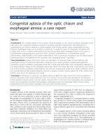
Figure 1 Restaging of the tumor. (a) formalin-fixed, paraffin-embedded
biopsy of t he invasive cervix c ancer; (b) corresponding CT scan of t he
pelvis; (c) formalin-fixed, paraffin-embedded biopsy of the para-aortic lymph
node metastases and her abdomen showing bladder invasion; and (d)
corresponding CT scan of the enlarged para-aortic lymph nodes.
Braun et al. Journal of Medical Case Reports 2011, 5:316
/>Page 3 of 5
stimulatory effect on the immune syste m and hence an
antineoplastic influence. Effects in preventing high-grade
cervical lesions to date have been noted only for bivalent
and quadrivalent vaccines against HPV type 16 or 18
and HPV types 6, 11, 16, and18, with vaccine efficacies
well above 90% [1]. Remarkably, and in sharp contrast
to the mentioned vaccine efficacy, the non-specific
approaches used in complementary alternative medicine,
as described in our present case, are deemed rather
inefficacious.
HPV type 16 or 18 infections are responsible for
approximately 60-80% of all invasive cancers, varying
according to the patient’s socioeconomic status [6]. Of
all new HPV infections, both oncogenic and non-onco-
genic type infections last between eight and five months,
respectively, and the large majority of initially HPV-
infected women show clearance within two years [7].
Pre-invasive surrogate lesions of squamous cervical can-
cerwouldbethoseofgradeIIandIII,withthelowest
potential of regression being that for grade III cervical
intra-epithelial neoplasia [8]. Since our patient refused
histopathological verification of the first cytological
abnormalities in 1993, we were unable to determine
whether a single, persistent HPV type 18 infection gave
rise to her cervical cancer, which was diagnosed in
2003. The assumption that this was the case is highly
likely to be true, since progression from HPV infection
to invasive cancer is believed to take place during the
course of several years, although we cannot exclude
HPV type 18 reinfection after initial clearance. Cervical
precursor lesions of oncogenic HPV infections, such as
HPV type 18 in our case, are known to persist longer
and progress more often than non-oncogenic type infec-
tions [9]. The likelihood of regression, stable dysplasia
or progression from moderate cervical dysplasia (CIN II,
which could have been the underlying disease in our
patient, who had Pap IIID and Pap IV) is known to be
almost equal. Because progression lead times are usually
in the two- to five-year range [10], even if we ta ke into
account a potential reinfection as well as some time for
progression from the first invasion to the bulky disease
(on which we have no firm information available), in
our case a gradual escape of the tumor from the host’s
immune surveillance may explain the rather slow pro-
gression to bulky cervix cancer over a ten-year period.
This ten-year period of uninfluenced tumor growth
also allowed for systemic spread and a pattern of distant
metastasis that, to the best of our knowledge, has not
thus far been reported in the literature, but suggests a
much more complex homing pattern of disseminated
tumor cells. Overall, cervical cancer has a low propen-
sity for distant hem atogenous metastatic spread. The
first clinical sign of metastasis to para-aortic lymph
nodes, that is, beyond the true pelvis, was assessed after
10 years on the basis of a CT scan. Furthermore, the
most common sites of dist ant metastasis are the lung,
liver, bone and, rarely, the peritoneum. Si ngle reports
would add the orbit [5] and bone marrow [11]. Our
patient’s liver and spleen metastasis as well as carcinosis
peritonei shortly before her death are rarely seen, but
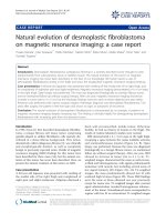
Figure 2 CT scans. (a) local tumor progression in her pelvis and vesical invasion and hemorrhage; (b) left ureter obstruction; (c) ileus through
descending colon and rectosigmoid obstruction; (d) malignant pleural effusion; (e) spleen metastasis; (f) liver metastasis.
Braun et al. Journal of Medical Case Reports 2011, 5:316
/>Page 4 of 5
further contribute to our knowledge of viable tumor cell
spread in cervical cancer.
Conclusion
In summary, we have presented an unusual case of
untreated, presumably HPV type 18-induced cervical
dysplasia with progression to invasive and finally meta-
static cervical cancer that demonstrated a ten-year lead
time between the diagnosis of dysplasia and invasive
cancer. This case serves as a caveat against the promo-
tion and use of complementary alternative medicine as
pseudo-i mmunologic approaches outside evidence-based
medicine paths. It also highlights the individualized
demands in diagnosis, treatment and palliative care of
advanced cancer patients who express their will to
refuse evidence-based treatment recommendations.
Consent
Written informed consent for publication could not be
obtained despite all reasonable attempts to trace the
patient’ s family. Every effort was made t o protect the
identity of our patient, and there is no reason to believe
that any of her relatives would object to publication.
Author details
1
Department of Obstetrics and Gynecology, Innsbruck Medical University,
Anichstrasse 35, AT-6020 Innsbruck, Austria.
2
Institute of Virology, University
of Cologne, Fürst-Prückler-Strasse 65, D-50935 Cologne, Germany.
3
Department of Obstetrics and Gynecology, Medical Services Hospital,
Bressanone, Italy.
Authors’ contributions
IS, PW, SF, AS and CM cared for the patient during her time in the hospital
(LKH Innsbruck and LKH Brixen) and assisted in data collection and the
preparation of the manuscript. SB, DR and AGZ were the major contributors
in writing the manuscript. EMH performed the histological examination of
the tumor tissues. UW performed the HPV testing. All authors read and
approved the final manuscript.
Competing interests
The authors declare that they have no competing interests.
Received: 26 September 2010 Accepted: 18 July 2011
Published: 18 July 2011
References
1. FUTURE II Study Group: Quadrivalent vaccine against human
papillomavirus to prevent high-grade cervical lesions. N Engl J Med 2007,
356:1915-1927.
2. Mayrand MH, Duarte-Franco E, Rodrigues I, Walter SD, Hanley J, Ferenczy A,
Ratnam S, Coutlée F, Franco EL, Canadian Cervical Cancer Screening Trial
Study Group: Human papillomavirus DNA versus Papanicolaou screening
tests for cervical cancer. N Engl J Med 2007, 357:1579-1588.
3. Runowicz CD: Molecular screening for cervical cancer: time to give up
pap tests? N Engl J Med 2007, 357:1650-1653.
4. Kreuter A, Brockmeyer NH, Hochdorfer B, Weissenborn SJ, Stücker M,
Swoboda J, Altmeyer P, Pfister H, Wieland U: Clinical spectrum and
virologic characteristics of anal intraepithelial neoplasia in HIV infection.
J Am Acad Dermatol 2005, 52:603-608.
5. McCulley TJ, Yip CC, Bullock JD, Warwar RE, Hood DL: Cervical carcinoma
metastatic to the orbit. Ophthal Plast Reconstr Surg 2002, 18:385-387.
6. Herrero R, Hildesheim A, Bratti C, Sherman ME, Hutchinson M, Morales J,
Balmaceda I, Greenberg MD, Alfaro M, Burk RD, Wacholder S, Plummer M,
Schiffman M: Population-based study of human papillomavirus infection
and cervical neoplasia in rural Costa Rica. J Natl Cancer Inst 2000,
92:464-474.
7. Franco EL, Villa LL, Sobrinho JP, Prado JM, Rousseau MC, Désy M, Rohan TE:
Epidemiology of acquisition and clearance of cervical human
papillomavirus infection in women from a high-risk area for cervical
cancer. J Infect Dis 1999, 180:1415-1423.
8. Sawaya GF, Smith-McCune K: HPV vaccination: more answers, more
questions. N Engl J Med 2007, 356:1991-1993.
9. Schlecht NF, Platt RW, Duarte-Franco E, Costa MC, Sobrinho JP, Prado JC,
Ferenczy A, Rohan TE, Villa LL, Franco EL: Human papillomavirus infection
and time to progression and regression of cervical intraepithelial
neoplasia. J Natl Cancer Inst 2003, 95:1336-1343.
10. Holowaty P, Miller AB, Rohan T, To T: Natural history of dysplasia of the
uterine cervix. J Natl Cancer Inst 1999, 91:252-258.
11. Janni W, Hepp F, Strobl B, Rack B, Rjosk D, Kentenich C, Schindlbeck C,
Hantschmann P, Pantel K, Sommer H, Braun S: Patterns of disease
recurrence influenced by hematogenous tumor cell dissemination in
patients with cervical carcinoma of the uterus. Cancer 2003, 97:405-411.
doi:10.1186/1752-1947-5-316
Cite this article as: Braun et al.: Fatal invasive cervical cancer secondary
to untreated cervical dysplasia: a case report. Journal of Medical Case
Reports 2011 5:316.
Submit your next manuscript to BioMed Central
and take full advantage of:
• Convenient online submission
• Thorough peer review
• No space constraints or color figure charges
• Immediate publication on acceptance
• Inclusion in PubMed, CAS, Scopus and Google Scholar
• Research which is freely available for redistribution
Submit your manuscript at
www.biomedcentral.com/submit
Braun et al. Journal of Medical Case Reports 2011, 5:316
/>Page 5 of 5