Báo cáo y học: "Neuroendocrine tumors of the gallbladder: a case report and review of the literature" docx
Bạn đang xem bản rút gọn của tài liệu. Xem và tải ngay bản đầy đủ của tài liệu tại đây (2.11 MB, 5 trang )
CAS E REP O R T Open Access
Neuroendocrine tumors of the gallbladder: a case
report and review of the literature
Silvia Mezi
1*
, Vincenzo Petrozza
2
, Orazio Schillaci
3
, Valentina La Torre
4
, Barbara Cimadon
1
, Martina Leopizzi
2
,
Errico Orsi
4
and Filippo La Torre
4
Abstract
Introduction: Primary gallbladder neuroendocrine tumors are extremely rare, representing 0.2% of all
neuroendocrine tumors. The diagnosis is incidental in most cases.
Case presentation: We describe the case of a 57-year-old Caucasian man who underwent laparoscopic
cholecystectomy for the evaluation of a gallblad der polyp that had been incidentally detected by ultasonography.
Histologically, his lesion was composed of monomorphic cells that contained small round nuclei and that were
organized in small nodular, trabecular, and acinar structures. His cells were positive for chromogranin A and
synaptophysin, and a diagnosis of “typical” carcinoid of the gallbladder was made. His post-operative computerized
axial tomography,
111
In-pentetreotide scintigraphy, and hormone-specific marker results were negative. He is
disease-free 45 months after surgical treatment.
Conclusions: Characteristic pathological findings of the gallbladder neuroendoc rine tumors predict the prognosis.
Whereas classical carcinoids of the gallbladder only rarely have a metastatic or invasive phenotype, the “atypical”
variants are more aggressive and are associated with a poorer prognosis. Given the difficulty in distinguishing
between benign and malignant lesions in the pre-surgical setting, we tend to consider each polypoid-like lesion of
the gallbl adder to be a high-risk lesion if it is larger than 1 cm and, as a result, to emphasize the nee d for
cholecystectomy in all cases, relying on the pathological and immunohistochemistry analyses for the final
diagnosis.
Introduction
Carcinoids are rare neuroendocrine tumors (NETs)
derived from enterochromaffin or Kul chitsky cells,
which are widely distributed in the body [1,2]. Conse-
quently, NETs can be found in any location of the body,
although the sites most commonly affected are the gas-
trointestinal and bronchopulmonary tracts, representing
approximately 67% and 25% of cases, respectively [3].
NETs are histologically varied entities and can range
from indolent, unrecognized neoplasms to highly active,
metastatic secretory tumors [4]. Prognostic factors
include primary tumor site, hi stological differentiation,
tumor size , angioinvasion, infiltrative growth, and pro-
duction of hormones [5]. Although the incidence of
NETs has increased over the past 30 years, survival has
also improved (reviewed by Zuetenhorst and Taal [2]).
According to American epidemiological data, gallblad-
der (GB) NETs are rare, representing only 0.2% of all
NETs [6]. Approximately half of the cases reported in
the literature as GB carcinoid tumors appear to be
endocrine cell carcinomas, which are histologically and
clinically distinct entities [6]. Whereas classical carci-
noids of the GB only rarely have a metastatic or invasive
phenotype, the “ atypical” variants are more aggressive
and are associated with a poorer prognosis [6-9]. Here,
we describe a case of incidental GB carcinoid tumor in
a 57-year-old man.
Case presentation
A 57-year-old Caucasian man with a seven-year history
of hepatitis B virus infection was admitted to our hospi-
tal for the treatment of a GB polyp. The abdominal
ultrasonography (US) revealed the presence of a well-
* Correspondence:
1
Department of Radiology, Oncology and Human Pathology, Division of
Oncology B, “Sapienza” University of Rome, Rome, Italy
Full list of author information is available at the end of the article
Mezi et al. Journal of Medical Case Reports 2011, 5:334
/>JOURNAL OF MEDICAL
CASE REPORTS
© 2011 Mezi et al; licensee BioMed Central Ltd. This is an Open Access article distributed under the terms of the Creative Commons
Attribution License ( which pe rmits unrestricted use, distribution, and reproduction in
any medium, provided the original work is properly cited.
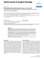
defined polypoid mass of approximately 12 × 8 mm in
his GB fossa (Figure 1). No evidence of biliary dilatation
was noted, and there was no ascites. No image of stones
was documented (Figure 1).
On examination, there was no pertinent medical or
surgical history, and our patient was asymptomatic and
showed no evidence of jaundice. An abdominal exami-
nation revealed no tenderness or abnormal mass. The
results of laboratory assessments (complete blood count
and serum chemistry panel) on admission were normal.
On the basis of these data, a pre-operative diagnosis of
a single polypoid lesion of the GB (PLG) of larger than
1 cm was made and a la paroscopic cholecystectomy was
performed.
On gross inspection, the GB measured 6 cm and no
evidence of stones in our patient’ slumenwasfound.
However, a poly poid, yellowish lesion, measuring 11 × 8
mm, was found between the body a nd the neck of his
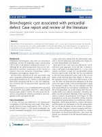
GB. Histologically, his tumor was composed of mono-
morphic cells containing small, round nuclei and eosi-
nophilic cytoplasm. His cells were organized in small
nodular, trabecular, or acinar structures surrounded by
a richly vascularized stroma but showed no mitotic
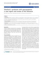
structures (Figure 2). Immunohistochemical studie s
revealed that his cells were negative for cytokeratin,
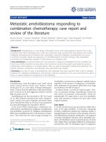
vimentin, and CD-31 and CD-34 (Figure 3). His staining
resultswerepositivefortumorcellgranulesofsynapto-
physin and chromogranin A (CgA) (Figure 4). Histologi-
cally, his GB lesion presented as an NET, and the final
diagnosis of “typical carcinoid” was made.
Post-operatively, our patient underwent a total body
comp uted tomography (CT) scan and bone scintigraphy
and the results were normal. The results of his
111
In-
pentetreotide scintigraphy, which is used to detect cells
with somatostatin receptors, were also normal. His
blood levels of glucagon, serotonin, vasoactive intestinal
peptide, somatostatin, and gastrin were normal, as were
Figure 1 Abdominal ultrasound image of a polypoid mass
between the neck and body of a gallbladder.
Figure 2 Hematoxylin-and-eosin section of a carcinoid tumor.
An organ-like growth pattern and rosettes with large cells,
prominent nucleoli, and coarse “salt and pepper” chromatin are
shown. Magnifications: ×2.5 (A), ×10 (B).
Figure 3 Tumor cells did not express cytokeratin (A), vimentin
(B), CD-31 (C), or CD-34 (D), as revealed by
immunohistochemistry. Endothelial cells positive for CD-31 (C) and
CD-34 (D). Magnification: ×20.
Mezi et al. Journal of Medical Case Reports 2011, 5:334
/>Page 2 of 5
his 24-hour urinary levels of 5-hydroxyindoleacetic acid
(5-HIAA) and CgA. After an une ventful recovery, our
patient was discharg ed in good condition, and he is dis-
ease-free 45 months after surgical treatment.
Discussion
Primary GB carcinoids are extremely rare. The first
case of a carcinoid tumor of the GB was reported in
1929, and 43 cases of carcinoid tumors have been
reported to date. Ap proximately half of the reporte d
cases of GB carcinoid tumors appear to be endocrine
cell carcinomas [3-10]. At present, 278 cases of GB
NETs are reported in the Surveillance, Epidemiology,
and End Results (SEER) database. Only five well-differ-
entiated NETs are registered in SEER, indicating that
the entity of “benign” NETs is very rare in the G B [1].
Neuroendocrine cells derive from local multipotent
gastrointestinal stem cells rather than, as initially
guessed, by migration by the neural crest. GB NETs
may develop from endocrine cells induced by intestinal
metaplasia of the b ody and fundus as well as from pre-
existing endocrine cells in the neck of the GB [1-11].
The age at presentation of GB NETs ranges from 38 to
81 years, and there is a markedly higher incidence in
women[10].Carcinoidsyndromeisveryrare(<1%),
and most GB carcinoids are diagnosed incidentally
during a histological examination of GB specimens at
autopsy, after chole cystectomy for acute or chronic
cholecystitis, or after surgery for another suspected
biliary p athology [6-8,12-17].
The case reported here was initially diagnosed as a
polyp after an ultrasound examination. PLGs are readily
detected by US [18] with high specificity (95.8%) [19].
The lifetime prevalence of GB polyps ranges from 1% to
4%. PLGs are “incidentally detected” in approximately 4%
to 7% of patients undergoing US of the GB [20], and PLG
is one of the most common diseases in biliary surgery.
The majority of GB polyps are non- neoplastic and
most commonly include cholesterol polyps (60%) or
inflammatory ones (10%). Adenomyomas represent the
second most common type of GB polyps (25%). This
type of lesion is associated with an increased incidence
of GB cancer, and the GB should be removed surgically.
Adenonomatous polyps represent a minority. They can
progress to cancer, and this risk is related to their size:
polyps larger than 1 cm are considered high-risk lesions.
The fifth class of GB polyps consist s of rare lesions that
include heterotopic gastric glands, neurofibromas, carci-
noid tumors, leiomyomas, and fibromas.
The specificity of abdominal US in PLG detection i s
high [19], but the sensitivity of US was reported to b e
low[21].EndoscopicUS(EUS)maybecomethestan-
dard to define PLGs. Studies have shown a correlation
between EUS characteristics and the actual histology of
PLGs. EUS is considered to be superior to all types of
imaging for GB lesions, particular ly for early GB cancer
because of the higher operatin g frequency (7.5 to 12
MHz) that can provide high-resolution images of small
lesions and a diagnostic sensitivity for GB malignancy of
90% [21]. High-resolution US (HRUS) has demonstrated
a diagnostic sensi tivity of as high as 90% and an accu-
racy of 62.9% for staging the depth of cancer invasion
[22]. Both EUS and HRUS minimize the changes of not
identifying pre-malignant lesion. If the polyps are severe
or appear malignant or if large or irregular lesions are
found, a CT scan should be performed in order to avoid
missing a GB carcinoma. Pre-operative suspici on and a
differential diagnosis of GB cancer are very important
for selecting the optimal treatment. CT could be used
not only to distinguish an early GB carcinoma from a
PLG but also to assess the tissue around the malignant
PLG and regional lymph node metastases [19]. Although
imaging such as US, EUS, or CT has been widely used,
it is still difficult to differentiate cancer from non-neo-
plastic lesions before an operation. Hence, differentiating
a pre-cancerous lesion from early GB cancer is essential.
The risk of malignancy is between 45% and 67% in
polyps from 1 to 1.5 cm in size [21].
Operative indications for PLGs included a maximal
diameter of 1 cm, a wide-base lesion, lesions tending to
become e nlarged in a short period, patien t age of more
than 50 years, a single p olypoid lesion, coexisting GB
stones, and a PLG associated with irregular thickening
of the local GB wall.
Figure 4 Tumor cells stained positive for chromogranin A (A,
B) and for synaptophysin (C, D). Magnifications: ×2.5 (A, C), ×10
(B, D).
Mezi et al. Journal of Medical Case Reports 2011, 5:334
/>Page 3 of 5
Our patient’s histological results after chol ecystectomy
were suggestive of an NET tumor. The determination of
the histological type of the tumor and differential diag-
nosis from GB adenocarcinoma are often difficult. The
identification of neuroendocrine cells and the immuno-
histochemical expression of marker proteins as well as
other c ell type-specific amines and peptides are neces-
sary to define a GB NET. Our patient’s immunohisto-
chemistry test results were negative for cytokeratin,
vimentin, and CD-31 and CD-34, allowing us to exclude
a likely diagnosis of adenocarcinoma, sarcoma, or vascu-
lar tumor, respectively. The combination of the high his-
tological diff erentiation, the tumor size, the absence of
angioinvasion and infiltrative growth, and the immuno-
histochemical staining supported the final diagnosis of a
“typical” rather than of an “atypical” NET tumor.
When feasible, surgical treatment, with the goal of
complete resection, is the g old standard for typical car-
cinoids of the GB. F or pre-invasive and early-detected
cancer (T1s and T1), simple cholecystectomy is probably
an adequate therapy. For advanced lesions, a more
aggressive radical surgery, including radical cholecystect-
omy and regio nal lymphadenectomy combined with a
hepatic resection in order to obtain adequa te free mar-
gins, is needed [1]. Additional therapies in an adjuvant
setting are not required for typi cal carcinoids according
to the low metastatic po tential of the neoplasia as well
as to the general insensitivity to traditional radiotherapy
and chemotherapy in low-grade cancer disease.
For many years, sieric CgA and urinary 5-HIAA, each
of which has a specifi city of nearly 100% but a low sen-
sit ivity, have been the gold standard for detecting carci-
noids and conducting follow-up [23].
111
In-pentetreotide
has a high affinity for somatostatin subtype 2 and 5
receptors, w hich are present on the cell membranes of
carcinoid tumor cells, making
111
In-pentetreotide scinti-
graphy a good technique for imaging carcinoid tumors
[24]. Standard bone scinti graphy has a higher sensitivity
for the detection of bone metastases in patients with
carcinoid tumors [25]. Post-operative specific tumor
markers, tot al body CT,
111
In-pentetreotide scintigraphy,
and bone scintigraphy tests in our patient were all nor-
mal, indicating the lack of metastases and the successful
surgical treatment of a “ typical” carcinoid of the GB.
Indeed, in one study, 82.4% of GB carcinoids rem ained
localized and only 11.8% of patients demonstrated dis-
tant metastases [3]. The same source reported a five-
year survival of 60.8% ± 14.8%. Modlin and colleagues
[1] reported a median survival of 9.8 months among 278
cases of GB NETs reported in SEER. The five-year sur-
vival rates for tumors classified as carcinoids-neuroen-
docrine carcinoma or small-cell cancer were 36.9% and
0%, respectively [1].
Conclusions
Considering the difficulties i n making a pre-operative
differential diagnosis between a benign “typical” carci-
noid and the more aggressive “ atypical ” variants or
between NET, adenocarcinoma, and benign lesions of
the GB, we emphasize the need for surgical manage-
ment for any suspected polypoid lesion, relying on the
pathologist and immunohistochemistry analyses for the
final diagnosis. We underli ne the need t o distinguish
between different forms of NETs of the GB with differ-
ent metastatic potential, prognosis, and clinical course.
Consent
Written informed consent was obtained from the patient
for publication of this case report and any accompany-
ing images. A copy of the written consent is available
for review by the Editor-in-Chief of this journal.
Abbreviations
5-HIAA: 5-hydroxyindoleacetic acid; CgA: chromogranin A; CT: computed
tomography; EUS: endoscopic ultrasonography; GB: gallbladder; HRUS: high-
resolution ultrasonography; NET: neuroendocrine tumor; PLG: polypoid lesion
of the gallbladder; SEER: Surveillance, Epidemiology, and End Results; US:
ultrasonography.
Acknowledgements
English language assistance for the preparation of this manuscript was
provided by Rod McNab, of Wolters Kluwer Medical Communications
(Auckland, New Zealand). This assistance was funded by Novartis Farma SpA
(Origgio, Italy)
Author details
1
Department of Radiology, Oncology and Human Pathology, Division of
Oncology B, “Sapienza” University of Rome, Rome, Italy.
2
Department of
Surgical Science and Biotechnology, Division of Pathology, Polo Pontino,
“Sapienza” University of Rome, Rome, Italy.
3
Department of Biopathology and
Diagnostic Imaging, Division of Nuclear Medicine, University “Tor Vergata ” ,
Rome, Italy, and IRCCS NEUROMED, Rome, Italy.
4
Department of Surgical
Science, Division of DEA, “Sapienza” University of Rome, Rome, Italy.
Authors’ contributions
SM drafted and wrote the manuscript and was involved in data
interpretation. EO was involved in the conception and design of the study.
FLT and VLT were involved in the care of our patient. VP was involved in
histological diagnosis, pathological findings, immunohistochemical studies,
and figures and contributed to writing the manuscript according to his
specialty. ML was involved in immunohistochemical studies and figures. OS
provided scintigraphic images and was responsible for critical revision of CT
images. BC was involved in administrative support. All authors read and
approved the final manuscript.
Competing interests
The authors declare that they have no competing interests.
Received: 27 October 2010 Accepted: 29 July 2011
Published: 29 July 2011
References
1. Eltawil KM, Gustafsson BI, Kidd M, Modlin IM: Neuroendocrine tumors of
the gallbladder: an evaluation and reassessment of management
strategy. J Clin Gastroenterol 2010, 44:687-695.
2. Zuetenhorst JM, Taal BG: Metastatic carcinoid tumors: a clinical review.
Oncologist 2005, 10:123-131.
Mezi et al. Journal of Medical Case Reports 2011, 5:334
/>Page 4 of 5
3. Modlin IM, Lye KD, Kidd M: A 5-decade analysis of 13,715 carcinoid
tumors. Cancer 2003, 97:934-959.
4. Modlin IM, Lye K, Kidd M: Carcinoid tumors. In Endocrine Surgery. Edited by:
Schwartz AE, Pertsemlidis D, Gagner M. New York: Marcel Dekker, Inc;
2003:611-639.
5. Capella C, Heitz PU, Hofler H, Solcia E, Kloppel G: Revised classification of
neuroendocrine tumours of the lung, pancreas and gut. Virchows Arch
1995, 425:547-560.
6. Nishigami T, Yamada M, Nakasho K, Yamamura M, Satomi M, Uematsu K,
Ri G, Mizuta T, Fukumoto H: Carcinoid tumor of the gall bladder. Intern
Med 1996, 35:953-956.
7. Kaiho T, Tanaka T, Tsuchiya S, Miura M, Saigusa N, Yanagisawa S,
Takeuchi O, Kitakata Y, Saito H, Shimizu A, Miyazaki M: A case of classical
carcinoid tumor of the gallbladder: review of the Japanese published
works. Hepatogastroenterology 1999, 46:2189-2195.
8. Mizukami Y, Nagashima T, Ikuta K, Chikamatsu E, Kurachi K, Kanemoto H,
Yagi T, Ohhira S, Nimura Y: Advanced endocrine cell carcinoma of the
gallbladder: a patient with 12-year survival. Hepatogastroenterology 1998,
45:1462-1467.
9. Soga J, Yakuwa Y, Osaka M: Carcinoid syndrome: a statistical evaluation
of 748 reported cases. J Exp Clin Cancer Res 1999, 18:133-141.
10. Modlin IM, Shapiro MD, Kidd M: An analysis of rare carcinoid tumors:
clarifying these clinical conundrums. World J Surg 2005, 29:92-101.
11. Laitio M: Goblet cells, enterochromaffin cells, superficial gastric-type
epithelium and antral-type glands in the gallbladder. Beitr Pathol 1975,
156:343-358.
12. Anjaneyulu V, Shankar-Swarnalatha G, Rao SC: Carcinoid tumor of the gall
bladder. Ann Diagn Pathol 2007, 11:113-116.
13. Deehan DJ, Heys SD, Kernohan N, Eremin O: Carcinoid tumour of the gall
bladder: two case reports and a review of published works. Gut 1993,
34:1274-1276.
14. Khetan N, Bose NC, Arya SV, Gupta HO: Carcinoid tumor of the
gallbladder: report of a case. Surg Today 1995, 25:1047-1049.
15. Konishi E, Nakashima Y, Smyrk TC, Masuda S: Clear cell carcinoid tumor of
the gallbladder. A case without von hippel-lindau disease. Arch Pathol
Lab Med 2003, 127:745-747.
16. Porter JM, Kalloo AN, Abernathy EC, Yeo CJ: Carcinoid tumor of the
gallbladder: laparoscopic resection and review of the literature. Surgery
1992, 112:100-105.
17. Ozawa K, Kinoshita M, Kagata Y, Matsubara O: A case of double carcinoid
tumors of the gallbladder. Dig Dis Sci 2003, 48:1760-1761.
18. Sugiyama M, Xie XY, Atomy Y, Saito M: Differential diagnosis of small
polypoid lesions of the gallbladder: the value of endoscopic
ultrasonography. Ann Surg 1999, 229:498-504.
19. Sun XJ, Han Y, Wang JS, Ren H: Diagnosis and treatment of polypoid of
polypoid lesions of the gallbladder: report of 194 cases. Hepatobiliary
Pancreat Dis Int 2004, 3:591-594.
20. Corwin MT, Siewert B, Sheiman RG, Kane RA: Incidentally detected
gallbladder polyps: is follow-up necessary? Long term clinical and US
analysis of 346 patients. Radiology 2011, 258:277-282[http://radiology.
rsnajnls.org/content/258/1/277.long].
21. Chattopadhyay D, Lochan R, Balupuri S, Gopinath BR, Wynne KS: Outcame
of gall bladder polypoidal lesions detected by transabdominal
ultrasound scanning: a nine years experience. Word J Gastroenterol 2005,
11:2171-2173.
22. Barreto SG: Improving the preoperative diagnostic yield of gallbladder
cancers. Ann Surg 2010, 252:572.
23. Bajetta E, Ferrari L, Martinetti A, Celio L, Procopio G, Artale S, Zilembo N, Di
Bartolomeo M, Seregni E, Bombardieri E: Chromogranin a, neuron specific
enolase, carcinoembryonic antigen, and hydroxyindole acetic acid
evaluation in patients with neuroendocrine tumors. Cancer 1999,
86:858-865.
24. Taal BG, Hoefnagel CA, Valdes Olmos RA, Boot H: Combined diagnostic
imaging with 131i-metaiodobenzylguanidine and 111in-pentetreotide in
carcinoid tumours. Eur J Cancer 1996, 32A:1924-1932.
25. Zuetenhorst JM, Hoefnageli CA, Boot H, Valdes Olmos RA, Taal BG:
Evaluation of (111)in-pentetreotide, (131)i-mibg and bone scintigraphy
in the detection and clinical management of bone metastases in
carcinoid disease. Nucl Med Commun 2002, 23:735-741.
doi:10.1186/1752-1947-5-334
Cite this article as: Mezi et al.: Neuroendocrine tumors of the
gallbladder: a case report and review of the literature. Journal of Medical
Case Reports 2011 5:334.
Submit your next manuscript to BioMed Central
and take full advantage of:
• Convenient online submission
• Thorough peer review
• No space constraints or color figure charges
• Immediate publication on acceptance
• Inclusion in PubMed, CAS, Scopus and Google Scholar
• Research which is freely available for redistribution
Submit your manuscript at
www.biomedcentral.com/submit
Mezi et al. Journal of Medical Case Reports 2011, 5:334
/>Page 5 of 5