Báo cáo y học: "Henoch-Schönlein purpura in an older man presenting as rectal bleeding and IgA mesangioproliferative glomerulonephritis: a case report" ppt
Bạn đang xem bản rút gọn của tài liệu. Xem và tải ngay bản đầy đủ của tài liệu tại đây (1.86 MB, 5 trang )
CAS E REP O R T Open Access
Henoch-Schönlein purpura in an older man
presenting as rectal bleeding and IgA
mesangioproliferative glomerulonephritis:
a case report
Wisit Cheungpasitporn
*
, Teeranun Jirajariyavej, Charles B Howarth and Raquel M Rosen
Abstract
Introduction: Henoch-Schönlein purpura is the most common systemic vasculitis in children. Typical presentations
are palpable purpura, abdomi nal pain, arthritis, and hematuria. This vasculitic syndrome can present as an
uncommon cause of rectal bleeding in older patients. We report a case of an older man with Henoch-Schönlein
purpura. He presented with rectal bleeding and acute kidney injury secondary to IgA mesangioproliferative
glomerulonephritis.
Case presentation: A 75-year-old Polish man with a history of diverticulosis presented with a five-day history of
rectal bleeding. He had first noticed colicky left lower abdominal pain two months previously. At that time he was
treated with a 10-day course of ciprofloxacin and metronidazole for possible diverticulitis. He subsequently
presented with rectal bleeding to our emergency department. Physical examination revealed generalized palpable
purpuric rash and tenderness on his left lower abdomen. Laboratory testing showed a mildly elevated serum
creatinine of 1.3. Computed tomography of his abdomen revealed a diffusely edematous and thickened sigmoid
colon. Flexible sigmoidoscopy showed severe petechiae throughout the colon. Colonic biopsy showed small vessel
acute inflammation. Skin biopsy resulted in a diagnosis of leukocytoclastic vasculitis. Due to worsening kidney
function, microscopic hematuria and new onset proteinuria, he underwent a kidney biopsy which demonstrated
IgA mesangioproliferative glomerulonephritis. A diagnosis of Henoch-Schönlein purpura was made. Intravenous
methylprednisolone was initially started and transitioned to prednisone tapering orally to complete six months of
therapy. There was marked improvement of abdominal pain. Skin lesions gradually faded and gastrointestinal
bleeding stopped. Acute kidney injury also improved.
Conclusion: Henoch-Schönlein purpura, an uncommon vasculitic syndrome in older patients, can present with
lower gastrointestinal bleeding, extensive skin lesions and renal involvement which responds well to systemic
steroid therapy. A history of diverticulosis can mislead physicians to the diagnosis of diverticular bleeding which is
more common in this age group. The clinical manifestations of the disease, including characteristic skin rash,
abdominal pain, joint inflammation and renal involvement raised the suspicious of Henoch-Schönlein purpura.
* Correspondence:
Department of Internal Medicine, Bassett Medical Center, Cooperstown, NY
13326, USA
Cheungpasitporn et al. Journal of Medical Case Reports 2011, 5:364
/>JOURNAL OF MEDICAL
CASE REPORTS
© 2011 Cheungpasitporn et al; licensee BioMed Central Ltd. This is an Open Access article distributed under the terms of the Creative
Commons Attribution License ( which permits unrestricted use, distribution, and
reprodu ction in any medium , provided the original work is properly cited.
Introduction
Henoch-Schönlein purpura (HSP) is a predominantly
pediatric vasculitic syndrome. Ninety percent of cases
occur in the pediatric age group between the ages of 3
and 15 years. HSP occurs unc ommonly in adults with
an incidence rate of 0.1 to 1.2 per million in adults over
20-years old [1]. The classic tetrad of HSP includes
palpable purpura without thrombocytopenia and coagu-
lopathy, arthritis, abdominal pain and renal involvement.
The extensive lower gastrointestinal hemorrhage due
to colitis associated with vasculitis is an uncommon
presentation of HSP and can be associated with an
increased risk of renal involvement [2]. Conversely,
colonic diverticular diseases such as diverticulitis and
diverticular bleeding commonly present in older patients
as left lower abdominal pain and rectal bleeding, respec-
tively [3]. A documented history of diverticulosis in
patients who present with gastrointestinal bleeding may
mis lead physicians to the wrong diagnosis and manage-
ment. We report a case of Henoch-Schönlein purpura
in an older man that presented as rectal bleeding and
acute kidney injury secondary to IgA mesangioprolifera-
tive glomerulonephritis.
Case Presentation
A 75-year-old Polish man with a history of kidney stones
and colonic diverti culosis presented with bright red
bleeding from his rectum for the previous five days to
our emergency department. About two months prior, he
had developed lower abdominal pain, left-sided more
than right-sided. He was seen in Ur gent Care and the
diagnosis of urolithiasis was made as he had 6 to 10 red
blood cells per high power field (RBCs/HPF) on urine
analysis. He was referred to a urologist for further evalua-
tion. Renal ultrasound was performed and showed
benign-appearing bilateral renal cysts without renal
stones or hydronephrosis. A cystoscopy was suggested,
but not pursued. During the same perio d of time, he also
noticed a generalized skin rash, more pronounced on his
lower extremities. He was asymptomatic from the rash at
that point with no itching or pain. No respiratory infec-
tions had occurred before the onset of the rash. He was
seen by his family physician for follow up of his abdom-
inal pain and was treated with a 10-day course of cipro-
floxacin and metronidazole for possible diverticulitis as
the patient had a known finding of diverticulosis on
abdominal computed tomography in the past.
He reported rectal bleeding and worsening left lower
abdominal pain for five days prior to presenting to the
emergency department for evaluation. He had had swol-
len bilateral proximal interphalangeal (PIP) joints of his
hands in the past two years; however, there was no cur-
rently active joint pain. He denied having Raynaud’ s
disease, sun sensitivity, pleurisy, urethritis, o ral aphthae,
alopecia, or acute eye problems. He also denied recent
history of non-steroidal anti-inflammatory drugs and
angiotensin-converting enzyme inhibitor s use, food alle r-
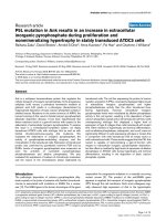
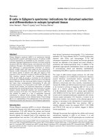
gies, an d vaccinations or insect bites. On physical exami-
nation, there was a generalized, pal pable, purpuric rash
on his trunk and both extremities, more pronounced on
his lower extremities and buttocks (Figures 1 and 2).
Abdominal examination showed mild tenderness of his
left lower abdomen without guarding or rebound. There
was bilateral pedal edema without significant joint swel-
ling. Laboratory testing showed a mildly elevated serum
creatinine of 1.3. Urine analysis was remarkable for
microscopic he matur ia; dys morphi c RBCs 20 to 25, and
new onset proteinuria; urine protein-to-creatinine ratio
was 1.53. The C-reactive protein was s lightly elevated at
1.3. Additional blood tests included anti-nuc lear antibody
(ANA), cryoglobul ins, hepatitis B and C antibodies, anti-
double stranded DNA antibodies, complement l evels,
serum protein electrophoresis, and myelop ero xidase and
PR3 antibodies that were negative. Abdominal computed
tomography with contrast showed a diffusely edematous
and thickened sigmoid colon and probably t he rectum
with surrounding inflammation. The possibilities o f
infectious colitis, ischemic colitis, and vasculitis such as
small vessel and drug-induced vasculitis were raised.
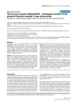
Emergency flexible sigmoidoscopy was performed and
showed severe petechiae starting just above the anal
verge, throughout the examined part of the colon, and
much more pronounced in the rectal area (Figure 3).
Colonic biopsy demonstrated small vessel, acute inflam-
mation in colonic mucosa with superficial hemorrhage
and patchy, acute cryp titis. Skin biopsy revealed leukocy-
toclastic vasculitis inv olving the small vessels. A direct
Figure 1 Skin lesion of the patient on lower extremities.
Generalized palpable purpuric rash on both lower extremities.
Cheungpasitporn et al. Journal of Medical Case Reports 2011, 5:364
/>Page 2 of 5
immunofluorescent technique showed rare colloid bodies
with antibodies to IgG and IgA, trace granular basement
membrane staining with antibod ies to C3 and trace base-
ment membrane staining with antibodies to Ig M. With
these clinical and laboratory results he was diagnosed
with a case of adult onset HSP and was initially treated
with intravenous methylprednisolone 125 mg every 8
hours for one day and intravenous isotonic fluids. There
was marked improvement of abdominal pain. Skin
lesions gradually faded away and rectal bleeding resolved.
After nephrology evaluation of acute kidney injury, along
with proteinuria and hematuria, he underw ent a kidney
biopsy which demonstrated IgA mesangioproliferative
glomerulonephritis with less than 50% crescents. Oral
prednisone tapering was continued to comp lete the six
months of steroid therapy. Acute kidney injury and
proteinuria were markedly improved after one month of
treatment. He was doing well without another episode of
rectal bleeding.
Discussion
HSP, a vasculitic syndrome characterized by skin rash,
abdominal colic, joint pain and glomerulonephritis, was
first described in 1801 by Dr. William Hebe rden [4].
Thesyndromeismainlyadiseaseofearlychildhood
with most cases presenting by 10 years of age. It is
uncommoninadultsovertheageof20.Menare
affected more than women with a ratio of 1.2: 1 to 1.8:1
[5]. A recent history of respiratory tract infection is
reported in 90% of cases. Other precipitating factors,
reported in the adult onset of HSP, include medicati ons
(non-steroidal anti-inflammatory drugs, angiotensin-con-
verting enzyme inhibitors, and antibiotics such as vanco-
mycin and cefuroxime), food allergies, vaccinations, and
insect bites.
The clinical manifestations of H SP may develop over
the course of days to weeks and may vary in their order
of presentation; however, renal involvement usually pre-
sents late. The purpuric skin lesions are typically located
on the lower extremities but may also be seen on the
hands, arms, trunk, and buttocks.
Gastrointestinal disease occurs in up to 70% of
patients varying from colicky abdominal pain, nausea
and vomiting to intestinal hemorrhage, intussusceptions,
pancreatitis and hydrops of the gall bladder. More than
30% of patients experience diffuse pain described as
‘ bowel angina’ typicall y occurring after meals and
accompanied by bloody diarrhea. Renal involvement is
usually noted within a few days to one month after the
onset of systemic symptoms. Renal manifestations occur
Figure 2 Skin lesion of the patient on lower extremities.
Generalized palpable purpuric rash on lower extremities.
Figure 3 Sigmoidoscopic findings of the patient. This slide demonstrates vasculitic colitis in HSP.
Cheungpasitporn et al. Journal of Medical Case Reports 2011, 5:364
/>Page 3 of 5
more commonly and tend to be more severe in adults
including end-stage renal disease [6]. Urinary abnormal-
ities are present in 25% to 50% of patients. Hematuria is
the most common symptom and the earliest sign of
renal involvement. Although early studies suggested that
renal involvement could not be predicted from the
severity of extra-renal involvement, a recent study
showed that a recent infectious history, pyrexia, spread
of purpura to the trunk, and biological markers of
inflammation were predictive factors for renal involve-
ment [7]. The risk of renal involvement is also increased
when HSP patients present with bloody stools [2] as in
our patient.
Joint involvement occurs in 60% to 84% of cases and
generally affects ankles and knees. In adults, involve-
ment of the small joints is more common [8]. Our
patient did not experience active joint symptoms which
is an atypical presentation.
The diagnosis of HSP is based on clinical signs and
symptoms. Laboratory studies generally show a mild
leukocytosis, a normal platelet count, and occasionally
eosinophilia. Serum complement components are nor-
mal. IgA levels are elevated in about one-half of
patients. In patients with unusual presentations, a biopsy
of an affected organ (for example, skin or kidney) that
demonstrates leukocytoclastic vasculitis with a predomi-
nance of IgA deposition confirms the diagnosis of HSP.
A kidney biopsy can be done to establish the diagno-
sis, but this invasive procedure is g enerally reserved for
patients in whom the diagnosis is uncertain or who
have more severe renal involvement such as marked
proteinuria and/or impaired renal function during the
acute episode. The percentage of glomeruli showing
crescents is the most important prognostic finding.
The long term prognosis of HSP i s almost entirely
determined by the behavior of the nephritis. The short
term outcome of renal disease in HSP is favorable in
most patients, with complete recovery reported in 94%
of children and 89% of adults [9]. Recurrence of HSP is
common, occurring in up to one-third o f patients and
more likely in patients with renal involvement. Among
adults, the reported rates of end-stage renal disease
range from 10% to 30% at 15 years.
There is n o specific treatment for HSP. The majority
of cases are mild and need onlysupportivemeasures.
Although there is evidence suggesting that corticoster-
oids enhance the rate of resolution of the arthritis and
abdominal pain, they do not seem to prevent recurrence
of disease. Aggressive therapy with corticosteroids or
cyclophosphamide has not been proven to be beneficial
in reversing the renal disease except among patients
with crescentic nephritis [10]. However, some experts
recommend a six-month course of corticosteroids for
patients with the nephrotic syndrome and those with a
reduced glomerular filtration rate. Renal transpl antation
can be performed in those patients who progress to
end-stage renal disease.
Our patien t underwent a kidney biopsy because of
marked proteinuria and acute kidney injury. The biopsy
showed IgA mesangioproliferative glomerulonephritis
with less than 50% crescents indicati ng a good prognosis.
He was initially treated with intravenous methylpredniso-
lone and was transitioned to prednisone tapering orally
to complete the six-months of steroid therapy. There was
marked improvement of abdominal pain and gastroin-
testinal bleeding. Skin lesions gradually faded. Acute kid-
ney injury and proteinuria also improved.
Conclusion
HSP is a vasculitic syndrome that can present with
extensive skin lesions, lower gastrointestinal bleeding
and renal involvement even in very old patients and
responds well to systemic steroid therapy. A history of
diverticulosis can mislead physicians to a diagnosis of
diverticular bleeding which is more common in this age
group. Physicians should be suspicious of H SP in
patients who present with clinical manifestations of the
disease comprising characteristic skin rash, abdominal
colic, joint pain and renal involvement.
Consent
Written informed consent was obtained from the patient
for publication of this case report and any accompany-
ing images. A copy of the written consent is available
for review by the Editor-in-Chief of this journal.
Abbreviations
ANA: anti-nuclear antibody ; Anti PR-3 Ab: anti-proteinase-3 antibodies; HSP:
Henoch-Schönlein purpura; Ig: Immunoglobulin; PIP: proximal
interphalangeal; RBC/HPF: red blood cells per high power field.
Acknowledgements
We acknowledge Dr. Donald A Raddatz, chief of Rheumatology at Bassett
Medical Center, who evaluated the patient and provided us with
recommendations and Dr. William W. LeCates, Program Director of the
Internal Medicine Residency Program at Bassett Medical Center, who always
encourages us to learn from patients.
Authors’ contributions
WC, CBH, and RMR were involved in the diagnosis and treatment of the
patient. WC and TJ drafted the manuscript. CBH and RMR revised the
manuscript. All authors read and approved the final manuscript.
Competing interests
The authors declare that they have no competing interests.
Received: 5 April 2011 Accepted: 10 August 2011
Published: 10 August 2011
References
1. Watts RA, Carruthers DM, Scott DG: Epidemiology of systemic vasculitis:
changing incidence or definition? Semin Arthritis Rheum 1995, 25:28-34.
2. Lanzkowsky S, Lanzkowsky L, Lanzkowsky P: Henoch Schonlein Purpura.
Paediatr Rev 1992, 13:130-137.
Cheungpasitporn et al. Journal of Medical Case Reports 2011, 5:364
/>Page 4 of 5
3. Parks TG: Natural history of diverticular disease of the colon. Clin
Gastroenterol 1975, 4:53-69.
4. Ballinger S: Henoch-Schönlein purpura. Curr Opin Rheumatol 2003,
15:591-594.
5. Trapani S, Micheli A, Grisolia F, Resti M, Chiappini E, Falcini F, De Martino M:
Henoch Schonlein purpura in childhood: epidemiological and clinical
analysis of 150 cases over a 5-year period and review of literature. Semin
Arthritis Rheum 2005, 35:143-153.
6. Ly MN, Breza TS Jr: Henoch-Schonlein purpura in an adult. Skin Med 2003,
2:262-264.
7. Tancrede-Bohin E, Ochorrisky S, Vignon-Pennamen MD, Flaseul B, Morel P,
Rybojad M: Schonlein-Henoch purpura in adult patients. Predictive
factors for IgA glomerulonephritis in a retrospective study of 57 cases.
Arch Dermatol 1997, 133:438-442.
8. Han Y, Naparstek Y: Schonlein-Henoch syndrome in adults and children.
Semin Arthritis Rheum 1991, 21:103-109.
9. Blanco R, Martínez-Taboada VM, Rodríguez-Valverde V, García-Fuentes M,
González-Gay MA: Henoch-Schönlein purpura in adulthood and
childhood: two different expressions of the same syndrome. Arthritis
Rheum 1997, 40:859-864.
10. Austin HA 3d, Balow JE: Henoch-Schönlein nephritis: Long-term
prognostic features and the challenge of therapy. Am J Kidney Dis 1983,
2:512-520.
doi:10.1186/1752-1947-5-364
Cite this article as: Cheungpasitporn et al.: Henoch-Schönlein purpura in
an older man presenting as rectal bleeding and IgA
mesangioproliferative glomerulonephritis: a case report. Journal of
Medical Case Reports 2011 5:364.
Submit your next manuscript to BioMed Central
and take full advantage of:
• Convenient online submission
• Thorough peer review
• No space constraints or color figure charges
• Immediate publication on acceptance
• Inclusion in PubMed, CAS, Scopus and Google Scholar
• Research which is freely available for redistribution
Submit your manuscript at
www.biomedcentral.com/submit
Cheungpasitporn et al. Journal of Medical Case Reports 2011, 5:364
/>Page 5 of 5