Báo cáo y học: "Teratoma of the lumbosacral region: a case report" pps
Bạn đang xem bản rút gọn của tài liệu. Xem và tải ngay bản đầy đủ của tài liệu tại đây (1.14 MB, 4 trang )
CAS E REP O R T Open Access
Teratoma of the lumbosacral region: a case
report
Mohd Faheem
1*
, Hasan H Syed
1
, Dinesh Kardam
1
, Veena Maheshwari
2
, Roobina Khan
2
and Atul Sharma
1
Abstract
Introduction: Teratoma is a tumor that usually arises from one or more germ layers. They are most commonly
found in the sacrococcygeal region and have a female preponderance. We present a very rare case of a boy with a
benign cystic teratoma in the lumbosacral region.
Case presentation: A 16-year-old Indian boy presented to our hospital with a history of a lump in the lower back
region since birth. Initially, it was small, but its size increased gradually over time to a size of 15 cm × 15 cm at
presentation. There were no other associate d abnormalities. Investigations revealed the lump to be a benign cystic
teratoma. The patient underwent surgery, and the whole tumor, from its base to the vertebrae, was excised.
Bisection of the tumor revealed that it contained hair and pultaceous material consistent with a teratoma, which
was later confirmed by histopathologic examination.
Conclusion: Benign cystic teratomas should be diagnosed and managed aggressively because they generally have
a greater tendency to progress toward malignancy. After extensively searching the case report database, we arrived
at the conclusion that this was a rare case of a benign cystic teratoma in the lumbosacral region in a boy.
Introduction
Teratomas a re germ cell tumors primarily composed of
multiple types of cells derived from one or more of the
three germ layers [1]. The term “teratoma,” which lit-
erally means “ monster” in Greek, was coined by
Virchow. Teratomas can be categorized into two types:
mature and immature. Mature teratomas can further be
classified as solid or cystic (dermoid cysts). A dermoid
cyst is lined with epithelium that contains tissues and
cells normally present in the skin layer, including hair
follicles and sebaceous and sweat glands. The most
common locations are t he sacrococcygeal region (57%),
followed by the gonads (29%), the mediastinal region
(7%), the retroperitoneum (3%), the cervical area, and
the cranium [2-4]. The “ sacrococcygeal” term is a mis-
nomer b ecause teratomas almost always arise from the
coc cyx and not from the sacral region. Teratomas show
a female preponde rance at a ratio of four to one [5,6].
However, the occurrence of a lumbosacral teratoma in a
male patient is fairly rare. Hence, the present case report
is intended to highlight this extremely rare occurrence
regarding the tumor site.
Case report
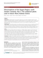
A 16-year-old Indian boy was brought to our hospital
with swelling in the midline lower back that had been
present since birth (Figure 1). The swelling had gradu-
ally increased to its size at presentation and was asso-
ciated with mild physical discomfort. Apart from these
findings, there was no significant history as far as the
patient’s swelling was concerned.
The initial examination revealed a cystic, non-mobile,
non-tender mass approximately 15 cm × 15 cm in size
attached to the back in the midline in the lumbosacral
region. However, the patient’s blood counts, urine analy-
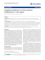
sis, and liver function test results were normal. Further-
more, the radiographs of the lumbosacral region showed
a well-defined swelling 15 cm × 20 cm in size with a
smooth margin from the L3 vertebra to the S3 vertebra
(Figure 2). On the basis of our clinical suspicion of a
cystic tumor, fine-needle aspiration cytology (FNAC)
was performed to confirm the diagnosis. The results
were positive for a mature cystic teratoma. Accordingly,
the patient was prepared for surgery, and MRI was
* Correspondence:
1
Department of Surgery, Jawahar Lal Nehru Medical College, Aligarh Muslim
University, Aligarh, India- PIN 202002
Full list of author information is available at the end of the article
Faheem et al. Journal of Medical Case Reports 2011, 5:370
/>JOURNAL OF MEDICAL
CASE REPORTS
© 2011 Faheem et al; licensee BioMed Central Ltd. This is an Open Access art icle distribut ed under the terms of the Cre ative Comm ons
Attribution License ( which permits unrestricted u se, distribution, and reproductio n in
any medium, provide d the original work i s properly cited.
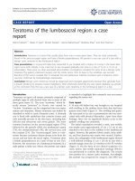
performed to establish the extent of the tumor. MRI of the
lumbosacral spine revealed a well-defined l esion in the
midline extending to the right gluteal region in the subcu-
taneous p lane from approximately the L3-L4 to the S4
vertebrae and crossing the midline. It was further observed
that the tumor was hyperintense o n T1-weighted images
and hypointense on T2-weighted i mages, which was
suggestive of fat contents. There was no obvious commu-
nication with the spinal cord (Figure 3).
The tumor was excised by creating an elliptical inci-
sion over the cyst. A whitish yellow, well-encapsulated,
non-mobile mass was observed. The tumor was carefully
dissected to allow us to reach the base, which was found
to be attac hed to the L5 lumbar verte bra. The attach-
ments, along with a small piece of lumbar vertebra,
were also removed to minimize the chance of
recurrence.
Discussion
A t eratoma is an encapsulated tumor with components
resembling normal derivatives of all three germ layers
[2]. Teratomas usually arise as masses in the sacrococcy-
geal region [7]. Their predilection for this area is most
likely related to the large number of pluripotent cells
usually found in the caudal region of the embryo, which
is closely associated with the distal sacrum and coccyx.
Being encapsulated, teratomas are usually benign,
Figure 1 Photograph showing the teratoma in the lumbosacral
region.
Figure 2 Radiograph showing the well-defined ou tline of the
teratoma.
Figure 3 MRI scan showing the teratoma at the level of the
lumbosacral region.
Faheem et al. Journal of Medical Case Reports 2011, 5:370
/>Page 2 of 4
although sometimes malignant transformation may
occur, mainly into squamous cell carc inoma [1,8,9]. It is
therefore recommended that they be excised as soon as
possible. A mature teratoma is typically benign and is
found more commonly in females, but immature terato-
mas are typically malignant and are found more often in
males.
The other differential diag noses considered in this
case were lumbosacral lipomeningomyelocele, congenital
lipo ma, and sacrococcygeal teratoma. Lipomeningomye-
loceles commonly occur in the lumbosacral area, but
the MRI examination of our patient revealed no com-
munication with the spinal cord, so this possibility was
ruled out [10]. Similarly, congenital lipoma was also
excluded from the differential diagnosis based on
FNAC, which did not show any fat cells [11]. A sacro-
coccygeal teratoma almost always arises from the coccyx
and not from the sacral area, so this possibility was
ruled out on the basis of the findings suggested by the
clinical examination and MRI [12].
The diagnosis of a teratoma is based mainly on his-
topathologic examination, although MRI is also helpful
in determining its connection with the vertebral col-
umn or its extension into t he spinal cord. Prenatally,
teratomas are usually diagnosed on the basis of obste-
tric ultrasonography in utero [7]. They appear as a
mixture of cystic and solid components. Recently, pre-
natal MRI has also been used in the imaging of
antenatal fetal anomalies. Mothers carrying fetuses
with cystic teratomas may develop polyhydramnios,
which may lead to pre-term labor secondary to uterine
distension. Volume reduc tion amniocentesis and toco-
lytics may be required to treat symptomatic polyhy-
dramnios and prevent pre-term delivery [7]. In this
case, the mother of the patient had not undergone any
prenatal ultrasonography since she was illiterate and
was not aware of the importance of prenatal ultrasono-
graphy in diagnosing neural tube defect in utero so she
did not turn up for ultrasonography. She did not
develop any difficulties during labor.
Evidence indicates that if the base is not excised along
with its attachment to underlying bone, a teratoma may
recur because it might contain totipotent cells. There-
fore, complete excision is imperative [5,13]. However, in
our patient, the base of the teratoma was found to be
attached to the L5 vertebra, a small chip of which was
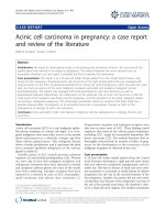
removed along with its attachment. Furthermore, the
excised specimen, which w as sent for histopathologic
examination, also revealed it to be a benign cystic t era-
toma (Figure 4).
The site of the teratoma in our patient was the L5
vertebra, which is extremely rare [14-17]. A study at
the SMS Medical College, Jaipur, India, revealed only
onecaseofthistypeofteratomaarisingfromthe
lumbosacral region (also in a female) among 75 cases
of teratomas studied over a span of 22 years (Table 1)
[13].
Conclusion
Teratomas are usually benign but sometimes may occur
as malignant tumors. To avoid any diagnostic dilemma,
it is significant to understand the rare presentation with
regard to the tumor site and the possibility of malig-
nancy. The case history and the very rare site of the
tumor described in this report will help clinicians in
diagnosing such cases and will help in enhancing clinical
knowledge and experie nce for better treatment and
patient care.
Consent
Written informed consent was obtained from the patient
for publication of this case report and any
Table 1 Anatomic sites and sex distribution of
teratomas
a
Site Patients, n (%) Men, n Women, n
Sacrococcygeal 49 (65.3) 12 37
Ovarian 10 (13.3) - 10
Testicular 5 (6.7) 5 -
Oral cavity 3 (4.0) 1 2
Retroperitoneal 2 (2.7) - 2
Cervical 2 (2.7) 2 -
Nasopharyngeal 1 (1.3) - 1
Lumbosacral 1 (1.3) - 1
Perineal 1 (1.3) 1 -
Gastric 1 (1.3) 1 -
Total 75 22 (29%) 53 (71%)
a
Data are from [13].
Figure 4 Slide showing stratified squamous epithelium within
the sebaceous gland.
Faheem et al. Journal of Medical Case Reports 2011, 5:370
/>Page 3 of 4
accompanying images. A copy of the written consent is
available for review by the Editor-in-Chief of this
journal.
Author details
1
Department of Surgery, Jawahar Lal Nehru Medical College, Aligarh Muslim
University, Aligarh, India- PIN 202002.
2
Department of Pathology, Jawahar Lal
Nehru Medical College, Aligarh Muslim University, Aligarh, India-PIN 202002.
Authors’ contributions
MF was a major contributor to the writing of the manuscript. HHS analyzed
and interpreted the patient data. VM and RK performed the histologic
examination. DK and AS helped in the writing of the manuscript. All authors
read and approved the final manuscript.
Competing interests
The authors declare that they have no competing interests.
Received: 28 October 2010 Accepted: 12 August 2011
Published: 12 August 2011
References
1. Kumar V, Abbas AK, Fausto N: The Female Genital Tract. Pathologic Basis of
Disease. 7 edition. St Louis: Elsevier; 2006, 1099-1110.
2. Teratoma, Cystic. [ />overview].
3. Barksdale EM Jr, Obokhare L: Teratomas in infants and children. Curr Opin
Pediatr 2009, 21:344-349.
4. Azizkhan R, Caty MG: Teratomas in children. Curr Opin Pediatr 1996,
8:287-292.
5. Legbo JN, Opara WE, Legbo JF: Mature sacrococcygeal teratoma: case
report. Afr Health Sci 2008, 8:54-57.
6. Sacrococcygeal Teratoma. [ />article/1760982/1906534].
7. Krishan S, Solanki R, Sethi SK: Sacrococcygeal teratoma: role of ultrasound
in antenatal diagnosis and management. JHK Coll Radiol 2004, 7:35-39.
8. Shanbhogue LKR, Bianchi A, Doig CM, Gough DCS: Management of
benign sacrococcygeal teratoma: reducing mortality and morbidity.
Pediatr Surg Int 1990, 5:41-44.
9. Terenziani M, D’Angelo P, Bisogno G, Boldrini R, Cecchetto G, Collini P,
Conte M, De Laurentis T, Ilari I, Indolfi P, Inserra A, Pira L, Siracusa F,
Spreafico F, Tamaro P, Lo Curto M: Teratoma with a malignant somatic
component in pediatric patients: the Associazione Italiana Ematologia
Oncologia Pediatrica (AIEOP) experience. Pediatr Blood Cancer 2010,
54:532-537.
10. AANS: Tethered Spinal Cord Syndrome.[ />20Information/Conditions%20and%20Treatments/Tethered%20Spinal%
20Cord%20Syndrome.aspx].
11. Pierre-Kahn A, Zerah M, Renier D, Cinalli G, Sainte-Rose C, Lellouch-
Tubiana A, Brunelle F, Le Merrer M, Giudicelli Y, Pichon J, Kleinknecht B,
Nataf F: Congenital lumbosacral lipomas. Childs Nerv Syst 1997, 13:298-335.
12. Mahour GH: Sacrococcygeal teratomas. CA Cancer J Clin 1988, 38:362-367.
13. Sharma AK, Sharma CS, Gupta AK, Sarin YK, Agarwal LD, Zaffar M: Teratoma
in pediatric age group: experience with 75 cases. Indian Pediatr 1993,
30:689-694.
14. Reid SA, Mickle JP: Myelomeningocele occurring within a lumbosacral
teratoma: case report. Neurosurgery 1985, 17:338-340.
15. Bucy PC, Haymond HE: Lumbosacral teratoma associated with spina
bifida occulta: report of a case with review of the literature. Am J Pathol
1932, 8:339-346.
16. Sharma MC, Jain D, Sarkar C, Bhatnagar V, Rishi A, Suri V, Garg A:
Lumbosacral Wilms’ tumor as a component of immature teratoma
associated with spinal dysraphism: a rare case and short literature
review. Fetal Pediatr Pathol 2009, 28:201-208.
17. Ibrahim AE, Myles L, Lang DA, Ellison DW: Case of the month: June 1998.
2 year old boy with lumbosacral mass. Brain Pathol 1998, 8:817-818.
doi:10.1186/1752-1947-5-370
Cite this article as: Faheem et al.: Teratoma of the lumbosacral region: a
case report. Journal of Medical Case Reports 2011 5:370.
Submit your next manuscript to BioMed Central
and take full advantage of:
• Convenient online submission
• Thorough peer review
• No space constraints or color figure charges
• Immediate publication on acceptance
• Inclusion in PubMed, CAS, Scopus and Google Scholar
• Research which is freely available for redistribution
Submit your manuscript at
www.biomedcentral.com/submit
Faheem et al. Journal of Medical Case Reports 2011, 5:370
/>Page 4 of 4