Introduction to the Cardiovascular System - part 6 docx
Bạn đang xem bản rút gọn của tài liệu. Xem và tải ngay bản đầy đủ của tài liệu tại đây (348.08 KB, 18 trang )
epinephrine are different because epineph-
rine binds to ␣-adrenoceptors as well as to -
adrenoceptors. Increasing concentrations of
epinephrine result in further cardiac stimula-
tion along with ␣-adrenoceptor mediated acti-
vation of vascular smooth muscle leading to
vasoconstriction. This increases arterial blood
pressure (pressor response) owing to both an
increase in cardiac output and an increase in
systemic vascular resistance.
Circulating norepinephrine affects the
heart and systemic vasculature by binding to

1
, 
2
, ␣
1
, and ␣
2
adrenoceptors; however, the
affinity of norepinephrine for 
2
and ␣
2
-
adrenoceptors is relatively weak. Therefore,
the predominant affects of norepinephrine
are mediated through 
1
and ␣
1
-adrenocep-
tors. If norepinephrine is injected intra-
venously, it causes an increase in mean arter-
ial blood pressure (systemic vasoconstriction)
and pulse pressure (owing to increased stroke
volume) and a paradoxical decrease in heart
rate after an initial transient increase in heart
rate (Fig. 6-9; Table 6-3). The transient in-
crease in heart rate is due to norepinephrine
binding to 
1
-adrenoceptors in the sinoatrial
node, whereas the secondary bradycardia is
due to a baroreceptor reflex (vagal-mediated),
which is in response to the increase in arterial
pressure.
High levels of circulating catecholamines,
caused by a catecholamine-secreting adrenal
tumor (pheochromocytoma), causes tachy-
cardia, arrhythmias, and severe hypertension
(systolic arterial pressures can exceed 200 mm
Hg).
Other actions of circulating catecholamines
include (1) stimulation of renin release with
subsequent elevation of angiotensin II (AII)
and aldosterone, and (2) cardiac and vascular
smooth muscle hypertrophy and remodeling.
These actions of catecholamines, in addition to
the hemodynamic and cardiac actions already
described, make them a frequent therapeutic
target for the treatment of hypertension, heart
failure, coronary artery disease, and arrhyth-
mias. This has led to the development and use
of many different types of ␣ and -adrenocep-
NEUROHUMORAL CONTROL OF THE HEART AND CIRCULATION 131
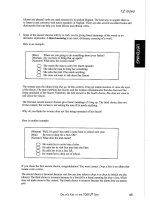
How would the changes in arterial pressure and heart rate shown in Figure 6-8 be dif-
ferent if

1
-adrenoceptors were blocked before the administration of low-dose epi-
nephrine?

1
-adrenoceptor activation is responsible for the tachycardia and increased cardiac
output produced by epinephrine. Blocking 
1
-adrenoceptors would abolish this re-
sponse. Epinephrine also binds to vascular 
2
-adrenoceptors to cause vasodilation;
therefore arterial pressure would fall during epinephrine infusion in the presence of

1
-adrenoceptor blockade because the decrease in systemic vascular resistance would
not be offset by an increase in cardiac output.
PROBLEM 6-2
How would the norepinephrine-induced changes in arterial pressure and heart rate
shown in Figure 6-9 be different in the presence of bilateral cervical vagotomy?
Bilateral cervical vagotomy would prevent vagal slowing of the heart and denervate
the aortic arch baroreceptors. Heart rate (and inotropy) would increase owing to nor-
epinephrine binding to 
1
-adrenoceptors on the heart that is now unopposed by the
vagus. This, along with aortic arch denervation, would enhance the pressor response of
norepinephrine.
PROBLEM 6-3
Ch06_117-140_Klabunde 4/21/04 11:26 AM Page 131
tor antagonists to modulate the effects of cir-
culating catecholamines as well as the norepi-
nephrine released by sympathetic nerves.
Renin-Angiotensin-Aldosterone
System
The renin-angiotensin-aldosterone system
plays an important role in regulating blood vol-
ume, cardiac and vascular function, and arterial
blood pressure. Although the pathways for
renin and angiotensin formation have been
found in a number of tissues, the most impor-
tant site for renin formation and subsequent
formation of circulating angiotensin is the kid-
ney. Sympathetic stimulation of the kidneys
(via 
1
-adrenoceptors), renal artery hypoten-
sion, and decreased sodium delivery to the dis-
tal tubules (usually caused by reduced
glomerular filtration rate secondary to reduced
renal perfusion) stimulate the release of renin
into the circulation. The renin is formed within,
and released from, juxtaglomerular cells as-
sociated with afferent and efferent arterioles of
renal glomeruli, which are adjacent to the mac-
ula densa cells of distal tubule segments that
sense sodium chloride concentrations in the
distal tubule. Together, these components are
referred to as the juxtaglomerular apparatus.
Renin is an enzyme that acts upon an-
giotensinogen, a circulating substrate syn-
thesized and released by the liver, which un-
dergoes proteolytic cleavage to form the de-
capeptide angiotensin I. Vascular endothe-
lium, particularly in the lungs, has an enzyme,
angiotensin-converting enzyme (ACE),
that cleaves off two amino acids to form the
octapeptide, angiotensin II.
Angiotensin II has several important func-
tions that are mediated by specific angiotensin
II receptors (AT
1
) (Figure 6-10). It
1. Constricts resistance vessels, thereby in-
creasing systemic vascular resistance and
arterial pressure.
2. Facilitates norepinephrine release from
sympathetic nerve endings and inhibits
norepinephrine re-uptake by nerve end-
ings, thereby enhancing sympathetic
adrenergic affects.
3. Acts upon the adrenal cortex to release al-
dosterone, which in turn acts upon the kid-
neys to increase sodium and fluid reten-
tion, thereby increasing blood volume.
4. Stimulates the release of vasopressin from
the posterior pituitary, which acts upon the
kidneys to increase fluid retention and
blood volume.
5. Stimulates thirst centers within the brain,
which can lead to an increase in blood vol-
ume.
6. Stimulates cardiac and vascular hypertrophy.
132 CHAPTER 6
60
80
100
140
180
100
120
60
FIGURE 6-9 Effects of intravenous administration of a moderate dose of norepinephrine on arterial pressure and
heart rate. Norepinephrine increases mean arterial pressure and arterial pulse pressure; heart rate transiently increases
(
1
-adrenoceptor stimulation), then decreases owing to baroreceptor reflex activation of vagal efferents to the heart.
Mean arterial pressure rises because norepinephrine binds to vascular ␣
1
-adrenoceptors, which increases systemic
vascular resistance.
Ch06_117-140_Klabunde 4/21/04 11:26 AM Page 132
Angiotensin II is continuously produced
under basal conditions, and this production
can change under different physiologic condi-
tions. For example, when a person exercises,
circulating levels of angiotensin II increase.
An increase in renin release during exercise
probably results from sympathetic stimulation
of the kidneys. Changes in body posture like-
wise alter circulating AII levels, which are in-
creased when a person stands. As with exer-
cise, this results from sympathetic activation.
Dehydration and loss of blood volume (hypo-
volemia) stimulate renin release and an-
giotensin II formation in response to renal
artery hypotension, decreased glomerular fil-
tration rate, and sympathetic activation.
Several cardiovascular disease states are as-
sociated with changes in circulating an-
giotensin II. For example, secondary hyper-
tension caused by renal artery stenosis is
associated with increased renin release and
circulating angiotensin II. Primary hyperal-
dosteronism, caused by an adrenal tumor
that secretes large amounts of aldosterone, in-
creases arterial pressure through its effects on
renal sodium retention. This increases blood
volume, cardiac output, and arterial pressure.
In this condition, renin release and circulating
angiotensin II levels are usually depressed be-
cause of the hypertension. In heart failure,
circulating angiotensin II increases in re-
sponse to sympathetic activation and de-
creased renal perfusion. Therapeutic manipu-
lation of the renin-angiotensin-aldosterone
system has become important in treating hy-
pertension and heart failure. ACE inhibitors
and AT
1
receptor blockers effectively decrease
arterial pressure, ventricular afterload, blood
volume, and hence ventricular preload, and
they inhibit and reverse cardiac and vascular
remodeling that occurs during chronic hyper-
tension and heart failure.
Note that local, tissue-produced an-
giotensin may play a significant role in cardio-
vascular pathophysiology. Many tissues and
organs, including the heart and blood vessels,
can produce renin and angiotensin II, which
have actions directly within the tissue. This
may explain why ACE inhibitors can reduce
arterial pressure and cause cardiac and vascu-
lar remodeling (e.g., diminish hypertrophy)
even in individuals who do not have elevated
NEUROHUMORAL CONTROL OF THE HEART AND CIRCULATION 133
Renin
A
II
Arterial
Pressure
Aldosterone
↑
Renal
Sodium & Fluid
Retention
Angiotensinogen
Sympathetic
Stimulation
Hypotension
Sodium
Delivery
ACE
Systemic
Vasoconstriction
Blood
Volume
A
I
Kidney
Cardiac
Output
Cardiac &
Vascular
Hypertrophy
Adrenal
Cortex
↓
↑
↑
Thirst
FIGURE 6-10 Formation of angiotensin II and its effects on renal, vascular, and cardiac function. Renin is released by
the kidneys in response to sympathetic stimulation, hypotension, and decreased sodium delivery to distal tubules.
Renin acts upon angiotensinogen to form angiotensin I (AI), which is converted to angiotensin II (AII) by angiotensin-
converting enzyme (ACE). AII has several important actions: it stimulates aldosterone release, which increases renal
sodium reabsorption; directly stimulates renal sodium reabsorption; stimulates thirst; produces systemic vasocon-
striction; and causes cardiac and vascular smooth muscle hypertrophy. The overall systemic effect of increased AII is
increased blood volume, venous pressure, and arterial pressure.
Ch06_117-140_Klabunde 4/21/04 11:26 AM Page 133
circulating levels of angiotensin II. In hyper-
tension and heart failure, for example, tissue
ACE activity is often elevated, and this may be
the primary target for the pharmacologic ac-
tions of ACE inhibitors.
Atrial Natriuretic Peptide
Atrial natriuretic peptide (ANP) is a 28-amino
acid peptide that is synthesized, stored, and
released by atrial myocytes in response to
atrial distension, angiotensin II stimulation,
endothelin, and sympathetic stimulation (-
adrenoceptor mediated). Therefore, elevated
levels of ANP are found during conditions
such as hypervolemia and congestive heart
failure, both of which cause atrial distension.
ANP is involved in the long-term regula-
tion of sodium and water balance, blood vol-
ume, and arterial pressure (Figure 6-11).
Most of its actions are the opposite of
angiotensin II, and therefore ANP is a
counter-regulatory system for the renin-
angiotensin-aldosterone system. ANP de-
creases aldosterone release by the adrenal
cortex; increases glomerular filtration rate;
produces natriuresis and diuresis (potassium
sparing); and decreases renin release, thereby
decreasing angiotensin II. These actions re-
duce blood volume, which leads to a fall in
central venous pressure, cardiac output, and
arterial blood pressure. Chronic elevations of
ANP appear to decrease arterial blood pres-
sure primarily by decreasing systemic vascular
resistance.
The mechanism of systemic vasodilation
may involve ANP receptor-mediated eleva-
tions in vascular smooth muscle cGMP (ANP
activates particulate guanylyl cyclase). ANP
also attenuates sympathetic vascular tone.
This latter mechanism may involve ANP act-
ing upon sites within the central nervous sys-
tem as well as through inhibition of norepi-
nephrine release by sympathetic nerve
terminals.
A new class of drugs that are neutral en-
dopeptidase (NEP) inhibitors may be useful
in treating heart failure. By inhibiting NEP,
the enzyme responsible for the degradation of
ANP, these drugs elevate plasma levels of
ANP. NEP inhibition is effective in some
models of heart failure when combined with
134 CHAPTER 6
↓
Aldosterone
↓
Angiotensin II
↓
Renin
Release
Natriuresis
Diuresis
↑
GFR
↓
↓
CO
↓
↓
SVR
Degradation
Atrial distension
Sympathetic
stimulation
Angiotensin II
Endothelin
NEP
Blood
Volume
CVP
↓
ANP
Arterial
Pressure
FIGURE 6-11 Formation and cardiovascular/renal actions of atrial natriuretic peptide (ANP). ANP, which is released
from cardiac atrial tissue in response to atrial distension, sympathetic stimulation, increased angiotensin II, and en-
dothelin, functions as a counter-regulatory mechanism for the renin-angiotensin-aldosterone system. ANP decreases
renin release, angiotensin II and aldosterone formation, blood volume, central venous pressure, and arterial pressure.
NEP, neutral endopeptidase; GFR, glomerular filtration rate; CVP, central venous pressure; CO, cardiac output; SVR,
systemic vascular resistance.
Ch06_117-140_Klabunde 4/21/04 11:26 AM Page 134
an ACE inhibitor. The reason for this is that
NEP inhibition, by elevating ANP, reinforces
the effects of ACE inhibition.
Brain-type natriuretic peptide (BNP), a 32-
amino acid peptide hormone related to ANP,
is synthesized and released by the ventricles in
response to pressure and volume overload,
particularly during heart failure. BNP appears
to have actions that are similar to those of
ANP. Recently, circulating BNP has been
shown to be a sensitive biomarker for heart
failure.
Vasopressin (Antidiuretic Hormone)
Vasopressin (arginine vasopressin, AVP; anti-
diuretic hormone, ADH) is a nonapeptide
hormone released from the posterior pituitary
(Figure 6-12). AVP has two principal sites of
action: the kidneys and blood vessels. The
most important physiologic action of AVP is
that it increases water reabsorption by the
kidneys by increasing water permeability in
the collecting duct, thereby permitting the
formation of concentrated urine. This is the
NEUROHUMORAL CONTROL OF THE HEART AND CIRCULATION 135
A 56-year old male patient is found to have an arterial pressure of 190/115 mm Hg.
Two years earlier he was normotensive. Diagnostic tests reveal bilateral renal artery
stenosis. Describe the mechanisms by which this condition elevates arterial pressure.
Bilateral renal artery stenosis reduces the pressure within the afferent arterioles,
which causes release of renin. This, in turn, increases circulating angiotensin II, which
stimulates aldosterone release. Activation of the renin-angiotensin-aldosterone system
causes sodium and fluid retention by the kidneys and an increase in blood volume,
which increases cardiac output. Increased vasopressin (stimulated by angiotensin II)
contributes to the increase in blood volume. Increased angiotensin II increases systemic
vascular resistance by binding to vascular AT
1
receptors and by enhancement of sympa-
thetic activity. These changes in cardiac output and systemic vascular resistance lead to
a hypertensive state.
CASE 6-1
Angiotensin II
Hyperosmolarity
Decreased atrial receptor firing
Sympathetic stimulation
Vasoconstriction
Pituitary
Renal Fluid
Reabsorption
Increased
Blood Volume
Increased
Arterial Pressure
Vasopressin
FIGURE 6-12 Cardiovascular and renal effects of arginine vasopressin (AVP). AVP release from the posterior pituitary
is stimulated by angiotensin II, hyperosmolarity, decreased atrial receptor firing (usually in response to hypovolemia),
and sympathetic activation. The primary action of AVP is on the kidney to increase water reabsorption (antidiuretic
effect), which increases blood volume and arterial pressure. AVP also has direct vasoconstrictor actions at high con-
centrations.
Ch06_117-140_Klabunde 4/21/04 11:26 AM Page 135
antidiuretic property of AVP, and it leads to an
increase in blood volume and arterial blood
pressure. This hormone also constricts arterial
blood vessels; however, the normal physio-
logic concentrations of AVP are below its va-
soactive range. Studies have shown, neverthe-
less, that in severe hypovolemic shock, when
AVP release is very high, AVP contributes to
the compensatory increase in systemic vascu-
lar resistance.
Several mechanisms regulate the release
of AVP. Specialized stretch receptors within
the atrial walls and large veins (cardiopul-
monary baroreceptors) entering the atria de-
crease their firing rate when atrial pressure
falls (as occurs with hypovolemia). Afferents
from these receptors synapse within the hy-
pothalamus, which is the site of AVP synthe-
sis. AVP is transported from the hypothala-
mus via axons to the posterior pituitary, from
where it is secreted into the circulation. Atrial
receptor firing normally inhibits the release
of AVP. With hypovolemia and decreased
central venous pressure, the decreased firing
of atrial stretch receptors leads to an increase
in AVP release. AVP release is also stimulated
by enhanced sympathetic activity accompany-
ing decreased arterial baroreceptor activity
during hypotension. An important mecha-
nism regulating AVP release involves hypo-
thalamic osmoreceptors, which sense extra-
cellular osmolarity. When osmolarity rises, as
occurs during dehydration, AVP release is
stimulated. Finally, angiotensin II receptors
located within the hypothalamus regulate
AVP release; an increase in angiotensin II
stimulates AVP release.
Heart failure causes a paradoxical increase
in AVP. The increased blood volume and atrial
pressure associated with heart failure suggest
that AVP secretion should be inhibited, but it
is not. It may be that sympathetic and renin-
angiotensin system activation in heart failure
override the volume and low pressure cardio-
vascular receptors (as well as the osmoregula-
tion of AVP) and cause the increase in AVP se-
cretion. This increase in AVP during heart
failure may contribute to the increased sys-
temic vascular resistance and to renal reten-
tion of fluid.
In summary, the importance of AVP in car-
diovascular regulation is primarily through its
effects on volume regulation, which in turn af-
fects ventricular preload and cardiac output
through the Frank-Starling relationship.
Increased AVP, by increasing blood volume,
increases cardiac output and arterial pressure.
The vasoconstrictor effects of AVP are proba-
bly important only when AVP levels are very
high, as occurs during severe hypovolemia.
INTEGRATION OF NEUROHUMORAL
MECHANISMS
Autonomic and humoral influences are neces-
sary to maintain a normal arterial blood pres-
sure under the different conditions in which
the human body functions. Neurohumoral
mechanisms enable the body to adjust to
changes in body posture, physical activity, or
environmental conditions. The neurohumoral
mechanisms act through changes in systemic
vascular resistance, venous compliance, blood
volume, and cardiac function, and through
these actions they can effectively regulate ar-
terial blood pressure (Table 6-4). Although
each mechanism has independent cardiovas-
cular actions, it is important to understand
that each mechanism also has complex inter-
actions with other control mechanisms that
serve to reinforce or inhibit the actions of the
other control mechanisms. For example, acti-
vation of sympathetic nerves either directly or
indirectly increases circulating angiotensin II,
aldosterone, adrenal catecholamines, and
arginine vasopressin, which act together to in-
crease blood volume, cardiac output, and ar-
terial pressure. These humoral changes are
accompanied by an increase in ANP, which
acts as a counter-regulatory system to limit the
effects of the other neurohumoral mecha-
nisms.
Finally, it is important to note that some
neurohumoral effects are rapid (e.g., auto-
nomic nerves and catecholamine effects on
cardiac output and pressure), whereas others
may take several hours or days because
changes in blood volume must occur before
alterations in cardiac output and arterial pres-
sure can be fully expressed.
136 CHAPTER 6
Ch06_117-140_Klabunde 4/21/04 11:26 AM Page 136
SUMMARY OF IMPORTANT
CONCEPTS
• Autonomic regulation of the heart and vas-
culature is primarily controlled by special
regions within the medulla oblongata of the
brainstem that contain the cell bodies of
sympathetic and parasympathetic (vagal)
efferent nerves.
• The hypothalamus plays an integrative role
by modulating medullary neuronal activity
(e.g., during exercise).
• Sensory information from peripheral
baroreceptors (e.g., carotid sinus barore-
ceptors) synapse within the medulla at the
nucleus tractus solitarius, which modulates
the activity of the sympathetic and vagal
neurons within the medulla.
• Preganglionic parasympathetic efferent
nerves exit the medulla as the tenth cranial
nerve and travel to the heart within the left
and right vagus nerves. Preganglionic fibers
synapse within ganglia located within the
heart; short postganglionic fibers innervate
the myocardial tissue. Preganglionic sym-
pathetic efferent nerves exit from the
spinal cord and synapse within paraverte-
bral or prevertebral ganglia before sending
out postganglionic fibers to target tissues in
the heart and blood vessels.
• Sympathetic activation increases heart rate,
inotropy, and dromotropy through the re-
lease of norepinephrine, which binds pri-
marily to postjunctional cardiac 
1
-adreno-
ceptors. Norepinephrine released by sym-
pathetic nerves constricts blood vessels by
binding to postjunctional ␣
1
and ␣
2
-
adrenoceptors. The release of norepineph-
rine from sympathetic nerve terminals is
modulated by prejunctional ␣
2
-adrenocep-
tors, 
2
-adrenoceptors and muscarinic (M
2
)
receptors.
• Parasympathetic activation decreases heart
rate, inotropy, and dromotropy, and it pro-
duces vasodilation in specific organs
through the release of acetylcholine, which
binds to postjunctional muscarinic (M
2
) re-
ceptors.
• Baroreceptors are mechanoreceptors that
respond to stretch induced by an increase
in pressure or volume. Arterial barorecep-
tor activity (e.g., carotid sinus and aortic
arch receptors) tonically inhibits sympa-
thetic outflow to the heart and blood ves-
sels, and it tonically stimulates vagal out-
flow to the heart. Decreased arterial
pressure, therefore, decreases the firing of
arterial baroreceptors, which leads to reflex
activation of sympathetic influences acting
on the heart and blood vessels and with-
drawal of the vagal activity to the heart.
• Peripheral chemoreceptors (e.g., carotid
bodies) and central chemoreceptors (e.g.,
medullary chemoreceptors) respond to de-
creased pO
2
and pH or increased pCO
2
of
the blood. Their primarily function is to
regulate respiratory activity, although
NEUROHUMORAL CONTROL OF THE HEART AND CIRCULATION 137
TABLE 6-4 EFFECTS OF NEUROHUMORAL ACTIVATION ON BLOOD VOLUME,
CARDIAC OUTPUT AND ARTERIAL PRESSURE
INCREASED BLOOD VOLUME CARDIAC OUTPUT ARTERIAL PRESSURE
Sympathetic Activity ↑↑ ↑
Vagal Activity — ↓↓
Circulating Epinephrine ↑↑↓↑*
Angiotensin II ↑↑ ↑
Aldosterone ↑↑ ↑
Atrial Natriuretic Peptide ↓↓ ↓
Arginine Vasopressin ↑↑ ↑
↑ = increase; ↓ = decrease. *dependent upon plasma epinephrine concentration.
Ch06_117-140_Klabunde 4/21/04 11:26 AM Page 137
chemoreceptor activation generally leads
to activation of the sympathetic nervous
system to the vasculature, which increases
arterial pressure. Heart rate responses de-
pend upon changes in respiratory activity.
• Reflexes triggered by changes in blood vol-
ume, cerebral and myocardial ischemia,
pain, pulmonary activity, muscle and joint
movement, and temperature alter cardiac
and vascular function.
• Sympathetic activation of the adrenal
medulla stimulates the release of cate-
cholamines, principally epinephrine. This
hormone produces cardiac stimulation (via

1
-adrenoceptors), and it either decreases
(via vascular 
2
-adrenoceptors) or in-
creases (via vascular ␣
1
and ␣
2
-adrenocep-
tors) systemic vascular resistance, depend-
ing upon the plasma concentration.
• The renin-angiotensin-aldosterone system
plays a major role in regulating renal excre-
tion of sodium and water, and therefore it
strongly influences blood pressure through
changes in blood volume. Renin is released
by the kidneys in response to sympathetic
stimulation, hypotension, and decreased
sodium delivery to distal tubules. Renin
acts upon angiotensinogen to form an-
giotensin I, which is converted to an-
giotensin II (AII) by angiotensin-convert-
ing enzyme (ACE). AII has the following
actions: (1) it stimulates aldosterone re-
lease from the adrenal cortex, which in-
creases renal sodium reabsorption; (2) it
acts on renal tubules to increase sodium re-
absorption; (3) it stimulates thirst; (4) it
produces systemic vasoconstriction; (5) it
enhances sympathetic activity; and (6) it
produces cardiac and vascular hypertrophy.
The overall systemic effect of increased AII
is increased blood volume, venous pres-
sure, and arterial pressure.
• Atrial natriuretic peptide (ANP), which is
released by the atria primarily in response
to atrial stretch, functions as a counter-reg-
ulatory mechanism for the renin-an-
giotensin-aldosterone system. Therefore,
increased ANP reduces blood volume, ve-
nous pressure, and arterial pressure.
• Arginine vasopressin (AVP; antidiuretic
hormone), which is released by the poste-
rior pituitary when the body needs to re-
duce renal loss of water, enhances blood
volume and increases arterial and venous
pressures. At high plasma concentrations,
AVP constricts resistance vessels.
Review Questions
Please refer to the appendix for the answers
to the review questions.
For each question, choose the one best
answer:
1. The cell bodies for the preganglionic vagal
efferents innervating the heart are found
in which region of the brain?
a. Cortex
b. Hypothalamus
c. Medulla
d. Nucleus tractus solitarius
2. Norepinephrine released by sympathetic
nerves
a. Binds preferentially to 
2
-adreno-
ceptors on cardiac myocytes.
b. Constricts blood vessels by binding
to ␣
1
-adrenoceptors.
c. Inhibits its own release by binding
to prejunctional 
2
-adrenoceptors.
d. Decreases renin release in the kid-
neys.
3. Stimulating efferent fibers of the right va-
gus nerve
a. Decreases systemic vascular resis-
tance.
b. Increases atrial inotropy.
c. Increases heart rate.
d. Releases acetylcholine, which binds
to M
2
receptors.
4. A sudden increase in carotid artery pressure
a. Decreases carotid sinus barorecep-
tor firing rate.
b. Increases sympathetic efferent nerve
activity to systemic circulation.
c. Increases vagal efferent activity to
the heart.
d. Results in reflex tachycardia.
138 CHAPTER 6
Ch06_117-140_Klabunde 4/21/04 11:26 AM Page 138
5. Which of the following can cause tachy-
cardia?
a. Face submersion in cold water
b. Increased blood pCO
2
c. Increased firing of carotid sinus
baroreceptors
d. Vasovagal reflex
6. Infusion of a low-to-moderate dose of epi-
nephrine following pharmacologic block-
ade of -adrenoceptors will
a. Decrease mean arterial pressure.
b. Have no significant cardiovascular
effects.
c. Increase heart rate.
d. Increase systemic vascular resis-
tance.
7. In an experimental protocol, intravenous
infusion of acetylcholine was found to de-
crease mean arterial pressure and increase
heart rate. These results can best be ex-
plained by
a. Direct action of acetylcholine on
muscarinic receptors at the sinoatrial
node.
b. Increased firing of carotid sinus
baroreceptors.
c. Reflex activation of sympathetic
nerves.
d. Reflex systemic vasodilation.
8. An increase in circulating angiotensin II
concentrations
a. Depresses sympathetic activity.
b. Increases blood volume.
c. Inhibits aldosterone release.
d. Inhibits the release of atrial natri-
uretic peptide.
9. Atrial natriuretic peptide
a. Enhances renal sodium retention.
b. Increases renin release.
c. Inhibits the release of aldosterone.
d. Increases blood volume and cardiac
output.
SUGGESTED READINGS
Berne RM, Levy MN. Cardiovascular Physiology. 8th
Ed. Philadelphia: Mosby, 2001.
Melo LG, Pang SC, Ackermann U. Atrial natriuretic
peptide: regulator of chronic arterial blood pressure.
News Physiol Sci 2000;15:143–149.
Mendolowitz D. Advances in parasympathetic control of
heart rate and cardiac function. News Physiol Sci
1999;14:155–161.
Rhoades RA, Tanner GA. Medical Physiology. 2nd Ed.
Philadelphia: Lippincott Williams & Wilkins, 2003.
Touyz CB, Dominiczak AF, Webb RC, Johns DB.
Angiotensin receptors: signaling, vascular pathophys-
iology, and interactions with ceramide. Am J Physiol
2001;281:H2337–H2365.
NEUROHUMORAL CONTROL OF THE HEART AND CIRCULATION 139
Ch06_117-140_Klabunde 4/21/04 11:26 AM Page 139
Ch06_117-140_Klabunde 4/21/04 11:26 AM Page 140
CD-ROM CONTENTS
LEARNING OBJECTIVES
INTRODUCTION
DISTRIBUTION OF CARDIAC OUTPUT
LOCAL REGULATION OF BLOOD FLOW
Tissue Factors
Endothelial Factors
Smooth Muscle (Myogenic)
Mechanisms
Extravascular Compression
Autoregulation of Blood Flow
Reactive and Active Hyperemia
SPECIAL CIRCULATIONS
Coronary Circulation
Cerebral Circulation
Skeletal Muscle Circulation
Cutaneous Circulation
Splanchnic Circulation
Renal Circulation
Pulmonary Circulation
Summary of Special Circulations
SUMMARY OF IMPORTANT CONCEPTS
REVIEW QUESTIONS
SUGGESTED READINGS
chapter
7
Organ Blood Flow
AnginaCD CONTENTS
LEARNING OBJECTIVES
Understanding the concepts presented in this chapter will enable the student to:
1. Describe the distribution of cardiac output among major organs when a person is at rest.
2. Describe how each of the following tissue factors influences blood flow: adenosine, inor-
ganic phosphate, potassium ion, carbon dioxide, hydrogen ion, tissue partial pressure of
oxygen, and paracrine hormones such as histamine, prostaglandins, and bradykinin.
3. Describe how each of the following endothelial factors influences local blood flow: nitric
oxide, endothelial-derived hyperpolarizing factor, endothelin-1, and prostacyclin.
4. Explain how extravascular compression alters blood flow in the heart and contracting
skeletal muscle.
5. Define autoregulation of blood flow, reactive hyperemia and active (functional) hyperemia.
6. Describe and contrast the local regulatory mechanisms that may be involved in autoregula-
tion, reactive hyperemia, and active hyperemia in major vascular beds of the body (coro-
nary, cerebral, skeletal muscle, cutaneous, splanchnic, renal, and pulmonary circulations).
7. Compare and contrast autonomic control of blood flow in major vascular beds of the body.
8. Describe the specialized vascular anatomy in the following organs: brain, heart, intestines
and liver, skin, kidneys, and lungs.
141
Ch07_141-170_Klabunde 4/21/04 11:43 AM Page 141
bradykinin, and prostaglandins). A paracrine
hormone is a substance released by one cell
that acts on another nearby cell by diffusing
through the interstitial fluid. This is in con-
trast to endocrine hormones that circulate
in the blood to reach distant target cells or au-
tocrine substances that affect the same cell
from which they are released.
Increases or decreases in metabolism alter
the release of some of these vasoactive sub-
stances; thus, metabolic activity is closely cou-
pled to blood flow in most organs of the body.
For example, an increase in tissue metabo-
lism, as occurs during muscle contraction or
during changes in neuronal activity in the
brain, leads to an increase in blood flow.
Extensive evidence shows that the actively
metabolizing cells surrounding arterioles re-
lease vasoactive substances that cause vasodi-
lation. This is termed the metabolic theory
of blood flow regulation. These vasoactive
substances, which are linked to tissue metab-
olism, ensure that the tissue is adequately sup-
plied by oxygen and that products of metabo-
lism (e.g., CO
2
, H
ϩ
, lactic acid) are removed.
Several substances have been implicated in
metabolic regulation of blood flow. Their rela-
tive importance depends on the tissue in
which they are formed as well as different
conditions that might cause their release.
1. Adenosine is a potent vasodilator in most
organs (although adenosine constricts renal
vessels). It is formed by the action of 5’-nu-
cleotidase, an enzyme that dephosphory-
lates cellular adenosine monophosphate
(AMP). The AMP is derived from hydroly-
sis of intracellular adenosine triphosphate
(ATP) and adenosine diphosphate (ADP).
Adenosine formation increases during hyp-
oxia and increased oxygen consumption,
both of which lead to increased ATP hy-
drolysis. Small amounts of ATP hydrolysis
can lead to large increases in adenosine for-
mation because intracellular concentra-
tions of ATP are about a thousand-fold
greater than adenosine concentrations.
Experimental evidence supports the idea
that adenosine formation is a particularly
important mechanism for regulating coro-
nary blood flow when myocardial oxygen
consumption increases or during hypoxic
conditions.
2. Inorganic phosphate is released by the
hydrolysis of adenine nucleotides (ATP,
ADP, and AMP). Inorganic phosphate may
have some vasodilatory activity in contract-
ing skeletal muscle, but its importance is
far less than that of adenosine, potassium,
and nitric oxide in regulating skeletal mus-
cle blood flow.
3. Carbon dioxide formation increases dur-
ing states of increased oxidative metabo-
lism. CO
2
concentrations in the tissue and
vasculature can also increase when blood
flow is reduced, which reduces the washout
of CO
2
. As a gas, CO
2
readily diffuses from
parenchymal cells to the vascular smooth
muscle of blood vessels, where it causes va-
sodilation. Considerable evidence indicates
that CO
2
plays a significant role in regulat-
ing cerebral blood flow through the forma-
tion of H
ϩ
.
4. Hydrogen ion increases when CO
2
in-
creases or during states of increased anaer-
obic metabolism (e.g., during ischemia or
hypoxia) when acid metabolites such as lac-
tic acid are produced. Increased H
ϩ
causes
local vasodilation, particularly in the cere-
bral circulation.
5. Potassium ion is released by contracting
cardiac and skeletal muscle. Muscle con-
traction is initiated by membrane depolar-
ization, which results from a cellular influx
of Na
ϩ
and an efflux of K
ϩ
. Normally, the
Na
ϩ
/K
ϩ
-ATPase pump is able to restore
the ionic gradients (see Chapter 2); how-
ever, the pump does not keep up with
rapid depolarizations (i.e., there is a time
lag) during muscle contractions, and a
small amount of K
ϩ
accumulates in the ex-
tracellular space. Small increases in extra-
cellular K
ϩ
around blood vessels cause hy-
perpolarization of the vascular smooth
muscle cells, possibly by stimulating the
electrogenic Na
ϩ
/K
ϩ
-ATPase pump and
increasing K
ϩ
conductance through potas-
sium channels. Hyperpolarization leads to
smooth muscle relaxation. Potassium ion
appears to play a significant role in caus-
144 CHAPTER 7
Ch07_141-170_Klabunde 4/21/04 11:43 AM Page 144
Fig. 7-3), the upper limit of the autoregulatory
range is reached and the vessels undergo no
further constriction with increases in perfu-
sion pressure; therefore, flow increases as
pressure increases. The autoregulatory re-
sponse can be modulated by neurohumoral
influences and disease states. For example,
sympathetic stimulation and chronic hyper-
tension can shift the cerebral autoregulatory
range to the right as described later in this
chapter.
Autoregulation may involve both metabolic
and myogenic mechanisms. If the perfusion
pressure to an organ is reduced, the initial fall
in blood flow leads to a fall in tissue pO
2
and
the accumulation of vasodilator metabolites.
These changes cause the resistance vessels to
dilate in an attempt to restore normal flow. A
reduction in perfusion pressure may also be
sensed by the smooth muscle in resistance
vessels, which responds by relaxing (myogenic
response), leading to an increase in flow.
Under what conditions does autoregulation
occur, and why is it important? In hypotension
caused by blood loss, despite baroreceptor re-
flexes that lead to constriction of much of the
systemic vasculature, blood flow to the brain
and myocardium will not decline appreciably
(unless the arterial pressure falls below the
autoregulatory range). This is because of the
strong capacity of these organs to autoregulate
and their ability to escape sympathetic vaso-
constrictor influences. The autoregulatory re-
sponse helps to ensure that these critical or-
gans have an adequate blood flow and oxygen
delivery even in the presence of systemic hy-
potension.
Other situations occur in which systemic
arterial pressure does not change, but in
which autoregulation is very important never-
theless. Autoregulation can occur when a dis-
tributing artery (e.g., coronary artery) to an
organ becomes partially occluded. This arte-
rial stenosis increases resistance and the pres-
sure drop along the vessel length. This re-
duces pressure in small distal arteries and
arterioles, which are the primary vessels for
regulating blood flow within an organ. These
resistance vessels dilate in response to the re-
duced pressure and blood flow caused by the
upstream stenosis. This autoregulatory re-
sponse helps to maintain normal blood flow in
the presence of upstream stenosis, and it is
particularly important in organs such as the
brain and heart in which partial occlusion of
large arteries can lead to significant reductions
148 CHAPTER 7
0
100 200
Blood Flow
A
B
B
A
Resistance
Flow
(ml/min)
Pressure
(mm Hg)
Time (min) Perfusion Pressure (mm Hg)
No Autoregulation
No Autoregulation
Autoregulation
Autoregulatory
Range
FIGURE 7-3 Autoregulation of blood flow. The left panel shows that decreasing perfusion pressure from 100 mm Hg
to 70 mm Hg at point A results in a transient decrease in flow. If no autoregulation occurs, resistance remains un-
changed and flow remains decreased. With autoregulation (red line), the initial fall in pressure leads to a decrease in
vascular resistance, which causes flow to increase to a new steady-state level despite the reduced perfusion pressure
(point B). The right panel shows steady-state, autoregulatory flows plotted against different perfusion pressures.
Points A and B represent the control flow and autoregulatory steady-state flow, respectively, from the left panel. The
autoregulatory range is the range of pressures over which flow shows little change. Below or above the autoregula-
tory range, flow changes are approximately proportional to the changes in perfusion pressure. The autoregulatory
range as well as the flatness of the autoregulatory response curve varies among organs.
Ch07_141-170_Klabunde 4/21/04 11:43 AM Page 148
vascular tone and thereby return flow to nor-
mal levels. The longer the period of occlusion,
the greater the metabolic stimulus for vasodi-
lation leading to increases in peak flow and
duration of hyperemia. Maximal vasodilation,
as indicated by a maximal peak hyperemic
flow, may occur following less than one
minute of complete arterial occlusion, or it
may require several minutes of occlusion de-
pending on the vascular bed and its metabolic
activity. For example, in the beating heart
(high metabolic activity), maximal reactive hy-
peremic responses are seen with coronary oc-
clusions of less than one minute, whereas in
resting skeletal muscle (low metabolic activ-
ity), several minutes of ischemia are necessary
to elicit a maximal vasodilator response.
Myogenic mechanisms may also contribute to
reactive hyperemia in some tissues because
arterial occlusion decreases the pressure in
arterioles, which can lead to myogenic-
mediated vasodilation.
Several examples of reactive hyperemia ex-
ist. The application of a tourniquet to a limb,
and then its removal, results in reactive hy-
peremia. During surgery, arterial vessels are
often clamped for a period of time; release of
the arterial clamp results in reactive hyper-
emia. Transient coronary artery occlusions
(e.g., coronary vasospasm) result in subse-
quent reactive hyperemia within the myo-
cardium supplied by the coronary vessel.
Active hyperemia is the increase in organ
blood flow that is associated with increased
metabolic activity of an organ or tissue. With
increased metabolic activity, vascular resis-
tance decreases owing to vasodilation and vas-
cular recruitment (particularly in skeletal mus-
cle). Active hyperemia occurs during muscle
contraction (also termed exercise or func-
tional hyperemia), increased cardiac activity,
increased mental activity, and increased gas-
trointestinal activity during food absorption.
In Figure 7-5, the left panel shows the ef-
fects of increasing tissue metabolism for 2
minutes on mean blood flow in a rhythmically
contracting skeletal muscle. Within seconds of
initiating contraction and the increase in
metabolic activity, blood flow increases. The
vasodilation is thought to be owing to a com-
bination of tissue hypoxia and the generation
of vasodilator metabolites such as potassium
ion, carbon dioxide, nitric oxide, and adeno-
sine. This increased blood flow (i.e., hyper-
emia) is maintained throughout the period of
increased metabolic activity and then subsides
as normal metabolism is restored. The ampli-
tude of the active hyperemia is closely related
to the increase in metabolic activity (e.g., oxy-
gen consumption) as shown in the right panel.
At high levels of metabolic activity, the vascu-
lature becomes maximally dilated, resulting in
a maximal increase in blood flow. Active hy-
peremia is important because it increases oxy-
150 CHAPTER 7
0246
Metabolic Activity
Flow
Steady-State Flow
Time (min)
Active or Functional
Hyperemia
Increased
Metabolism
FIGURE 7-5 Active hyperemia. The left panel shows that increasing metabolism for 2 minutes transiently increases
blood flow (active or functional hyperemia). The right panel shows that the steady-state increase in blood flow dur-
ing active hyperemia is directly related to the increase in metabolic activity until the vessels become maximally dilated
and flow can no longer increase.
Ch07_141-170_Klabunde 4/21/04 11:43 AM Page 150
epicardial arteries give off smaller branches
that dive into the myocardium and become
the microvascular resistance vessels that regu-
late coronary blood flow. The resistance ves-
sels give rise to a dense capillary network so
that each cardiac myocyte is closely associated
with several capillaries. The high capillary-to-
fiber density ensures short diffusion distances
to maximize oxygen transport into the cells
and removal of metabolic waste products
(e.g., CO
2
, H
ϩ
) (see Chapter 8).
Coronary veins are located adjacent to
coronary arteries. These veins drain into the
coronary sinus located on the posterior as-
pect of the heart. Blood flow from the coro-
nary sinus empties into the right atrium. Some
drainage also occurs directly into the cardiac
chambers through the anterior cardiac veins
and thebesian vessels.
Coronary blood flow is not steady as in
most other organs. When flow is measured
within a coronary artery, it is found to de-
crease during cardiac systole and increase
during diastole (Fig. 7-7). Therefore, most of
the blood flow to the myocardium occurs dur-
ing diastole. The reason that coronary flow is
influenced by the cardiac cycle is that during
systole, the contraction of the myocardium
compresses the microvasculature within the
ventricular wall, thereby increasing resistance
and decreasing flow. During systole, blood
flow is reduced to the greatest extent within
the innermost regions of the ventricular wall
(i.e., in the subendocardium) because this is
where the compressive forces are greatest.
(This results in the subendocardial regions be-
ing more susceptible to ischemic injury when
coronary artery disease or reduced aortic
pressure is present.) As the ventricle begins to
relax in early diastole, the compressive forces
are removed and blood flow is permitted to
increase. Blood flow reaches a peak in early
diastole and then falls passively as the aortic
pressure falls toward its diastolic value.
Therefore, it is the aortic pressure during di-
astole that is most crucial for perfusing the
coronaries. This explains why increases in
heart rate can reduce coronary perfusion. At
high heart rates, the length of diastole is
greatly shortened, which reduces the time for
coronary perfusion. This is not a problem
when the coronary arteries are normal, be-
cause they dilate with increased heart rate and
metabolism; however, if the coronaries are
diseased and their vasodilator reserve is lim-
ited, increases in heart rate can limit coronary
flow and lead to myocardial ischemia and
anginal pain.
The mechanical forces affecting coronary
flow are greatest within the left ventricle be-
cause this chamber develops pressures that are
several-fold greater than those developed by
152 CHAPTER 7
Aortic
Pressure
(mm Hg)
Coronary
Flow
(ml/min/100g)
120
80
0
200
0
0.8
Time (sec)
Diastole
Systole
Diastole
FIGURE 7-7 Pulsatile nature of coronary blood flow measured in the left coronary artery. Flow is lower during systole
because of mechanical compression of intramuscular coronary vessels. Flow is maximal early in diastole, and then it
falls as aortic pressure declines.
Ch07_141-170_Klabunde 4/21/04 11:43 AM Page 152
the right ventricle. The right ventricle, and to a
lesser extent the atria, show some effects of
contraction and relaxation on blood flow within
their musculature, but it is much less apparent
than that observed in the left ventricle.
Mean coronary blood flow (averaged over
several cardiac cycles) can range from 80
mL/min per 100 g of tissue at resting heart
rates to over 400 mL/min per 100 g during ex-
ercise (see Table 7-1). Therefore, the coronary
vasculature normally has a relatively high va-
sodilator reserve capacity.
Coronary blood flow is primarily regulated
by tissue metabolism. Adenosine has been
shown to be important in dilating the coronary
vessels when the myocardium becomes hyp-
oxic or when cardiac metabolism increases dur-
ing increased cardiac work. Experimental stud-
ies have shown that inhibiting adenosine
formation, enhancing its breakdown to inosine,
or blocking vascular adenosine receptors im-
pairs coronary vasodilation under these condi-
tions. In addition, nitric oxide has been shown
to be important in coronary vessels, particularly
in producing flow-dependent vasodilation.
Coronary vessels are innervated by both
sympathetic and parasympathetic nerves.
Unlike most other vascular beds, activation of
sympathetic nerves to the heart causes only
transient vasoconstriction (␣
1
-adrenoceptor
mediated) followed by vasodilation. The va-
sodilation occurs because sympathetic activa-
tion of the heart also increases heart rate and
inotropy through 
1
-adrenoceptors, which
leads to the production of vasodilator metabo-
lites that inhibit the vasoconstrictor response
and cause vasodilation. This is termed func-
tional sympatholysis. If 
1
-adrenoceptors are
blocked experimentally, sympathetic stimula-
tion of the heart causes coronary vasoconstric-
tion. Parasympathetic stimulation of the heart
(i.e., vagal nerve activation) elicits modest coro-
nary vasodilation owing to the direct effects of
released acetylcholine on the coronaries.
However, if parasympathetic activation of the
heart results in a significant decrease in myo-
cardial oxygen demand, local metabolic mech-
anisms increase coronary vascular tone (i.e.,
cause vasoconstriction). Therefore, parasympa-
thetic activation of the heart generally results in
a decrease in coronary blood flow, although the
direct effect of parasympathetic stimulation of
the coronary vessels is vasodilation.
Coronary blood flow is crucial for the nor-
mal function of the heart. Because of the high
oxygen consumption of the beating heart (see
Chapter 4) and the fact that the heart relies on
oxidative metabolism (see Chapter 3), coro-
nary blood flow (oxygen delivery) and the
metabolic activity of the heart need to be
tightly coupled. This is all the more important
because, as discussed in Chapter 4, the beat-
ing heart extracts more than half of the oxygen
from the arterial blood; therefore, there is rel-
atively little oxygen extraction reserve. In
coronary artery disease, chronic narrowing of
the vessels or impaired vascular function re-
duces maximal coronary blood flow (i.e., there
is reduced vasodilator reserve). When this oc-
curs, coronary flow fails to increase adequately
as myocardial oxygen demands increase (Fig.
7-8). This leads to cardiac hypoxia and im-
paired contractile function.
The relationship between coronary blood
flow and the metabolic demand of the heart is
often discussed in terms of the myocardial
oxygen supply/demand ratio. The oxygen
supply is the amount of oxygen delivered to
the myocardium in the arterial blood, which is
the product of the coronary blood flow and
ORGAN BLOOD FLOW 153
Myocardial Oxygen Consumption
Blood
Flow
Normal
Coronaries
Diseased
Coronaries
Oxygen Deficit
FIGURE 7-8 Relationship between coronary blood flow
and myocardial oxygen consumption. Coronary blood
flow increases as myocardial oxygen consumption in-
creases. However, if the coronary vessels are diseased
and have increased resistance owing to stenosis, blood
flow (and therefore oxygen delivery) will be limited at
higher oxygen consumptions leading to an oxygen
deficit and myocardial hypoxia.
Ch07_141-170_Klabunde 4/21/04 11:43 AM Page 153
arterial oxygen content. If blood flow is in the
units of mL blood/min and arterial oxygen
content is expressed in mL O
2
/mL blood, oxy-
gen delivery has the units of mL O
2
/min. The
oxygen demand of the heart is the myocardial
oxygen consumption, which is the product of
coronary blood flow and the difference be-
tween the arterial and venous oxygen con-
tents. A decrease in the oxygen supply/de-
mand ratio causes tissue hypoxia, which can
result in chest pain (angina pectoris) (see
Angina on CD). This can occur by a decrease
in oxygen supply (decreased coronary blood
flow or arterial oxygen content), an increase in
myocardial oxygen consumption, or a combi-
nation of the two. One of the therapeutic goals
for people who have coronary artery disease
and anginal pain is to increase the oxygen sup-
ply/demand ratio either by improving coro-
nary flow (e.g., coronary bypass grafts or coro-
nary stent placement) or by decreasing
myocardial oxygen consumption by reducing
heart rate, inotropy, and afterload (see
Chapter 4).
Coronary artery disease is a leading cause
of death. Both structural and functional
changes occur when coronary arteries become
diseased. Atherosclerotic processes decrease
the lumen diameter, causing stenosis. This
commonly occurs in the large epicardial arter-
ies, although the disease also afflicts small ves-
sels. The large coronary arteries ordinarily
represent only a very small fraction of total
coronary vascular resistance. Therefore,
stenosis in these vessels needs to exceed a
60% to 70% reduction in lumen diameter (i.e.,
exceed the critical stenosis) to have significant
effects on resting blood flow and maximal flow
capacity (see Chapter 5 and Stenosis on CD).
In addition to narrowing the lumen and in-
creasing resistance to flow, atherosclerosis
causes endothelial damage and dysfunction.
This leads to reduced nitric oxide and prosta-
cyclin formation, which can precipitate coro-
nary vasospasm and thrombus formation,
leading to increased vascular resistance and
decreased flow. Loss of these endothelial fac-
tors impairs vasodilation, which decreases the
vasodilator reserve capacity. When coronary
flow is compromised by coronary artery dis-
ease either at rest or during times of increased
metabolic demand (e.g., during exercise), the
myocardium becomes hypoxic, which can im-
pair mechanical function, precipitate arrhyth-
mias, and produce angina.
When coronary oxygen delivery is limited
by disease, collateral vessels can play an im-
portant adjunct role in supplying oxygen to
the heart. Conditions of chronic stress (e.g.,
chronic hypoxia or exercise training) can cause
154 CHAPTER 7
A patient with known coronary artery disease (multiple vessel stenosis) is also hyper-
tensive. Explain why blood pressure-lowering drugs that produce reflex tachycardia
should be not be used in such a patient.
It is important to control arterial pressure in patients with coronary artery disease
because hypertension increases ventricular afterload and myocardial oxygen demand.
However, it is important to lower arterial pressure using drugs that do not cause a re-
flex tachycardia for two reasons. First, reflex tachycardia (baroreceptor-mediated) in-
creases myocardial oxygen demand and offsets the beneficial effects of reducing after-
load (see Chapter 4). Second, tachycardia further impairs coronary perfusion because
the duration of diastole relative to systole decreases at elevated heart rates. This re-
duces the time available for coronary perfusion during diastole, which is the time
when the greatest amount of coronary perfusion occurs. It is common in clinical prac-
tice to give either a -blocker or calcium-channel blocker to a patient with both coro-
nary artery disease and hypertension, because both types of drugs lower pressure and
prevent reflex tachycardia.
CASE 7-1
Ch07_141-170_Klabunde 4/21/04 11:43 AM Page 154
important because cerebral function relies on
a steady supply of oxygen and cannot afford to
be subjected to a reduction in flow caused by
a fall in arterial pressure. If mean arterial
pressure falls below 60 mm Hg, cerebral per-
fusion becomes impaired, which results in de-
pressed neuronal function, mental confusion,
and loss of consciousness. When arterial pres-
sure is above the autoregulatory range (e.g., in
a hypertensive crisis), blood flow and pres-
sures within the cerebral microcirculation in-
crease. This may cause endothelial and vascu-
lar damage, disruption of the blood-brain
barrier, and hemorrhagic stroke. With chronic
hypertension, the autoregulatory curve shifts
to the right (see Fig. 7-11), which helps to
protect the brain at higher arterial pressures.
However, this rightward shift then makes the
brain more susceptible to reduced perfusion
when arterial pressure falls below the lower
end of the rightward-shifted autoregulatory
range.
Metabolic mechanisms play a dominant
role in the control of cerebral blood flow.
Considerable evidence indicates that changes
in carbon dioxide are important for coupling
tissue metabolism and blood flow. Increased
oxidative metabolism increases carbon dioxide
production, which causes vasodilation. It is
thought that the carbon dioxide diffuses into
the cerebrospinal fluid, where hydrogen ion is
formed by the action of carbonic anhydrase;
the hydrogen ion then causes vasodilation. In
addition, carbon dioxide and hydrogen ion in-
crease when perfusion is reduced because of
impaired washout of carbon dioxide.
Adenosine, nitric oxide, potassium ion, and
myogenic mechanisms have also been impli-
cated in the local regulation of cerebral blood
flow.
156 CHAPTER 7
ICP increased by:
Increased ICP:
CPP = MAP – ICP
↑ ICP
MAP
CVP
•
intracranial bleeding
•
cerebral edema
•
tumor
•
collapses veins
•
decreases effective CPP
•
reduces blood flow
Rigid Cranium
Artery
Vein
FIGURE 7-10 Effects of intracranial pressure (ICP) on cerebral blood flow. ICP is the pressure within the rigid cranium
(gray area of figure). Increased ICP decreases transmural pressure (inside minus outside pressure) of blood vessels
(particularly veins), which can cause vascular collapse, increased resistance, and decreased blood flow. Therefore, the
effective cerebral perfusion pressure (CPP) is mean arterial pressure (MAP) minus ICP. CVP, central venous pressure.
0
100 200
Blood Flow
Perfusion Pressure (mm Hg)
Normal
Chronic Hypertension
Acute Sympathetic Stimulation
FIGURE 7-11 Autoregulation of cerebral blood flow.
Cerebral blood flow shows excellent autoregulation be-
tween mean arterial pressures of 60 mm Hg and 130
mm Hg. The autoregulatory curve shifts to the right
with chronic hypertension or acute sympathetic activa-
tion. This shift helps to protect the brain from the dam-
aging effects of elevated pressure.
Ch07_141-170_Klabunde 4/21/04 11:43 AM Page 156