Echocardiography A Practical Guide to Reporting - part 6 pot
Bạn đang xem bản rút gọn của tài liệu. Xem và tải ngay bản đầy đủ của tài liệu tại đây (294.07 KB, 16 trang )
• TOE is occasionally necessary to confirm normal leaflet motion in a
valve with an equivocal EOA.
MITRAL POSITION
1. Is there regurgitation?
• An easily seen jet is usually paraprosthetic, since normal transpros-
thetic regurgitation tends to be hidden by flow shielding (unless the
LA is very large).
• The intraventricular flow recruitment region of a paraprosthetic
regurgitant jet can usually be seen even when the intra-atrial jet is
invisible. This allows the regurgitation to be localised using the
sewing-ring as a clockface.
2. Severity of mitral prosthetic regurgitation
• Severe paraprosthetic regurgitation may be obvious from:
– a large region of flow acceleration within the LV
– a broad neck
– a hyperdynamic LV
– a dense continuous-wave signal, especially with early depressurisa-
tion (dagger shape).
• If there is doubt, TOE is necessary to evaluate jet width, the size
of the intra-atrial jet, and PV flow (looking for systolic flow rever-
sal).
3. Is there evidence of obstruction? (Table 6.5)
• Most information for the diagnosis of obstruction is found from
imaging and colour flow mapping.
• Measure V
max
and mean gradient, and compare with normal values
(Appendix 2).
• Pressure half-time does not reflect orifice area in normally function-
ing prosthetic mitral valves so the Hatle orifice area formula is not
Echocardiography: A Practical Guide for Reporting
70
Table 6.4 When to suspect aortic obstruction
• Thickened or immobile cusps or occluder
• Measurements outside normal values (see Appendix 2)
• Change in measurements by about 25% on serial studies
ch06 4/5/07 1:33 pm Page 70
Prosthetic valves
71
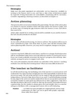
Figure 6.3 Normal transprosthetic regurgitation. (a) A thin jet of regurgitation
through a homograft aortic valve imaged in a parasternal long-axis view. (b) A tilting-disk
aortic valve imaged in an apical long-axis view, showing regurgitation related to the
major and minor orifices. (c) A bileaflet mechanical aortic valve in a parasternal short-
axis view, showing two jets from the upper and two from the lower pivotal point
(a)
(b) (c)
ch06 4/5/07 1:33 pm Page 71
valid. However, the pressure half-time lengthens significantly when the
valve becomes obstructed.
RIGHT-SIDED
• Tricuspid annuloplasty is performed if there is more than moderate
tricuspid regurgitation in the presence of left-sided disease. Tricuspid
replacement valves are not often implanted, and pulmonary replace-
ments are even less common.
1. Is there regurgitation?
• Regurgitation is easily seen after implantation of an annuloplasty ring
or with a pulmonary replacement.
• Tricuspid regurgitation may be partially shielded. Use multiple views
and look for flow reversal in the hepatic vein and a hyperdynamic RV.
2. Severity of regurgitation
• This is as for native tricuspid and pulmonary regurgitation.
Echocardiography: A Practical Guide for Reporting
72
Table 6.5 When to suspect mitral obstruction
• Thickened and immobile cusps or occluder
• Narrowed colour inflow
• Pressure half-time >200 ms with V
max
>2.5 m/s
• Change in measurements by about 25% from previous study
• Increase in PA pressure
Table 6.6 When to suspect tricuspid obstruction
1,2
• Thickened and immobile cusps or occluder
• Narrowed colour inflow
• Dilated IVC or RA
• Peak velocity >1.5 m/s (in the absence of severe tricuspid regurgitation)
• Mean gradient >5 mmHg
• Pressure half-time >240 ms
ch06 4/5/07 1:33 pm Page 72
3. Is there evidence of obstruction?
• Because of respiratory variability, measurements should be made over
several cycles for the tricuspid valve even if in sinus rhythm (Tables
6.6 and 6.7).
Prosthetic valves
73
Table 6.7 When to suspect pulmonary obstruction
3
• Cusp thickening or immobility
• Narrowing of colour flow
• V
max
>3 m/s (suspicious, not diagnostic)
• Increase in peak velocity on serial studies (more reliable)
• Impaired RV function
Checklist for reporting prosthetic valves
1. Valve position and type
2. Doppler forward flow values
3. LV dimensions and function (RV function for right-sided valves)
4. Pulmonary artery pressure
5. Any signs of obstruction?
6. Regurgitation: site and degree
REFERENCES
1. Connolly HM, Miller FA Jr, Taylor CL, et al. Doppler hemodynamic profiles of 82
clinically and echocardiographically normal tricuspid valve prostheses. Circulation
1993; 88:2722–7.
2. Kobayashi Y, Nagata S, Ohmori F, et al. Serial doppler echocardiographic evaluation
of bioprosthetic valves in the tricuspid position. J Am Coll Cardiol 1996; 27:1693–7.
3. Novaro GM, Connolly HM, Miller FA. Doppler hemodynamics of 51 clinically and
echocardiographically normal pulmonary valve prostheses. Mayo Clin Proc 2001;
76:155–60.
ch06 4/5/07 1:33 pm Page 73
ch06 4/5/07 1:33 pm Page 74
7
ENDOCARDITIS
The echocardiographic signs of endocarditis are as follows:
• vegetation
• local complication (Table 7.1)
• valve destruction.
1. Is there a vegetation?
• This is typically a mass attached to the valve and moving with a
different phase to the leaflet.
• However, sometimes it may be difficult to differentiate from other
types of masses (e.g. calcific or myxomatous degeneration). A term
should be chosen that will not lead to overdiagnosis of endocarditis
(Table 7.2).
• Note the size and mobility of the vegetation. Highly mobile masses
larger than 10 mm in length
1
have a relatively high risk of embolisa-
tion and may affect the decision for surgery.
2. Is there a local complication? (Table 7.1)
• A new paraprosthetic leak is a reliable sign of prosthetic endocarditis
provided there is a baseline postoperative study showing no leak.
Table 7.1 Local complications of endocarditis
• Abscess (Figure 7.1)
• Fistula
• Perforation
• Aneurysm of a leaflet
• Dehiscence of a replacement valve
ch07 4/5/07 1:34 pm Page 75
• An abscess usually suggests that surgery will be necessary.
3. Is there valve destruction?
• New or worsening regurgitation is a sign of endocarditis, even if no
vegetation is visible.
• Disruption of the edges of a cusp suggests endocarditis.
• Severe or progressive regurgitation suggest the need for early surgery.
Echocardiography: A Practical Guide for Reporting
76
Figure 7.1 Aortic abscess. Parasternal short-axis view showing cavities between
the PA and aorta and in the anterior aorta. The aortic valve cusps are thickened
because of endocarditis
Table 7.2 Terms suitable for describing a mass
• ‘Typical of a vegetation’
• ‘Consistent with a vegetation’
• ‘Consistent but not diagnostic of a vegetation’
• ‘Consistent with a vegetation but more in keeping with calcific
degeneration’
• ‘Most consistent with calcific degeneration’
ch07 4/5/07 1:34 pm Page 76
Endocarditis
77
4. Assess the LV
• Progressive systolic dilatation of the LV is one criterion for surgery.
• If there is acute severe aortic regurgitation, look for signs of a raised
LV end-diastolic pressure as an indication for urgent surgery:
– on M-mode, closure of the mitral valve at or before the Q wave
– on transmitral pulsed Doppler, an E deceleration time <150 ms
– diastolic mitral regurgitation.
5. Assess predisposing abnormality
See Table 7.3.
6. Is TOE necessary?
See Table 7.4.
Table 7.4 Indications for TOE in endocarditis
• Prosthetic valve
• Pacemaker
• Suspicion of abscess on transthoracic study
• Normal or equivocal TTE and continuing clinical suspicion of
endocarditis
Checklist for reporting endocarditis
1. Is there a vegetation, local complication, or evidence of valve destruction?
2. Grade of regurgitation?
3. Severity of predisposing disease (e.g., valve stenosis or VSD)
4. LV dimensions and function (or RV for tricuspid valve endocarditis)
Table 7.3 Predisposing abnormalities
• Valve disease
• Replacement heart valves
• Congenital disease (other than ASD)
• Hypertrophic cardiomyopathy
ch07 4/5/07 1:34 pm Page 77
REFERENCE
1. Thuny F, Disalvo G, Belliard O, et al. Risk of embolism and death in infective
endocarditis: prognostic value of echocardiography: a prospective multicenter study.
Circulation 2005; 112:69–75.
Echocardiography: A Practical Guide for Reporting
78
ch07 4/5/07 1:34 pm Page 78
8
AORTA
• The ascending thoracic aorta should be examined if the initial
minimum standard study shows:
– aortic dilatation
– significant aortic stenosis or regurgitation
– a bicuspid aortic valve.
• The whole of the thoracic aorta and also the abdominal aorta should
be examined in patients with:
– suspected aortic dissection (usually using TOE)
– a predisposition to aortic dilatation (e.g., Marfan syndrome,
Ehlers–Danlos syndrome type IV)
– a widened mediastinum on the chest X-ray
– trauma (usually using TOE).
AORTIC DILATATION
1. What is the diameter of the aorta?
• Measure the diameter at all levels (Figure 8.1) and compare with
normal ranges (Table 8.1).
• Aortic size is related to body habitus and age (Table 8.1); and see
Figures A1.3 and A1.4 in Appendix 1).
• A sinotubular junction diameter greater than the annulus diameter by
around 20% suggests early dilatation, even if the absolute values are
normal.
• Typical dilatation in Marfan syndrome affects predominantly annulus
and sinuses, causing a ‘pear-shaped’ aorta. Arteriosclerotic dilatation
typically affects the ascending aorta.
• Minimum thresholds for referral for surgery are given (Table 8.2).
ch08 4/5/07 1:34 pm Page 79
Echocardiography: A Practical Guide for Reporting
80
Table 8.1 Normal ranges for aortic diameter (cm)
1–5
Site Range Indexed to BSA
A Annulus 1.7–2.5 1.1–1.5
B Sinus of Valsalva 2.2–3.6 1.4–2.1
C Sinotubular junction 1.8–2.6 1.0–1.6
D Ascending 2.1–3.4
E Arch 1.4–2.9 0.8–1.9
F Descending 1.1–2.3 0.8–1.2
G Abdominal 1.0–2.2 0.6–1.3
Table 8.2 Thresholds for considering surgical referral in aortic dilatation
Arteriosclerotic dilatation 5.5 cm
a,6
Marfan and Ehlers–Danlos syndromes 4.5 cm
a,6,7
Bicuspid valve 5.0 cm (or 2.5 cm/m
2
)
8
Bicuspid valve if aortic valve replacement is 4.5 cm
8
independently indicated
The maximum diameter is used, regardless of level
a
Some recommend surgery at 6 cm in arteriosclerotic dilatation and 5.5 cm in Marfan
syndrome. Lower thresholds assume a young fit subject and a specialist surgical team with
excellent results. The decision for surgery also depends on the rate of increase in
diameter and on clinical factors.
2. How much aortic regurgitation?
See page 46.
3. Check for coarctation
• If there is a bicuspid aortic valve or unexplained aortic dilatation in
a young subject.
BEFORE AORTIC VALVE SURGERY
1. Dimensions of ascending aorta
See Table 8.1: A–D.
ch08 4/5/07 1:34 pm Page 80
Aorta
81
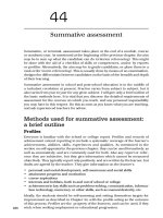
Figure 8.1 Levels for measuring the diameter of the aorta. Many normal ranges
are based on measurements taken from leading edge to leading edge, while current
guidelines for assessment recommend measuring from inner edge to inner edge.
Errors based on this discrepancy are likely to be small. (a) Parasternal long-axis view
of the annulus (level A in Table 8.1), sinus (level B), sinotubular junction (level C),
and ascending aorta (level D). (b) Suprasternal view of the arch (level E) (two
possible measurement sites). (c) Parasternal long-axis view showing the descending
thoracic aorta (level F) in short-axis. (d) Rotated view to show the descending
thoracic aorta in long-axis. (e) Abdominal aorta (level G) in a subcostal view
(a) (b)
(c) (d)
(e)
A
B
C
D
E
E
F
F
G
G
ch08 4/5/07 1:34 pm Page 81
2. Is there significant calcification in the aorta?
• Severe calcification may preclude implanting a stentless valve, may
affect the site of the trochars for the bypass machine, and may
occasionally preclude aortic valve replacement altogether.
DISSECTION
1. Is there a dissection flap?
• An intraluminal flap is the hallmark of dissection. Blooming from
calcium deposits or reverberation artifact can sometimes cause confu-
sion.
• TTE has limited diagnostic power in dissection. If the study is normal,
TOE is always necessary if the clinical suspicion is high (Table 8.3).
• Even if TOE is needed to delineate an intrathoracic flap, a trans-
thoracic study is better at showing the distal extent of the dissection
in the abdominal aorta.
2. What is the maximum aortic diameter?
3. How much aortic regurgitation?
4. Is there pericardial fluid?
• This suggests rupture into the pericardial sac, which is a common
cause of death in acute dissection. It may suggest the diagnosis even
if a flap cannot be imaged.
Echocardiography: A Practical Guide for Reporting
82
Table 8.3 Role of TOE in suspected dissection
• Detection of dissection flap
• Detection of mural haematoma
• Aortic diameters
• Entry tear
• Involvement of head and neck vessels
• Thrombosis of false lumen
ch08 4/5/07 1:34 pm Page 82
Aorta
83
5. LV function
• Impaired LV function on TTE can guide the decision for conservative
management, especially in dissections involving only the descending
thoracic aorta.
MARFAN AND EHLERS–DANLOS SYNDROMES
1. Aortic diameters at all levels
See Table 8.1: A–G.
2. How much aortic regurgitation?
3. Is there mitral or tricuspid prolapse or mitral annulus
calcification?
4. Is there coexistent PA dilatation?
See Table 8.4.
COARCTATION
1. Describe the coarctation
• From the suprasternal position, describe the site in relation to the left
subclavian artery and appearance (membrane, tunnel) using imaging
and colour flow.
• Measure the aortic dimensions above and below the coarctation.
Table 8.4 Normal PA dimensions
1
RV outflow diameter 1.8–3.4 cm
Pulmonary valve annulus 1.0–2.2 cm
Main PA 0.9–2.9 cm
Right pulmonary branch 0.7–1.7 cm
Left pulmonary branch 0.6–1.4 cm
ch08 4/5/07 1:34 pm Page 83
Echocardiography: A Practical Guide for Reporting
84
Checklist for reporting the aorta
1. Diameter at each level
2. Aortic regurgitation
Marfan and Ehlers–Danlos syndromes
1, 2, and
3. Mitral (and tricuspid) prolapse and annular calcification
4. PA diameter
Suspected dissection
1, 2, and
5. Dissection flap
6. Pericardial effusion
Coarctation
7. Site
8. Peak velocity
9. Aortic diameter above and below the coarctation and in the ascending aorta
10. Check for bicuspid aortic valve and associated LV hypertrophy
Figure 8.2 Coarctation. Continuous-wave recording from the suprasternal notch
ch08 4/5/07 1:34 pm Page 84
Aorta
85
2. Continuous-wave recording
• The most reliable feature on continuous-wave recording is forward
flow during diastole (Figure 8.2). Elevated flow velocities are usually
seen in systole, but may occasionally be absent or difficult to record
if there is a severe or complete coarctation with extensive collaterals.
Measure the peak velocity.
3. General
• Look for associated aortic root dilatation and bicuspid aortic valve.
• Check LV mass and LV function.
REFERENCES
1. Triulzi MO, Gillam LD, Gentile F. Normal adult cross-sectional echocardiographic
values: linear dimensions and chamber areas. Echocardiography 1984; 1:403–26.
2. Davidson WR Jr, Pasquale MJ, Fanelli C. A Doppler echocardiographic examination
of the normal aortic valve and left ventricular outflow tract. Am J Cardiol 1991;
67:547–9.
3. Unpublished work. Guy’s Hospital London. Guy’s Database, 1995.
4. Mintz GS, Kotler MN, Segal BL, Parry WR. Two dimensional echocardiographic recog-
nition of the descending thoracic aorta. Am J Cardiol 1979; 44:232–8.
5. Schnittger I, Gordon EP, Fitzgerald PJ, Popp RL. Standardized intracardiac measure-
ments of two-dimensional echocardiography. J Am Coll Cardiol 1983; 2:934–8.
6. Elefteriades JA. Natural history of thoracic aortic aneurysms: indications for surgery,
and surgical versus nonsurgical risks. Ann Thorac Surg 2002; 74(5):S1877–80; discus-
sion S1892–8.
7. Ergin MA, Spielvogel D, Apaydin A, et al. Surgical treatment of the dilated ascending
aorta: when and how? Ann Thorac Surg 1999; 67:1834–9; discussion 1853–6.
8. Bonow RO, et al. ACC/AHA 2006 guidelines for the management of patients with
valvular heart disease: a report of the American College of Cardiology/American Heart
Association Task Force on Practice Guidelines. J Am Coll Cardiol 2006; 48:e1–148.
9. Erbel R, Alfonso F, Boileau C, et al. Diagnosis and management of aortic dissection.
Eur Heart J 2001; 22:1642–81.
ch08 4/5/07 1:34 pm Page 85