Echocardiography A Practical Guide to Reporting - part 8 potx
Bạn đang xem bản rút gọn của tài liệu. Xem và tải ngay bản đầy đủ của tài liệu tại đây (547.21 KB, 16 trang )
• Perform a systematic study, allowing a specialist review if necessary.
• ‘AV valve’ refers to the tricuspid and mitral valves and ‘semilunar
valve’ to the pulmonary and aortic valves. Thus, ‘left AV valve’ makes
no assumption that this is truly the mitral valve.
• ‘Anatomic LV’ means the ventricle on the left of the heart; ‘morpho-
logic LV’ means the ventricle attached to the mitral valve.
• Discordant means incorrect connections – for example, LA attached
to morphologic RV or aorta leading from morphologic RV.
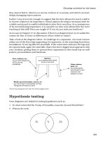
1. Are the atria correctly positioned?
• The morphologic LA and RA are distinguished by their appendages.
Since these may not be seen transthoracically, the relationship of the
abdominal vessels (‘situs’) is used.
• Are the IVC and abdominal aorta normally related in an abdominal
short-axis view?
• Follow the IVC (and SVC if visible) to the RA using subcostal views
and check that this is on the correct side of the heart.
2. Are the atria attached to the correct ventricles?
• This is usually appreciated best from the apical 4-chamber view.
• The morphologic RV is recognised because:
– its AV valve is more apical than the left-sided AV valve and it has
a septal attachment
– there is an offset between the AV valve and the semilunar valve
– there are more trabeculations than in the morphologic LV
– there is usually a moderator band.
3. Are the ventricles attached to the correct great arteries?
• The PA bifurcates early and the aorta has an arch giving off the head
and neck branch arteries.
Echocardiography: A Practical Guide for Reporting
102
Table 11.4 Findings suspicious of congenital disease
• Dilated or hypertrophied RV
• Pulmonary hypertension
• PA and aorta seen in cross-section in the same plane
• Hyperdynamic LV without aortic or mitral regurgitation
• Lack of off-setting between left and right AV valves (Figure 11.3)
• Dilated coronary sinus (Figure 11.4)
ch11 4/5/07 1:35 pm Page 102
Adult congenital disease
103
Figure 11.2 PDA. The defect from a suprasternal position (a) and on continuous-
wave (b) from a parasternal window in a large duct with normal pulmonary
pressures
(a)
(b)
ch11 4/5/07 1:35 pm Page 103
Echocardiography: A Practical Guide for Reporting
104
Figure 11.3 AVSD. (a) A normal 4-chamber view showing that the tricuspid valve
is offset or closer to the apex than the mitral valve. (b) Here there is lack of
offsetting, implying that there is a common AV valve. In addition, there is a large
ASD.
(a)
(b)
ch11 4/5/07 1:35 pm Page 104
Adult congenital disease
105
• Congenitally corrected transposition of the great arteries is suspected
if both great vessels can be imaged in transverse section from a
parasternal short-axis view or longitudinally in a subcostal view.
4. Assess the size and systolic function of both ventricles
(pages 5 and 89)
• It is usual for a morphologic RV connected to the systemic circula-
tion to be dilated.
5. Estimate PA pressure (page 94)
• Make sure to assess the AV valve jet from the ventricle connected to
the pulmonary circulation.
• If there is outflow obstruction from valve stenosis or a band, the
calculated pressures will reflect RV and not PA systolic pressure.
6. Are there any shunts at atrial or ventricular level?
• Measure size anatomically and estimate the shunt (page 138).
Figure 11.4 Dilated coronary sinus (arrow). This is usually caused by a left-sided
SVC
ch11 4/5/07 1:35 pm Page 105
7. Are the cardiac valves normal in appearance?
• Assess stenosis and regurgitation as for acquired valve disease.
• If there is no offsetting of the AV valves, the diagnosis is likely to be
an AVSD (Figure 11.3).
8. Is there aortic coarctation or a PDA?
See pages 83 and 100.
9. Are the origins of the coronary arteries correctly
positioned?
Echocardiography: A Practical Guide for Reporting
106
Table 11.5 TTE after device-closure of an ASD or PFO
• Position of device
• Is there any residual atrial shunt? A small central leak through the
device is normal and will disappear with time.
• Does the device obstruct the IVC or SVC?
• Is the device close to the mitral valve and is there new or worse mitral
regurgitation?
• RV size and function (size may start to fall soon after closure)
• Pericardial effusion (as a sign of perforation during the procedure)
Table 11.6 Transthoracic study after closure of a VSD or PDA
• Is there a residual shunt? If more than trivial, perform a shunt
calculation
• LV size and function
• Aortic regurgitation if closure of perimembranous VSD
• PA pressure
Table 11.7 Transthoracic study after coarctation repair or stenting
• Is there a residual, new or in-stent stenosis on imaging?
• On continuous-wave, an elevated systolic velocity is normal, but there
should be no diastolic forward flow
• Assess the aorta beyond the repair, looking for aneurysmal dilatation
• Assess the ascending aorta and aortic valve if bicuspid
ch11 4/5/07 1:35 pm Page 106
10. Is the coronary sinus dilated? (Figure 11.4)
• This is usually caused by a left-sided SVC (image from the medial edge
of the left supraclavicular fossa), occasionally by insertion of an
aberrant coronary artery.
POST-PROCEDURE STUDIES
• Checklists are given for the echocardiogram after:
– device-closure of an ASD or PFO (Table 11.5)
– closure of VSD or PDA (Table 11.6)
– coarctation repair (Table 11.7)
– Fallot repair (Table 11.8)
– Mustard–Senning procedures (Table 11.9)
• If the procedure is unknown, then perform a systematic examination
(page 100).
Adult congenital disease
107
Table 11.8 Transthoracic study after repair of tetralogy of Fallot
a
• Situs
• RV size and function, including RV pressure
• Is the VSD patch fully competent?
• Aortic dimensions (looking for aortic dilatation)
• Assess pulmonary valve for stenosis and regurgitation
• Assess branch PA flow on colour and pulsed Doppler if possible
• Assess the RV outflow tract for muscular hypertrophy
a
Over-riding aorta with VSD, RV outflow obstruction, with RV hypertrophy
Table 11.9 Mustard–Senning procedures
a
• Check unobstructed flow (V
max
<1.5 m/s) through the IVC and SVC
arms of the baffle to mitral valve, then LV, then PA (Figure 11.5a)
• Check unobstructed flow of pulmonary venous return (V
max
<1.5 m/s) to
the tricuspid valve, then RV, then aorta (Figure 11.5b)
• Check for baffle leak
• LV size and function (expected to be small)
• Systemic RV size and function (expected to be dilated)
a
For transposition of the great arteries
ch11 4/5/07 1:35 pm Page 107
Echocardiography: A Practical Guide for Reporting
108
Figure 11.5 Appearance after the Mustard procedure. There is unobstructed
flow from the great veins to the mitral valve (a) and from the pulmonary veins to
the RV (b)
(a)
(b)
ch11 4/5/07 1:35 pm Page 108
12
PERICARDIAL
EFFUSION
1. Pericardial or pleural?
See Table 12.1 and Figure 12.1.
2. Size and distribution
• Size (Table 12.2) is far less important than whether there are signs of
tamponade (Table 12.3).
Table 12.1 Differential diagnosis of pericardial and pleural effusions
Pericardial Pleural
Ends anterior to the descending Ends posterior to the descending
aorta aorta
Minor overlapping of LA May significantly overlap LA
Fluid between heart and diaphragm No fluid between heart and
on subcostal view diaphragm on subcostal view
Tamponade may be present No signs of tamponade
Rarely >4 cm May be >4 cm
If large, swinging of the heart Heart fixed
Table 12.2 Guideline to effusion size by maximum separation of the pericardial
layers
Small Moderate Large
<1 cm 1–3 cm >3 cm
ch12 4/5/07 1:35 pm Page 109
Echocardiography: A Practical Guide for Reporting
110
Figure 12.1 Pericardial versus pleural fluid. A pericardial effusion ends anterior to
the descending thoracic aorta (arrow); a pleural effusion ends posterior to the aorta.
A pleural effusion may extend over the LA; a pericardial effusion never does to any
significant degree
ch12 4/5/07 1:35 pm Page 110
Pericardial effusion
111
• Is the effusion generalised/posterior/apical/anterior?
• Is there enough fluid in the subcostal view for safe pericardiocentesis
(usually >2 cm)?
• What is the consistency of the fluid? Echodense collections may not
drain via a needle. Localised strands and masses are common if the
protein content is high, and do not usually represent a separate
pathology (e.g. metastasis).
Table 12.3 Echocardiographic evidence of tamponade
• Dilated IVC (>2 cm) with inspiratory collapse of <50% (Figure 12.2)
• Fall in aortic or early diastolic mitral velocity during inspiration >25%
(Figure 12.3)
1
• Prolonged and widespread diastolic RV collapse
• Note that collapse of the RA and RV outflow tract are non-specific
signs
Figure 12.2 Engorged inferior vena cava. There is little change in diameter
throughout the respiratory cycle or after a sniff
ch12 4/5/07 1:35 pm Page 111
3. Is tamponade present?
See Table 12.3.
4. General
• Is LV function poor? (Pericardiocentesis may cause circulatory
collapse.)
• If the effusion is small but there is respiratory variability of left-sided
velocities or the patient is significantly breathless, consider effusive–
constrictive pericarditis (a mixture of effusion and pericardial constric-
tion).
• If the pericardial region looks abnormal but there is no obvious
effusion, consider the causes listed in Table 12.4.
• In patients with unexplained hypotension after cardiac surgery, look
for localised effusion or haematoma, e.g. over the atria (usually
requires TOE).
Echocardiography: A Practical Guide for Reporting
112
Figure 12.3 Increased paradox. The subaortic peak velocity falls by >25% during
inspiration.
ch12 4/5/07 1:35 pm Page 112
Pericardial effusion
113
Table 12.4 Causes of an abnormal pericardial region
• Pericardial cyst
• Haematoma – usually after surgery or trauma
• Fat – this causes a layer of moderate echogenicity, usually anteriorly and
usually in obese subjects
• LV pseudo-aneurysm (page 25)
• Extrinsic mass
• Oesophageal hernia
Checklist for reporting pericardial effusion
1. Size and distribution
2. Evidence of tamponade
3. Is there enough fluid in the direction of proposed drainage
4. LV function
REFERENCE
1. Goldstein JA. Cardiac tamponade, constrictive pericarditis, and restrictive cardio-
myopathy. Curr Prob Cardiol 2004; 29:503–67.
ch12 4/5/07 1:35 pm Page 113
ch12 4/5/07 1:35 pm Page 114
13
MASSES
1. Describe the characteristics of the mass
See Table 13.1.
2. A mass attached to a valve
• This can be globular or thin (Table 13.2).
Table 13.1 Characteristics of the mass
• Site and attachment
• Size, shape
• Density (low intensity, dense, mixed)
• Mobility (fixed, mobile, free)
Table 13.2 Mass attached to a valve
Globular
• Vegetation
• Fibroelastoma
• Myxomatous tissue
Thin
• Ruptured chord
• Fibrin strand
ch13 4/5/07 1:35 pm Page 115
Echocardiography: A Practical Guide for Reporting
116
3. LA or RA mass
• A mass attached to the atrial septum is likely to be a myxoma.
• A fixed mass attached away from the septum is likely to be malig-
nant. An associated pericardial effusion makes an angiosarcoma likely.
• For an RA mass, check the IVC for tumour extension from a primary
in the kidney or ovaries. Thrombus from a DVT may also appear in
the IVC. Typically, a tumour will cause IVC dilatation, while a
thrombus will not.
• For a sessile LA mass, check the pulmonary veins and look for a
tumour mass outside the heart.
• LA thrombus is unlikely without a substrate (dilated LA, mitral
stenosis).
• For causes of a non-pathologic atrial structure, see Table 13.3.
4. LV or RV mass (Table 13.4)
• Characteristics of thrombus are given on page 24. If there is uncer-
tainty, use different views and consider transpulmonary contrast.
• Could the mass be normal? (Table 13.5)
Table 13.3 Non-pathologic RA ‘masses’
• Chiari membrane or net
• Eustachian valve
• Atrial septal aneurysm
• Atrial septal fat
• Pacemaker electrode
• Long central line
Table 13.4 Causes of LV or RV masses
• Thrombus
• Endomyocardial fibrosis
• Metastasis
• Primary tumour (e.g. rhabdomyosarcoma)
• Sarcoid
ch13 4/5/07 1:35 pm Page 116
5. Extrinsic masses
See Table 13.6.
6. Haemodynamic effect
• Assess the presence and degree of valve regurgitation or obstruction
to inflow, depending on the site of the mass.
Masses
117
Table 13.5 Normal LV or RV ‘masses’
• Trabeculation
• Prominent RV moderator band
• LV false tendon
• Prominent papillary muscle
Checklist for reporting a mass
1. Location and site of attachment
2. Size and density
3. Mobility
4. Involvement of adjacent veins
5. Haemodynamic effect
Table 13.6 Masses outside the heart
• Tumour
• Hiatus hernia
• Lymph node
• Haematoma
• Pericardial cyst
• Subdiaphragmatic masses (e.g. polycystic kidney)
ch13 4/5/07 1:35 pm Page 117