báo cáo khoa học: "mRNA detection of individual cells with the single cell nanoprobe method compared with in situ hybridization" doc
Bạn đang xem bản rút gọn của tài liệu. Xem và tải ngay bản đầy đủ của tài liệu tại đây (492.54 KB, 6 trang )
BioMed Central
Page 1 of 6
(page number not for citation purposes)
Journal of Nanobiotechnology
Open Access
Research
mRNA detection of individual cells with the single cell nanoprobe
method compared with in situ hybridization
Hironori Uehara*, Yuji Kunitomi, Atsushi Ikai and Toshiya Osada
Address: Department of Life Science, Graduate School of Bioscience and Biotechnology, Tokyo Institute of Technology, Nagatsuta, Midori-ku,
Yokohama 226-8501, Japan
Email: Hironori Uehara* - ; Yuji Kunitomi - ; Atsushi Ikai - ;
Toshiya Osada -
* Corresponding author
Abstract
Background: The localization of specific mRNA generates cell polarity by controlling the
translation sites of specific proteins. Although most of these events depend on differences in gene
expression, no method is available to examine time dependent gene expression of individual living
cells. In situ hybridization (ISH) is a powerful and useful method for detecting the localization of
mRNAs, but it does not allow a time dependent analysis of mRNA expression in single living cells
because the cells have to be fixed for mRNA detection. To overcome these issues, the extraction
of biomolecules such as mRNAs, proteins, and lipids from living cells should be performed without
severe damage to the cells. In previous studies, we have reported a single cell nanoprobe (SCN)
method to examine gene expression of individual living cells using atomic force microscopy (AFM)
without killing the cells.
Results: In order to evaluate the SCN method, we compared the SCN method with in situ
hybridization (ISH). First, we examined spatial β-actin mRNA expression in single living cells with
the SCN method, and then the same cells were subjected to ISH for β-actin mRNA. In the SCN
method, quantity of β-actin mRNA were analysed by quantitative PCR, and in ISH we used intensity
of ISH as a parameter of concentration of β-actin mRNA. We showed that intensity of ISH is higher;
quantity of β-actin mRNA detected by the SCN method increased more.
Conclusion: In this study, we compare the SCN method with the ISH. We examined β-actin
mRNA expression in single cells using both methods. We picked up β-actin mRNA from several
loci of a single living cell using an AFM nanoprobe, and identical cells were subjected to ISH. The
results showed a good correlation between the SCN method and ISH. The SCN method is suitable
and reliable to examine mRNAs at medium or higher expression level.
Background
In situ hybridization (ISH) is a powerful molecular tool
used to visualize nucleic acids, and it has attributed signif-
icantly to the advancement of the study of gene expression
in cells and tissues. ISH was invented by two groups in
1969 [1,2]. Around that time, only radioisotope (RI) was
available to label nucleic acids. But nowadays, non-RI ISH
can be preformed based on synthesis of nucleotides con-
taining certain functional groups and synthesis of a mod-
ified oligonucleotide by Digoxigenin (DIG) system [3-6].
Published: 10 October 2007
Journal of Nanobiotechnology 2007, 5:7 doi:10.1186/1477-3155-5-7
Received: 23 May 2007
Accepted: 10 October 2007
This article is available from: />© 2007 Uehara et al; licensee BioMed Central Ltd.
This is an Open Access article distributed under the terms of the Creative Commons Attribution License ( />),
which permits unrestricted use, distribution, and reproduction in any medium, provided the original work is properly cited.
Journal of Nanobiotechnology 2007, 5:7 />Page 2 of 6
(page number not for citation purposes)
Its primary advantage over the Northern blot and reverse
transcription polymerase chain reaction (RT-PCR) is its
ability to detect localization of specific mRNA to a partic-
ular cell or a particular region in a cell. So ISH are applied
for bacteria, culture cells, tissue section and whole mount
embryo [7-11]. However, ISH cannot examine time-lapse
change of identical cells because the cells have to be fixed.
We reported a single cell nanoprobe (SCN) method to
examine mRNA expression without killing cells in a previ-
ous report [12-14]. In the method, an atomic force micro-
scope (AFM) is used as a manipulator to obtain cell
components containing mRNA from the target living
cells. AFM has been applied for various biological samples
because it can be operated in solution [15-19]. An AFM
probe is inserted into the living cells to extract mRNAs.
Obtained mRNAs are subjected to RT-PCR and then to
nested PCR or quantitative PCR. Since the AFM has high
positional and loading force control, extraction of cell
components without severe damage to the cells is possi-
ble. By using the SCN method, we examined time-lapse
mRNA expression change and mRNA localization in sin-
gle living cells [12-14]. In those studies, we showed that
the SCN method has the possibility of compensating for
the disadvantages of ISH in the case of the single-cell
study.
Results and discussion
The purpose of this study is to evaluate the SCN method
and compare it with ISH. Thus it was necessary for us to
analyze the same cells with both methods (Fig. 1). First,
we picked up mRNAs from single living cells from 3–5 dif-
ferent regions around or far from the nucleus using the
SCN method. After the cell components containing
mRNA were picked up, the AFM probe that adsorbed the
mRNAs was subjected to PCR. The target cell was immedi-
ately fixed with 4% formaldehyde/PBS and subjected to
ISH. The interval time from first extraction of mRNA to
the cell fixation was about 15 minutes. After 30 cycles of
RT-PCR, quantitative real-time PCR was performed using
1 µl of RT-PCR reaction buffer as a template. The amounts
of initial β-actin mRNA were determined by a standard
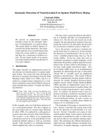
curve built using β-actin cDNA. Figure 2 shows some
examples of ISH and the amounts of β-actin mRNA
extracted with the SCN method. Each square (7 × 7 µm)
in Fig. 2 indicates the region of insertion with the AFM
probe. The number of each square is the order of insertion
with the AFM probes. β-actin mRNAs were detected
mainly in the vicinity of the nucleus. Usually β-actin
mRNA is known to exist mainly in the vicinity of the
nucleus, and these results agreed with the results of our
previous work and other studies by ISH [13,20]. Alkaline
phosphatase activity (dark intracellular staining) corre-
sponded to the distribution of endogenous β-actin mRNA
at the time of fixation of the cell. Our ISH results also
showed that dark staining was observed around the
nucleus predominately, indicating that β-actin mRNAs
existed mainly in the vicinity of the nucleus.
In order to analyze the correlation between both methods
more precisely, we attempted to standardize and evaluate
the ISH results. The darkness of ISH corresponded to β-
actin mRNA concentration. But this concentration was
not considered to have a linear correlation with the mRNA
concentration because it should be considered as absorp-
tion of light from a halogen lamp. To analyze the correla-
tion between darkness and β-actin mRNA concentration,
we applied a Lambert-Beer-like rule to these results.
[IISH: Intensity of ISH] = -Log
10
(I
n
/I
0
)
I
0
is the average of the background intensity. I
n
is the dark-
ness intensity of each point of the cell. In this equation,
we considered (I
n
/I
0
) as the transmission. Since the back-
ground intensity was stable, we calibrated the darkness of
each point on the cell according to the average of the back-
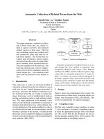
Experimental overview of the SCN method and ISHFigure 1
Experimental overview of the SCN method and ISH. The AFM probe was inserted into a cell to take mRNAs, and then
analyzed with RT-PCR, followed by quantitative PCR. The same cell was fixed by 4%folmaldehyde/PBS and subjected to ISH.
Journal of Nanobiotechnology 2007, 5:7 />Page 3 of 6
(page number not for citation purposes)
ground darkness. Although IISH does not have its own
unit, we could compare linearly the ratio between each
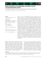
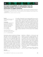
point. Figure 3 shows high magnification images of IISH
results with the amounts of β-actin mRNA picked up by
the SCN method. In this figure, IISH was divided into 8
classes within the range of -0.1 and 0.7. The center of each
image is the position of the center of the AFM probe
inserted. β-actin mRNA was not detected in the region of
Fig. 3(a) whose IISH was distributed from -0.1 to 0.1. We
could detect a very low β-actin mRNA quantity by the SCN
method as shown in Figures 3(b) and 3(c) which show
IISHs distributed from 0.1 to 0.4. When IISH was shown
to be mainly from 0.2 to 0.5, such as seen in Figures 3(d)
and 3(e), more β-actin mRNA was detected by the SCN
method. In addition, when IISH was very strong in the
center such as shown in Figure 3(f), a number of β-actin
mRNAs were detected by the SCN method. In this way,
when IISH became higher and the high intensity region
became larger, the amounts of β-actin mRNA detected
with the SCN method became higher. These results indi-
cated a good relationship between the results of the ISH
and the SCN method.
The table summarizes the comparison between the aver-
age of IISH and the SCN method. In this table, the aver-
ages of IISH were generated from the range of 1.4 × 1.4 µm
based on the position inserted by an AFM probe which
was centered. We used the AFM probes with square pyra-
mid shapes, whose height, horizontal length and 1/2 corn
angles were 3, 4 µm, and 35°, respectively. So if we
assume that the AFM probe is inserted into the cell by 1
µm, the range is 1.4 × 1.4 µm. In the table, when the aver-
age of IISH showed 0 to 0.1, β-actin mRNA was not
detected by the SCN method. When the average of IISH
was 0.1 to 0.25, β-actin mRNA was detected by the SCN
method in low probability (33%) and low quantity.
When the average of IISH was over 0.25, the probability
of β-actin mRNA detection was 100%. However, the aver-
age quantities of β-actin mRNA detected by the SCN
method were 50 and 120 molecules within the range of
0.25 to 0.4 and over 0.4, respectively. Based on this, as
IISH increased more, the probability and quantity of β-
actin mRNA detected by the SCN method increased more.
These results indicated a proportional relation between
the results of the ISH method and the SCN method.
Previously, we showed that detection probability of β-
actin mRNA by the SCN method changed according to the
distance from a nuclear membrane [13]. When the region
was 0–6 µm from the nuclear membrane, the probability
of detecting β-actin mRNA was 100%. As the distance
from the nuclear membrane became greater, the probabil-
ity decreased. In the region 6–9 µm from the nuclear
membrane, the probability was 61.5%, and in the region
9–18 µm away from the membrane, it was 11.1%. To
compare these previous results with the ISH results, we
calculated the average of IISH along with the distance
from the nuclear membrane. In the region of 0–6 µm
ISH result in higher resolutionFigure 3
ISH result in higher resolution. Black color indicates high
intensity of ISH. As black becomes white, the intensity of ISH
decreases. The numbers of each figure are the β-actin mRNA
quantity detected by the SCN method. Scale bar is 1 µm.
The results of the SCN method and ISHFigure 2
The results of the SCN method and ISH. (a-c) Each
square indicates the region of the AFM probe insertion.
Numbers in lower panels indicate β-actin mRNA quantities
detected by the SCN method, and dark intracellular staining
indicates distribution of β-actin mRNA detected by ISH. (d)
Negative control using sense RNA probe. Scale bar is 50 µm.
Journal of Nanobiotechnology 2007, 5:7 />Page 4 of 6
(page number not for citation purposes)
from the nuclear membrane, the average of IISH was 0.25,
as the distance increased more, the average of IISH
decreased (Figure 4). This tendency was similar with the
distance dependency of the detection possibility in the
SCN method. In the region of IISH > 0.25, we could detect
β-actin mRNA with the SCN method at 100% probability
(Table 1). This also shows that a good correlation between
ISH and the SCN method, and the SCN is suitable to
detect mRNA at medium or above expression.
Conclusion
We showed the correlation between ISH and the SCN
method. The SCN method can examine time-dependent
mRNA expression of single living cells, but it is limited to
the analysis of the fine localization of mRNA in the cells.
ISH can examine mRNA expression of the whole cells with
higher resolution, but time-lapse analysis cannot be done.
Besides, the SCN method is suitable and reliable to exam-
ine mRNAs at medium or higher expression level. By
using both methods, more accurate information about
mRNA expression of single cells is available.
Methods
Preparation of cells
Rat fibroblast-like VNOf06 cells derived from the vomer-
onasal organ [21] were grown in 35 mm Petri dishes in
Dulbecco's minimum essential medium (DMEM)/F12
supplemented with 100 U/mL penicillin, 100 µg/mL
streptomycin, and 10% heat-inactivated fetal bovine
serum (FBS). The cells were washed three times with
DMEM/F12 without FBS and used for the AFM experi-
ments.
The single cell nanoprobe (SCN) method
The details of the SCN method have been described in
previous studies [12-14].
Briefly, the AFM probe (NP, Digital Instruments, Santa
Barbara, CA) was positioned onto a target region of cells
under the observation of an inverted phase-contrast
microscope. The AFM probe was then inserted into the
target cell using the step motor of the AFM (NVB-100,
Olympus, Inc.), and held for about 30 s to allow the AFM
probe to bind the cell components containing mRNA
with physical adsorption. The AFM probe was lifted off
the cell and placed into a PCR tube.
PCR
The reagents and primers of RT-PCR and quantitative PCR
were used as previously described [12,13]. RT-PCR was
performed with a one-step RT-PCR kit (Qiagen, Valencia,
CA). First-strand cDNA synthesis was performed at 50°C
for 30 min, at which time the reaction was heated to 95°C
for 15 min to activate HotStrTaq DNA polymerase. The
amplification reaction was carried out for 30 cycles, and
each cycle was 94°C for 45 s, 55°C for 45 s, and 72°C for
1 min, followed by a final 10 min elongation at 72°C.
Quantitative PCR was performed with an Applied Biosys-
tems Prism 7000 and the SYBR Green 1 PCR Mastermix
(Qiagen, CA, USA) following previous studies [12,13].
ISH for
β
-actin mRNA of single cells
Digoxigenin (DIG) labeled RNA probe preparation
β-actin cDNA [224–987 bp] was prepared by RT-PCR. β-
actin cDNA was inserted into pGEM
(R)
-T Easy vector
(promega), and subcloned. The direction of the inserted
cDNA was examined by restriction enzyme and by its
sequence. Antisense DIG-labeled RNA probe was pre-
pared by SP6 and T7 RNA polymerase (stratagene) and 10
× DIG labeling mix (Roche). The efficiency of DIG labe-
ling was examined by dot-blotting.
Cell preparation for ISH
After picking up mRNA by the SCN method, the cells were
washed by PBS 3 times and fixed in 4% paraformalde-
hyde(PFA)/PBS for 30 min. From this point, all treat-
ments were performed under RNase free condition. After
PBS washing, the cells were treated by 1 µg/ml proteinase
K (Invitrogen) for 5 min at 37°C, washed in PBS, refixed
in 4% PFA/PBS for 10 min at RT, neutralized in 0.2% gly-
cine/PBS for 2 min, 0.2 N HCl at RT for 20 min and
washed with PBS two times.
Relation between ISH intensity and the distance from nuclear membraneFigure 4
Relation between ISH intensity and the distance
from nuclear membrane. The average of the ISH inten-
sity was calculated according to the distance from the
nuclear membrane. The detection probability of β-actin
mRNA with the SCN method changed according to the dis-
tance from the nuclear membrane. As the distance from the
nuclear membrane became greater, fewer positive results
were obtained with the SCN method.
Publish with Bio Med Central and every
scientist can read your work free of charge
"BioMed Central will be the most significant development for
disseminating the results of biomedical research in our lifetime."
Sir Paul Nurse, Cancer Research UK
Your research papers will be:
available free of charge to the entire biomedical community
peer reviewed and published immediately upon acceptance
cited in PubMed and archived on PubMed Central
yours — you keep the copyright
Submit your manuscript here:
/>BioMedcentral
Journal of Nanobiotechnology 2007, 5:7 />Page 5 of 6
(page number not for citation purposes)
Hybridization and detection by alkaline phosphatase reaction
Hybridization solution (60% formamide (deionized), 2 ×
SSC (1 × SSC is 150 mM NaCl, 15 mM), 10 mM EDTA, 25
mM NaH
2
PO
4
, 5% dextran sulfate and RNA probe (added
before use)) was add to the cells described above and
incubated overnight at 55°C. The RNA probe concentra-
tion was determined before the experiment and was
adjusted to be 0.1 ng/µl. After overnight incubation, the
cells were washed in the following order: 5 × SSC/50%
formamide 30 min 50°C two times, TNE buffer 5 min (10
mM Tris, 0.5 M NaCl, 1 mM EDTA pH7.5), 20 g/ml
RNase/TNE buffer 30 min 37°C, 2 × SSC 30 min 50°C
two times, 0.2 × SSC 30 min 50°C two times, blocking
solution (1% Blocking Reagent (Roche)/TBS (0.1 M Tris-
HCl pH7.5, 0.15 M NaCl)) 30 min. The cells were then
incubated in anti-DIG Fab fragment(Roche) diluted 1:500
with blocking solution for 60 min. After washing with
TNT buffer (0.2% Tween20/TBS) 15 min two times and
AP buffer (0.1 M Tris-HCl pH9.5, 0.1 M NaCl, 50 mM
MgCl
2
), the cells were stained by DIG Nucleic Acid Detec-
tion kit (Roche) for 6 hours using alkaline phosphatase
reaction of NBT/BCIP. After PBS washing, the cells were
embedded in PermaFluor Mountant Medium (Thermo,
USA) and obsreved by a phase-contrast microscope and a
bright-field microscope.
Competing interests
The author(s) declare that they have no competing inter-
ests.
Authors' contributions
HU conceived of the study and drafted the manuscript,
and carried out PCR and AFM. YK carried out ISH. AI and
TO participated in the design of the study and coordina-
tion. All authors read and approved the final manuscript.
References
1. John HA, Birnstiel ML, Jones KW: RNA-DNA hybrids at the cyto-
logical level. Nature 1969, 223:582-587.
2. Pardue Mary Lou, Gall Joseph G: Molecular hybridization of radi-
oactive DNA to the DNA of cytological preparations. Proc
Natl Acad Sci USA 1969, 64:600-604.
3. Agrawal Sudhir, Christodoulou Chris, Gait Michael J: Efficient
methods for attaching nonradioactive labels to the 5'-ends of
synthetic oligodeoxyribonucleotides. Nucleic Acids Res 1986,
14:6227-6245.
4. Chollet André, Kawashima Eric H: Biotin-labeled snthetic oligo-
deoxyribonucleotides: synthesis and uses as hybridization
probes. Nucleic Acids Res 1985, 13:1529-1541.
5. Muhlegger K, Huber E, von der Eltz H, Ruger R, Kesser C: Nonradi-
oactive labeling and detection of nucleic acids:IV. Synthesis
and properties of digoxigenin-modified 2'-deoxy-uridine-tri-
phosphates and a photoactivatable analog of digoxigenin
(photodigoxigenin). Biol Chem Hoppe-Seyler 1990, 371:953-965.
6. Muhlegger K, Batz HG, Bohm S, Eltz HVD, Holtke HJ, Kessler Ch:
Synthesis and use of new digoxigenin-labeled nucleotides in
nonradioactive labeling and detection of nucleic acids. Nucle-
osides, Nucleotides and Nucleic Acids 1989, 8:1161-1163.
7. Long RM, Singer RH, Meng X, Gonzalez I, Nasmyth K, Jansen R-P:
Mating Type Switching in Yeast Controlled by Asymmetric
Localization of ASH1 mRNA. Science 1997, 277:383-387.
8. Mingle Lisa A, Okuhama Nataly N, Shi Jian, Singer Robert H, Con-
deelis John, Liu Gang: Localization of all seven messenger RNAs
for the actin-polymerization nucleator Arp2/3 complex in
the protrusions of fibroblasts. J Cell Sci 2005, 118:2425-2433.
9. Wakabayashi Yoshihiro, Mori Yuji, Ichikawa Masumi, Yazaki Kazu-
mori, Hagino-Yamagishi Kimiko: A Putative Pheromone Recep-
tor Gene Is Expressed in Two Distinct Olfactory Organ in
Goats. Chem Senses 2002, 27:207-213.
10. Kanai-Azuma Masami, Kanai Yoshiakira, Gad Jacqueline M, Tajima
Youichi, Taya Choji, Kurohmaru Masamichi, Sanai Yutaka, Yonekawa
Hiromichi, Yazaki Kazumori, Tam Patrick PL, Hayashi Yoshihiro:
Depletion of definitive gut endoderm in Sox17-null mutant
mice. Development 2002, 129:2367-2379.
11. Kidokoro Tomohide, Matoba Shogo, Hiramatsu Ryuji, Fujisawa Masa-
hiko, Kanai-Azuma Masami, Taya Choji, Kurohmaru Masamichi,
Kawakami Hayato, Hayashi Yoshihiro, Kanai Yoshiakira, Yonekawa
Hiromichi: Influence on spatiotemporal patterns of a male-
specific Sox9 activation by ectopic Sry expression during
early phases of testis differentiation in mice. Developmental
Biology 2005, 278:511-525.
12. Osada Toshiya, Uehara Hironori, Kim Hyonchol, Ikai Atsushi:
mRNA analysis of single living cells. Journal of Nanobiotechnology
2003, 1(2):1-8.
13. Uehara Hironori, Osada Toshiya, Ikai Atsushi: Quantitative meas-
urement of mRNA at different loci within an individual living
cell. Ultramicroscopy 2004, 100(3–4):197-201.
14. Osada Toshiya, Uehara Hironori, Kim Hyonchol, Ikai Atsushi: Clini-
cal Laboratory implications of single living cell mRNA analy-
sis. Advances in Clinical Chemistry 2004, 38:239-57.
15. Sekiguchi H, Arakawa H, Taguchi H, Itoh T, Kokawa R, Ikai Atsushi:
Specific Interaction between GroEL and Denatured Protein
Measured by Non-pushing Force Spectroscopy. Biophys J 2003,
85:484-490.
16. Hertadi Rukman, Ikai Atsushi: Unfolding Mechanics of Holo and
Apo Calmodulins studied by Atomic Force Microscope. Pro-
tein Science 2002, 11:1532-1538.
17. Sekiguchi Hiroshi, Ikai Atsushi, Arakawa Hideo, Sugiyama Shigeru:
AFM analysis of interaction forces between bio-molecules
Table 1: Comparison between single cell nanoprobe method and ISH result (± S.D)
ISH intensity 0–0.1 0.1–0.25 0.25–0.4 0.4-
Single cell nanoprobe method β-actin mRNA detection probability 0%(n = 5) 33%(n = 6) 100%(n = 7) 100%(n = 3)
Average number of detected β-actin mRNA 0 5(± 5) 50(± 20) 120(± 65)
Publish with Bio Med Central and every
scientist can read your work free of charge
"BioMed Central will be the most significant development for
disseminating the results of biomedical research in our lifetime."
Sir Paul Nurse, Cancer Research UK
Your research papers will be:
available free of charge to the entire biomedical community
peer reviewed and published immediately upon acceptance
cited in PubMed and archived on PubMed Central
yours — you keep the copyright
Submit your manuscript here:
/>BioMedcentral
Journal of Nanobiotechnology 2007, 5:7 />Page 6 of 6
(page number not for citation purposes)
using ligand-functionalized polymers. e-J Surf Sci Nanotech 2006,
4:149-154.
18. Afrin Rehana, Ikai Atsushi: Force profiles of protein pulling with
or without cytoskeletal links studied by AFM. Biochem Biophys
Res Commun 2006, 348:238-244.
19. Maeda S, Sahara N, Saito Y, Murayama M, Yoshiike Y, Kim H, Miyasaka
T, Murayama S, Ikai A, Takashima A: Granular tau oligomers as
intermediates of tau filaments. Biochemistry 2007, 46:3856-3861.
20. Kislauskis EH, Zhu X-C, Singer RH, J : Beta-Actin messenger
RNA localization and protein synthesis augment cell motil-
ity. J Cell Biol 1997, 136:1263-1270.
21. Osada T, Ikai A, Costanzo RM, Matsuoka M, Ichikawa M: Continual
neurogenesis of vomeronasal neurons in vitro. J Neurobiol
1999, 40:226-233.