báo cáo khoa học: "Capillary electrophoresis for the characterization of quantum dots after non-selective or selective bioconjugation with antibodies for immunoassay" docx
Bạn đang xem bản rút gọn của tài liệu. Xem và tải ngay bản đầy đủ của tài liệu tại đây (3.52 MB, 15 trang )
BioMed Central
Page 1 of 15
(page number not for citation purposes)
Journal of Nanobiotechnology
Open Access
Research
Capillary electrophoresis for the characterization of quantum dots
after non-selective or selective bioconjugation with antibodies for
immunoassay
Mark Pereira and Edward PC Lai*
Address: Department of Chemistry, Ottawa-Carleton Chemistry Institute, Carleton University, Ottawa, ON K1S 5B6, Canada
Email: Mark Pereira - ; Edward PC Lai* -
* Corresponding author
Abstract
Capillary electrophoresis coupled with laser-induced fluorescence was used for the
characterization of quantum dots and their conjugates to biological molecules. The CE-LIF was
laboratory-built and capable of injection (hydrodynamic and electrokinetic) from sample volumes
as low as 4 μL via the use of a modified micro-fluidic chip platform. Commercially available quantum
dots were bioconjugated to proteins and immunoglobulins through the use of established
techniques (non-selective and selective). Non-selective techniques involved the use of EDCHCl/
sulfo-NHS for the conjugation of BSA and myoglobin to carboxylic acid-functionalized quantum
dots. Selective techniques involved 1) the use of heterobifunctional crosslinker, sulfo-SMCC, for
the conjugation of partially reduced IgG to amine-functionalized quantum dots, and 2) the
conjugation of periodate-oxidized IgGs to hydrazide-functionalized quantum dots. The migration
times of these conjugates were determined in comparison to their non-conjugated QD relatives
based upon their charge-to-size ratio values. The performance of capillary electrophoresis in
characterizing immunoconjugates of quantum dot-labeled IgGs was also evaluated. Together, both
QDs and CE-LIF can be applied as a sensitive technique for the detection of biological molecules.
This work will contribute to the advancements in applying nanotechnology for molecular diagnosis
in medical field.
Background
Quantum dots (QDs) are fluorescent nanoparticles that
receive increasing recognition as a viable alternative (to
conventional organic fluorophores) for molecular labe-
ling. Their quantum mechanical and electronic character-
istics give QDs unique optical properties that are
advantageous in the fields of bioanalytical, biomedical
and biophotonic research. Such optical properties include
size-tunable emission wavelengths, broad excitation
wavelengths, long fluorescence lifetimes, large Stokes
shifts, and high quantum yields [1-3]. Other advanta-
geous properties include resistance to photo- and chemi-
cal- degradation and their capability for performing
multiplexing experiments [3]. QDs are relatively large par-
ticles, with typical diameters ranging from 1–10 nm [1].
The inorganic core (typically a semiconductor) is respon-
sible for their fluorescent properties. This core is typically
surrounded by a shell (ZnS is common) for protection
from chemical- and photo-oxidation [2]. The shell also
provides a means of functionalizing the QD with carbox-
Published: 1 October 2008
Journal of Nanobiotechnology 2008, 6:10 doi:10.1186/1477-3155-6-10
Received: 3 May 2008
Accepted: 1 October 2008
This article is available from: />© 2008 Pereira and Lai; licensee BioMed Central Ltd.
This is an Open Access article distributed under the terms of the Creative Commons Attribution License ( />),
which permits unrestricted use, distribution, and reproduction in any medium, provided the original work is properly cited.
Journal of Nanobiotechnology 2008, 6:10 />Page 2 of 15
(page number not for citation purposes)
ylic acids or primary amines, for good solubility in aque-
ous solutions and relative ease of specific labeling
reactions [1].
QDs, often applied for the labeling of biological mole-
cules (proteins, peptides, antibodies, etc.), require specific
techniques for their conjugation [4-7]. The most popular
bioconjugation technique involves the use of a zero-
length crosslinker, 1-ethyl-3- [3-dimethylaminopro-
pyl]carbodiimide hydrochloride (EDCHCl) [1-4,6,7], in
the presence of a hydrophilic active group, N-hydroxysul-
fosuccinimide (sulfo-NHS) [8], for the formation of a sta-
ble amide bond between carboxylic acid-functionalized
QDs (QD-COOH) and any biomolecules containing a
primary amine [9] (Figure 1).
This method, while proven to yield exclusively QD-pro-
tein conjugates in a controlled manner, randomizes the
location on a protein to which conjugation can occur,
resulting in a non-selective bioconjugation [9]. Despite
high bioconjugation efficiencies, this can be detrimental
in the case where an immunoassay is to be performed
next. For instance, a labeled protein serving as an antigen
might lose its antigenicity (ability to bind an antibody)
when conjugated to a large QD. A similar concern can be
conveyed if an antibody were conjugated in a region close
to the antigen-binding site (the hypervariable region).
Either one of these variations can significantly reduce the
efficiency of immunoassay applications [9].
Other techniques make effective use of selective bioconju-
gation, targeting specific sites on the protein. These
include the use of a heterobifunctional crosslinker such as
sulfosuccinimidyl-4-(N-maleimidomethyl)cyclohexane-
1-carboxylate (sulfo-SMCC) [9-11]. In the case for anti-
bodies, as shown in Figure 2 below, sulfo-SMCC can form
stable amide bonds to amine-functionalized QDs (QD-
NH
2
) [9]. The resultant QDs, through sulfo-SMCC's male-
imide region, can next form stable a thioether bond with
a sulfhydryl-exposed antibody [9]. Mild reducing reagents
such as cysteamineHCl (or DTT) can selectively cleave the
disulfide bonds (hinge region) connecting the IgG heavy
chains, while leaving the other disulfide bonds that make
up the antigen binding site (hypervariable region) unaf-
fected, thus producing a partially reduced IgG (rIgG) [12].
In addition, the resulting exposed sulfhydryls (hinge
region) are sufficiently far away (from the hypervariable
region) for QD-bioconjugation to occur. The resulting
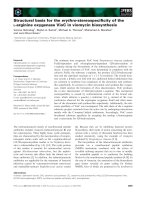
Non-selective bioconjugation reaction scheme of carboxylated QDs (QD-COOH) to amine-containing proteinsFigure 1
Non-selective bioconjugation reaction scheme of carboxylated QDs (QD-COOH) to amine-containing pro-
teins. This two-step reaction involves a) the activation of QD-COOH with EDC/sulfo-NHS, resulting in a semi-stable active
ester (QD-NHS), and b) the nucleophilic reaction between the QD-NHS and amine-containing protein, forming a QD-protein
conjugate via a stable amide bond.
Journal of Nanobiotechnology 2008, 6:10 />Page 3 of 15
(page number not for citation purposes)
quantum dot-conjugated half antibody (QD-rIgG) will
allow an immunoreaction to proceed readily.
Reductive amination is a bioconjugation technique popu-
lar in the labeling of glycoproteins. Taking advantage of
the polysaccharide chains within the Fc region of an anti-
body, it can allow bioconjugation to occur relatively far
away from the antigen binding site. Through oxidation
(using sodium periodate) of the carbohydrate hydroxyls,
the aldehydes formed are highly reactive toward primary
amines and hydrazides [9]. This makes QD-NH
2
or QD-
COOH (derivatized with adipic acid dihydrazide (ADH))
suitable candidates for conjugation [9]. In addition, selec-
tive bioconjugation can occur without a proceeding
reduction reaction, thus retaining the integrity of the anti-
body (Figure 3).
Capillary electrophoresis (CE) has seen increasing use in
the separation and characterization of inorganic nanopar-
ticles (Ag, Au, TiO
2
, Al
2
O
3
, Fe
2
O
3
) [13-17], polystyrene
microspheres [18], biomolecules (proteins, peptides) [19-
30], QDs [31], QD-conjugates with bovine serum albu-
min (BSA) and horse radish peroxidase (HRP) [7], and
QD-conjugates with Ulex europaeus (UEA-1) and anti-von
Willebrand factor (anti-vWF) [32]. CE has also been used
for immunoassays involving hepatitis B, prion protein,
alpha-fetoprotein, etc [24-30]. Recently, a CE-based
immunoassay involving QDs conjugated to anti-IgM anti-
bodies followed by immuno-conjugation to its compli-
mentary antigen IgG was performed with satisfactory
results [33]. Another recent advancement involved the
CE-characterization of QDs (of differing emission wave-
lengths) exclusively conjugated to biotin and streptavidin
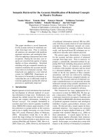
Selective bioconjugation reaction scheme of amino QDs (QD-amine) to free sulhydryl-containing IgG antibodiesFigure 2
Selective bioconjugation reaction scheme of amino QDs (QD-amine) to free sulhydryl-containing IgG antibod-
ies. The reaction involves a) the mild reduction of IgG with cysteamine to yield partially reduced IgG antibody fragments
(rIgG); b) the activation of QD-NH
2
by nucleophilic reaction with NHS-moiety of sulfo-SMCC, resulting in maleimide-function-
alized quantum dot (QD-maleimide); and c) the rIgG and QD-maleimide conjugation (QD-rIgG) via the formation of a
thioether bond.
Journal of Nanobiotechnology 2008, 6:10 />Page 4 of 15
(page number not for citation purposes)
[34]. Their work followed the characterization of the con-
jugates' affinity to each other via strong biotin-streptavi-
din interactions. However, present publications reporting
the use of QDs in CE-based immunoassays are very pre-
liminary, due in part to a QD-biomolecule conjugate's
(and immunoconjuagte's) complex charge-to-size ratio.
Thus, more research is required in its development as a
fast and efficient method for performing immunoassays.
In this paper, we report more preliminary results of cova-
lently bioconjugating QDs to various biomolecules (pro-
teins and immunoglobulins). These QD-conjugated
biomolecules are characterized via a laboratory-built cap-
illary electrophoresis instrument with laser-induced fluo-
rescence detection (CE-LIF) [35]. The instrumental
capabilities (comparable to commercial CE-LIF systems)
include the use of a micro-sample injection platform that
can load sample volumes as low as 4 μL [35]. We also dis-
cuss some of the challenges faced when performing bio-
conjugation through the various schemes described
above. The purpose is to validate a fast, selective, and
reproducible CE-LIF analysis method that can be efficient
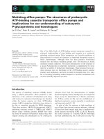
Selective bioconjugation reaction scheme of hydrazide QDs (QD-hydrazide) to aldehyde-containing IgG antibodies (IgG-CHO)Figure 3
Selective bioconjugation reaction scheme of hydrazide QDs (QD-hydrazide) to aldehyde-containing IgG anti-
bodies (IgG-CHO). The reaction involves a) mild periodate oxidation of glycosylated IgG, yielding IgG-CHO; b) synthesis of
QD-hydrazide via derivatization of QD-COOH with EDC/ADH; and c) conjugation of QD-hydrazide with IgG-CHO via for-
mation of hydrazone linkage to yield QD-IgG.
Journal of Nanobiotechnology 2008, 6:10 />Page 5 of 15
(page number not for citation purposes)
and robust. This work will evolve to perform QD-based
immunoassays using CE-LIF as an effective separation and
sensitive detection technique. The aim is to apply this
research in the area of infectious biological materials that
are generally present in relatively low concentrations and
small volumes.
Methods
Chemicals and reagents
Boric acid (certified A.C.S.), sodium meta-periodate (crys-
talline, A.C.S. grade), sodium hydroxide (reagent grade)
were purchased from Fisher Scientific (Ottawa, Ontario,
Canada). CdSe/ZnS carboxy-terminated QDs (Maple Red-
Orange, 620 nm) and CdSe/ZnS amine-terminated QDs
(Maple Red-Orange, 620 nm) were purchased from Evi-
dent Technologies (Troy, NY, USA). EDCHCl, Sulfo-NHS,
lysozyme (Lys), and MES buffered saline packs were pur-
chased from Pierce Biotechnology. Sodium acetate (rea-
gent grade) and hydroxylamine hydrochloride (reagent
grade) was purchased from Anachemia. EDTA (0.1 M vol-
umetric standard), ADH (= 98%), sulfo-SMCC (= 98%),
DL-DTT (1 M in water solution), anti-human albumin
(polyclonal IgG produced in rabbit), human serum albu-
min (HSA), cysteamine hydrochloride (Purum = 97.0%),
2-mercaptoethanol (14 M), 10× PBS concentrate, bovine
serum albumin (BSA), horse myoglobin (Myo) cyto-
chrome c (CytC), ethanolamine, and sodium cyanoboro-
hydride (5 M in 1 M sodium hydroxide) were purchased
from Sigma Aldrich. Coumarin 521 was purchased from
Exciton (Dayton, Ohio, USA). Micro-centrifuge tubes (50
kDa and 100 kDa MWCO) were purchased from Fisher
Scientific.
Preparation of buffer solutions and stock solutions
All buffer solutions were prepared and pH-adjusted using
sodium hydroxide (10 M, 5 M, and 1 M) and hydrochloric
acid (1 M and 0.5 M). All CE separation buffers were fil-
tered through a 0.45 μm membrane filter (Pall Corpora-
tion, Ann Arbor, MI, USA).
Carboxy- and amine- terminated QDs were used from
supply stock (11 μM) without any prior treatment.
Stock solutions of EDCHCl (20 mM) and sulfo-NHS (50
mM) were prepared by dissolution of dry reagents in 0.1
M MES (pH 5.2) buffered saline and used immediately
after preparation. Stock solutions of 2-mercaptoethanol
(1 M) and hydroxylamine hydrochloride (1 M) were pre-
pared and stored at room temperature.
Stock solutions of cysteamineHCl (100 mM) were pre-
pared by dissolution of dry reagent in 1× PBS (pH 7.2), 10
mM EDTA and used immediately after preparation. Stock
solutions of DTT (100 mM) were prepared by dilution of
a 1 M DTT stock solution and used within 3 days of prep-
aration.
Stock solutions of NaIO
4
(100 mM) were prepared by dis-
solution of dry reagents in 0.1 M sodium acetate (pH 5.5)
buffered saline. Preparation and storage was performed in
minimal lighting and used immediately after use. Sodium
cyanoborohydride (5 M in 1 N NaOH) was used as pre-
pared from supplier. Stock solution of ethanolamine (1
M) was prepared by dissolution of dry reagent in distilled
deionized water (ddw) and pH adjusted to 9.6.
Stock solutions (1 mg/mL) of bovine serum albumin
(BSA), myoglobin (Myo), cytochrome c (CytC), and lys-
ozyme (Lys), were prepared in 1× PBS (pH 7.2). Human
serum albumin (HSA) was prepared in ddw (11 mg/mL).
Anti-human albumin IgG (4 mg/mL) was prepared in 1×
PBS (pH 7.2).
Non-specific bioconjugation of whole IgG using EDCHCl/
sulfo-NHS
A mixture containing 2 mM EDCHCl, 5 mM sulfo-NHS,
and 1.1 μM carboxy-terminated QDs (QD-carboxyl) was
prepared in 0.1 M MES, pH 6.0 and incubated for 15 min-
utes at room temperature. The remaining unreacted EDC
was quenched with the addition of 2-mercaptoethanol (1
M) to a final concentration of approximately 20 mM and
the mixture was left to stand for 10 minutes. The activated
QDs were purified of unreacted reagents and byproducts
by dialysis using 100 kDa MWCO microcentrifuge tubes
and re-suspended in 1× PBS (pH 7.2) containing dis-
solved protein. The reaction proceeded for 2 hours with
gentle mixing. The reaction was quenched with addition
of hydroxylamine hydrochloride (1 M) to a final concen-
tration of approximately 10 mM. The bioconjugation mix-
ture was left to stand for 10 minutes at room temperature
prior to purification by dialysis using 100 kDa MWCO
microcentrifuge tubes. The mixture was analyzed by CE-
LIF and stored at 4°C.
Selective bioconjugation of reduced IgG (rIgG) using
cysteamineHCl or DTT and sulfo-SMCC
A mixture containing approximately 1 mg/mL rabbit anti-
human albumin IgG and cysteamineHCl (concentration
ranging from 0.1 mM to 100 mM) was incubated at 37°C
for 90 minutes in 0.1 M sodium phosphate (pH 7.0), 0.15
M, 0.01 M EDTA. The resulting partially reduced antibody
(rIgG) was purified of byproducts and unreacted com-
pounds via dialysis using a 50 kDa MWCO microcentri-
fuge tube with successive washings of 0.1 M sodium
phosphate (pH 6.8), 0.15 M NaCl, 0.01 M EDTA buffer.
The rIgG was temporarily stored at 4°C until use for QD
coupling.
Journal of Nanobiotechnology 2008, 6:10 />Page 6 of 15
(page number not for citation purposes)
Amine-functionalized QDs (QD-amine) were added to a
50 mM sodium phosphate (pH 7.2) solution containing
sulfo-SMCC (8.8 mM) and incubated at room tempera-
ture for 60 minutes with gentle mixing. The maleimide-
activated QDs (QD-maleimide) were purified of unre-
acted cross-linker via dialysis using 100 kDa MWCO
microcentrifuge tubes at room temperature with succes-
sive washings of 0.1 M sodium phosphate (pH 6.8), 0.15
M NaCl, 0.01 M EDTA buffer. The purified QD-maleimide
was used immediately.
The rIgG and QD-maleimide were combined and incu-
bated overnight at 4°C. Purification of QD-rIgG of "free"
rIgG in solution was performed via dialysis using 100 kDa
MWCO microcentrifuge tubes. The purified QD-rIgG was
washed several times with ddw. The purified QD-rIgG was
analyzed by CE-LIF and stored at 4°C.
Selective bioconjugation of whole IgG using EDC/ADH and
sodium meta-periodate
A mixture containing 20 μL QD-carboxyl (11 μM), 16 mg
EDCHCl, and 32 mg ADH were incubated in 1 mL 1× PBS
for 4 hours at room temperature with gentle mixing. The
hydrazide-functionalized QDs (QD-hydrazide) were puri-
fied from excess reagents via dialysis using a 100 kDa
MWCO microcentrifuge tube. The purified concentrate
was stored at 4°C until analysis by CE-LIF and IgG-CHO
coupling.
A 500 μL mixture containing approximately 1 mg/mL rab-
bit anti-human albumin IgG and sodium meta periodate
dissolved in 0.1 M sodium acetate buffered saline was
incubated in the absence of light for 1 hour at room tem-
perature with gentle mixing. The oxidized IgG (IgG-CHO)
was purified of excess reagents via dialysis using a 100 kDa
MWCO. The purified IgG-CHO was used immediately.
The IgG-CHO was combined with QD-hydrazide (50 μL
total volume) and incubated overnight (14 hrs) at room
temperature with gentle mixing. Stabilization of the
hydrazone linkages were performed via the addition of 5
μL sodium cyanoborohydride (5 M in 1 N NaOH) with
continued incubation for 1 hour. Unreacted aldehydes
were blocked via addition of 25 μL of 1 M ethanolamine
(pH 9.6) with continued incubation for 1 hour. Mixture
was removed of excess sodium cyanoborohydride and
ethanolamine via dialysis using 100 kDa MWCO. Mixture
was not purified of unreacted IgG or QD.
Immunoconjugation of QD-rIgG with corresponding
antigen
A 10 μL aliquot of immunogen HSA (11 mg/mL) was
added to a 300 μL solution of QD-rIgG (rabbit anti-
human albumin) and incubated for 15 minutes at room
temperature. The mixture was immediately analyzed be
CE-LIF and later stored at 4°C.
CE-LIF analysis
CE-LIF analysis of QDs, bioconjugates, and immunocon-
jugates were performed on a laboratory-built system
described previously. A fused silica capillary (51 mm id,
362 mm o.d., L
t
= 58.5 cm, L
d
= 52.1 cm, and L
dw
= 2 mm)
was flushed with 1.0 M NaOH, 0.1 M NaOH, DDW, and
run buffer. Prior to each use, the capillary was equilibrated
with the run buffer at an applied voltage of 25 kV for 10
min. Capillary temperature was maintained constant at
20.0°C by water from a PolyScience 1160A circulating
bath (Niles, IL, USA). Hydrodynamic injections were per-
formed by elevating the sample to 8 cm for 15 s. Micro-
sample injections were performed using the sample port
of a modified microfluidic chip as described previously
[34]. An Extreme DPSS 473 nm, 500 mW solid-state diode
laser (Seabrook, TX, USA) was used for fluorescence exci-
tation. The LIF intensity was detected using a Hamamatsu
model H7827-001 PMT (Bridgewater, NJ, USA) equipped
with a 620 ± 5 nm interference filter. Spectral response of
the PMT was 300–650 nm. The detector output signal was
acquired through the Peak Simple Chromatography Data
System.
Results and discussion
Use of EDCHCl/sulfo-NHS as a non-selective technique for
bioconjugation of QDs to proteins
This non-selective technique for bioconjugation involved
a two-step reaction using EDCHCl/sulfo-NHS to control
the conjugate formation. Bioconjugation of proteins to
carboxylated QDs have been performed with the use of
EDC alone [7]. Despite the simplicity of a one-step reac-
tion, the drawback involves a degree of uncontrollability
during bioconjugation, forming unlabeled protein-pro-
tein conjugates and QD-protein polymers that can ulti-
mately lead to precipitation. The use of sulfo-NHS was
included to prevent these unwanted conjugate by-prod-
ucts and yield exclusively QD-protein conjugates. How-
ever, the number of proteins bound to a single QD may
vary (depending on experimental conditions) and have
yet to be determined.
Figure 4 illustrates the CE separation of carboxylated QDs
(QD-COOH) (1) from their conjugation to BSA (QD-
BSA) (2). The QD-BSA was detected at a longer migration
time with respect to QD-COOH due to the inherent
increase in the net negative charge of the conjugate. This
was expected since the isoelectric point (pI) of BSA (~5.6)
is much lower than the CE buffer pH (9.2) and thus
expressing an increased number of negative charges that
will ultimately influence the net-charge of the conjugate.
The increase in peak width of the QD-BSA can be attrib-
uted to a number of factors, including the polydispersity
Journal of Nanobiotechnology 2008, 6:10 />Page 7 of 15
(page number not for citation purposes)
of QDs during synthesis, the binding ratio of BSA to QDs,
and the protein-capillary wall interactions that can take
place with protein functionalized-QDs.
Figure 5 illustrates the CE separation of QD-COOH (1)
with their conjugation to myoglobin (QD-Myo) (2). The
migration time of QD-Myo is also longer with respect to
QD-COOH. However the differences are not substantial
enough for baseline separation to occur. In comparison to
QD-BSA, there may be a weakened net negative charge
that is present on QD-Myo, since myoglobin has a pI
value measured at ~7.2. In addition, there is a considera-
ble size difference between BSA (MW~66 kDa) and Myo
(MW~16.7 kDa) that may likely influence the respective
conjugate's migration time. As both MW and pI can influ-
ence a protein's charge-to-size ratio, their conjugation to
polydisperse QDs (each with possibly different binding
ratios) will contribute to their respective migration times.
The chemistry of bioconjugating QD-COOH to proteins
using EDCHCl/sulfo-NHS was attractive due to its versa-
tility, as primary amines (lysine ε-amine and N-terminal
α-amine) are present on many proteins. This ultimately
led to the attempt of bioconjugating QD-COOH to pro-
teins of increasingly higher pI, using cationic proteins
such as cytochrome c and lysozyme. However, it was
observed that the pI of proteins can play a determining
factor in the efficiency of a bioconjugation. While the
reaction was efficient in conjugating anionic proteins
(BSA and myoglobin) to QD-COOH, it was unsuccessful
in conjugating to cationic proteins (cytochrome c and lys-
ozyme). It is suspected that the pI of cytochrome c (~10)
and lysozyme (~11) maintained the primary amines
(those accessible for conjugation) in a protonated state.
This protonated state would render these proteins poor in
a nucleophilic reaction with the NHS-activated QD-
COOH (QD-NHS), thus inhibiting bioconjugation. The
lack of a bioconjugation results in an eventual hydrolysis
reaction with QD-NHS leading to the formation of the
QD-COOH which can be identified using CE (data not
shown).
Another drawback for the use of EDCHCl/sulfo-NHS for
the formation of stable bioconjugates is the lack of specif-
icity on the protein of interest. As numerous amine func-
tional groups can be distributed throughout the surface of
Electropherogram of mixture containing QD-COOH (1) and BSA-conjugated QDs (QD-BSA) (2)Figure 4
Electropherogram of mixture containing QD-COOH (1) and BSA-conjugated QDs (QD-BSA) (2). CE buffer
electrolyte used was 50 mM borate, pH 9.2. Gravity injection performed by elevating inlet capillary 7 cm for 5 s. Applied volt-
age for CE separation was 20 kV. Capillary temperature maintained at 20°C. Excitation source and detection wavelength was
473 nm and 620 nm, respectively.
Journal of Nanobiotechnology 2008, 6:10 />Page 8 of 15
(page number not for citation purposes)
the protein, a bioconjugation involving such functional
groups via a EDCHCl/sulfo-NHS reaction would lead to a
randomization of crosslinking sites.
Use of selective (heterobifunctional crosslinker) technique
for bioconjugation of QDs to IgGs
The use of the heterobifunctional crosslinker sulfo-SMCC
allowed for straightforward activation of amine-function-
alized QDs (QD-NH
2
) via a nucleophilic reaction
between the active ester on the crosslinker and the amine
moiety of the QD. Despite the activated QD (QD-maleim-
ide) being relatively stable at physiological pH, tempera-
ture is an important factor to control as higher
temperatures (above room temperature) can accelerate
hydrolysis reactions. Hydrolysis of the maleimide moiety
will form maleamic acid that is unreactive towards free
Electropherogram of mixture containing QD-COOH (1) and myoglobin-conjugated QDs (QD-Myo) (2)Figure 5
Electropherogram of mixture containing QD-COOH (1) and myoglobin-conjugated QDs (QD-Myo) (2). CE
buffer electrolyte used was 50 mM borate, pH 9.2. Gravity injection performed by elevating inlet capillary 7 cm for 5 s. Applied
voltage for CE separation was 20 kV. Capillary temperature maintained at 20°C. Excitation source and detection wavelength
was 473 nm and 620 nm, respectively.
Reaction scheme illustrating hydrolysis of sulfo-SMCC activated of QD-NH
2
(QD-maleimide)Figure 6
Reaction scheme illustrating hydrolysis of sulfo-SMCC activated of QD-NH
2
(QD-maleimide). Hydrolyzed QD-
maleimide will contain maleamic acid moiety (QD-maleamic) unreactive towards free sulfhydryls.
Journal of Nanobiotechnology 2008, 6:10 />Page 9 of 15
(page number not for citation purposes)
sulfhydryls (Figure 6). Characterization of the hydrolyzed
QD-maleimide by CE detected a migration time similar to
that for QD-COOH (data not shown).
Due to the high-pH instability of QD-maleimide, CE char-
acterization was not performed. However, it can be
expected that the neutral charge present on the maleimide
would compel the QD-maleimide to migrate more slowly,
relative to the positively charged QD-amine prior to acti-
vation. The use of either cysteamineHCl (50–100 mM) or
DTT (1–10 mM) as the reducing agent for IgGs provided
similar results. However, both incubation time and tem-
perature are dramatically different (90 min at 37°C for
cysteamineHCl and 30 min at room temperature for
DTT). Furthermore, the use of 50 kDa MWCO centrifuge
filters allowed for retention of the partially-reduced IgG
(rIgG), while removing unused reagents and byproducts.
Combining of the QD-maleimide with rIgG at room tem-
perature for at least 2 hours (or at 4°C overnight) pro-
vided similar results shown in Figure 7 below.
Figure 7 illustrates overlapping electropherograms of QD-
NH
2
(1) and their conjugation to the reduced anti-human
albumin IgG (QD-rIgG) (2). The longer migration time
observed for QD-rIgG can lead to the assumption that the
rIgG exhibits a net negative charge in this CE separation
buffer. Thus, when conjugated to the positively charged
QD-NH
2
, the charge influence of the rIgG results in the
conjugate displaying a smaller net positive charge. It is
suspected that the IgG is comparable in acidity to the
smaller proteins (BSA and myoglobin) used, however
other factors including size and QD:biomolecule binding
ratios need to be taken into consideration. Similar electro-
pherograms were obtained when conjugating QD-NH
2
to
another IgG, anti-chicken lysozyme (data not shown).
This can be attributed to IgGs having MWs typically at 150
kDa. However, IgG can range in pI from 6.4 to 9.0, due
mainly to changes in their hypervariable region which can
contain various charged residues. Thus, changes in CE
separation buffer (particularly pH) could possibly influ-
ence the relative migration times of QDs conjugated to
different IgGs and hence aid in selectivity and resolution.
The observed migration time for the EOF was measured
slightly earlier than the QD-NH
2
(data not shown). This
was unexpected since these observations would suggest
QD-NH
2
expressing a net negative charge. However,
higher concentration borate buffers (greater than 200
mM) did measure the EOF at a later migration time than
Overlapping electropherograms illustrating QD-NH
2
(1) and QDs conjugated to reduced antibodies QD-rIgG (2)Figure 7
Overlapping electropherograms illustrating QD-NH
2
(1) and QDs conjugated to reduced antibodies QD-rIgG
(2). IgG used for conjugation was rabbit anti-human albumin. CE buffer electrolyte used was 50 mM borate, pH 9.2. Gravity
injection performed by elevating inlet capillary 7 cm for 5 s. Applied voltage for CE separation was 25 kV. Capillary tempera-
ture maintained at 20°C. Excitation source and detection wavelength was 473 nm and 620 nm, respectively
Journal of Nanobiotechnology 2008, 6:10 />Page 10 of 15
(page number not for citation purposes)
QD-NH
2
(data not shown). The reason for the unexpected
migration time for QD-NH
2
at different borate concentra-
tions may require further knowledge of the commercial-
ized QD coating/functionalization process.
Use of selective (hydrazone linkage) technique for
conjugation of IgGs to QDs
Conjugation of IgG-CHO with QD-NH
2
is possible using
reductive amination. However, the drawback is the degree
of uncontrollability of the resulting conjugate, as undesir-
able IgG-IgG crosslinking can occur through the presence
of primary amines on the IgGs surface. Thus, conjugating
IgG-CHO with QDs functionalized with hydrazides was
reasoned to be more selective as conjugation is occurring
exclusively on the polysaccharide chain. However, since
commercially obtainable QDs are typically functionalized
with carboxylic acids or amines, a derivatization was
required. Derivatization was performed on QD-COOH
and involved the use of EDCHCl in the presence of the
bis-hydrazide compound, ADH, yielding relatively stable
hydrazide-functionalized QDs (QD-hydrazide). The
drawback is that ADH, being is homobifunctional
crosslinker, can introduce undesirable side reactions. As
both functional groups on the crosslinker are identical,
they each have the potential of reacting with the same QD,
resulting in a closed ring structure that can essentially
inactivate that particular region of the QD. However, it is
suspected that the spacer arm of the crosslinker lacks the
length required to form such a ring structure. Another
more likely scenario involves the cross-reaction between a
derivatized QD (QD-hydrazide) with an underivatized
QD (QD-COOH). This uncontrolled reaction can lead to
the undesirable formation of a QD-QD polymer (Figure
8a), but is believed to be minimized when using ADH in
excessive quantities during the derivatization.
Figure 9 illustrates overlapping electropherograms of QD-
hydrazide (2) in comparison to QD-NH
2
(1) and QD-
COOH (3). Their characteristic migration times can be
attributed to the pKa of the functional group expressed on
the QD relative to the pH of the CE separation buffer
(9.2). Alkylated primary amines and carboxylic acids have
measured pKa ~10, and ~4.5, respectively. Thus, the effect
of the CE separation buffer pH allows the QD-NH
2
to
exhibit a net positive charge due to protonation of the pri-
mary amines. However, the QD-COOH will be com-
Possible unfavorable polymer formation during following bioconjugation stepsFigure 8
Possible unfavorable polymer formation during following bioconjugation steps. a) QD-hydrazide synthesis from
QD-COOH, and b) QD-IgG bioconjugation from QD-hydrazide and IgG-CHO.
Journal of Nanobiotechnology 2008, 6:10 />Page 11 of 15
(page number not for citation purposes)
pletely ionized, exhibiting a net negative charge. Figure 8
can show a distinct change in migration time between
QD-NH
2
and QD-COOH. Hydrazides have remarkably
low pKa values (~2.5), thus QD-hydrazides will be depro-
tonated during CE separation and exhibit a net-neutral
charge. Again, this can be observed in figure 8 as QD-
hydrazide migrates intermediate of the positively- and
negatively- charged QDs. The small differences in migra-
tion times between QDs with substantially different
charged residues on their surfaces can be attributed to the
very large size of the particles that greatly influence their
migration. Suppression of the EOF may improve resolu-
tion by means of increased electrophoretic contributions
from QD-biomolecule conjugates [33].
The use of QD-hydrazide (in contrast to QD-NH
2
) for bio-
conjugation with an oxidized IgG (IgG-CHO) increases
the selectivity of the reaction. However, there still remains
the potential to form undesirable conjugates. The is due to
not only the QD-hydrazide containing many reactive
sites, but also the IgG-CHO which can contain many
polysaccharide chains which can again contain many
reactive aldehydes. This can lead to the uncontrolled for-
mation of -QD-IgG-QD- polymers (Figure 8b). However,
this undesirable polymer formation can possibly be min-
imized with using QD-hydrazide in very limited quanti-
ties with respect to the IgG-CHO during bioconjugation.
Reduced reaction times, temperature, and mildly acidic
pH conditions may also prevent undesirable conjugates.
Figure 10 illustrates the CE separation of QD-hydrazide
(1) and their conjugation to whole anti-human albumin
IgG-CHO (QD-IgG) (2). The separation is not baseline
resolved but can be distinguished by the vertical line sep-
arating the two peaks. The QD-IgG, not purified by size-
exclusion or dialysis, retains a considerable amount of
unconjugated IgG in the sample. This resulted in signifi-
cant changes in EOF, peak shape, and resolution due to
protein-capillary wall adsorption. In addition, the lack of
baseline separation could be attributed to the whole IgG
exerting a reduced negative charge influence when conju-
gated to QD-hydrazide. To reduce the effects of protein-
capillary wall interaction, a 0.1% BSA additive was
included in the CE separation buffer. However, due to the
Overlapping electropherograms illustrating QD-NH
2
(1), QD-hydrazide (2), and QD-COOH (3)Figure 9
Overlapping electropherograms illustrating QD-NH
2
(1), QD-hydrazide (2), and QD-COOH (3). CE buffer elec-
trolyte used was 50 mM borate, pH 9.2. Gravity injection performed by elevating inlet capillary 7 cm for 5 s. Applied voltage for
CE separation was 28 kV. Capillary temperature maintained at 20°C. Excitation source and detection wavelength was 473 nm
and 620 nm, respectively.
Journal of Nanobiotechnology 2008, 6:10 />Page 12 of 15
(page number not for citation purposes)
similarities between BSA and the IgG immunogen,
human serum albumin (HSA), cross-reactivity may have
occurred. The cross-reactivity, leading to a non-specific
immunoconjugate (QD-IgG-BSA) may be observed in the
electropherogram as sharp spikes, unresolved from the
QD-IgG peak.
Characterization of immunoconjugates
Figure 11 illustrates overlapping electropherograms of the
conjugate QD-rIgG (1) in comparison when exposed to
an excess of immunogen, HSA specific for the antibody
(see Figure 12 for reaction scheme). There is a significant
change in migration time between the bioconjugate QD-
rIgG and the resulting immunoconjugate QD-rIgG-HSA
(2). The peak fronting observed for the QD-rIgG-HSA
overlaps with QD-rIgG and could possibly be due to an
incomplete immunochemical reaction. Although the
reaction was allowed to take place in the presence of
excess HSA, an incubation period of 15 minutes at room
temperature may not have been sufficient. The difference
in migration time between QD-rIgG and QD-rIgG-HSA
was ~25 s. Minimal changes in migration time between
successive runs were calculated (~1.8 s) and were attrib-
uted to the excess HA present in the sample. However,
these changes in migration time due to protein-capillary
wall adsorption were not significant in obscuring the
detection of an immunoconjugate peak.
Conclusion
In this paper we used CE-LIF to investigate the bioconju-
gation of QDs to proteins and immunoglobulins. The
electropherograms shown above demonstrate each QD-
biomolecule conjugate's electrophoretic behavior. The
electropherograms for the various QD-protein, QD-rIgG,
and QD-IgG conjugates displayed migration times rela-
tively longer in contrast to QDs prior to conjugation due
to increased net-negative charge influenced by the bio-
molecule. In addition, increased peak broadening was
observed with each of the QD-biomolecule conjugates.
QD polydispersity and protein/immunoglobulin proper-
ties (ie. size, pI, active functional groups for conjugation)
were principal contributors for the QD-biomolecule elec-
trophoretic behavior. Various methods for bioconjuga-
tion (selective and non-selective) were performed based
on the nature of the biomolecule (ie. functional groups
available). These bioconjugation techniques, while exten-
sively used with molecular labels, can also be applied for
QD labeling. However, due to QDs exhibiting fundamen-
tal differences with molecular labels, complications can
arise during bioconjugation that can be detrimental to a
Non-resolved electropherogram of mixture QD-hydrazide (1) and whole antibody-conjugated QDs (QD-IgG) (2)Figure 10
Non-resolved electropherogram of mixture QD-hydrazide (1) and whole antibody-conjugated QDs (QD-IgG)
(2). Sharp peaks (3) observed at the migration time of QD-IgG are attributed to buffer additive (BSA) cross-reacting with the
antigen-binding site of the IgG. IgG used for conjugation was rabbit anti-human albumin. CE buffer electrolyte used was 50 mM
borate (pH 9.2), 0.1% BSA. Gravity injection performed by elevating inlet capillary 7 cm for 5 s. Applied voltage for CE separa-
tion was 25 kV. Capillary temperature maintained at 20°C. Excitation source and detection wavelength was 473 nm and 620
nm, respectively.
Journal of Nanobiotechnology 2008, 6:10 />Page 13 of 15
(page number not for citation purposes)
CE separation. The large size of a QD as well as its vastly
functionalized surface can cause a multitude of biomole-
cules to conjugate with its surface. In addition, biomole-
cules, particularly proteins and immunoglobulins may
contain many functional groups that can actively partici-
pate in the conjugation process, leading to an uncon-
trolled polymerization. Another underlying matter is the
significant electrophoretic contribution that the QD gives
Overlapping electropherograms illustrating QD-rIgG (1) and antibody's respected immunogen QD-rIgG-HSA (2)Figure 11
Overlapping electropherograms illustrating QD-rIgG (1) and antibody's respected immunogen QD-rIgG-HSA
(2). IgG used for conjugation was rabbit anti-human albumin. Immunogen used was human serum albumin (HSA). CE buffer
electrolyte used was 50 mM borate, pH 9.2. Gravity injection performed by elevating inlet capillary 7 cm for 5 s. Applied volt-
age for CE separation was 25 kV. Capillary temperature maintained at 20°C. Excitation source and detection wavelength was
473 nm and 620 nm, respectively.
Immunochemical reaction between QD-rIgG and corresponding antigen human serum albumin (HSA)Figure 12
Immunochemical reaction between QD-rIgG and corresponding antigen human serum albumin (HSA).
Journal of Nanobiotechnology 2008, 6:10 />Page 14 of 15
(page number not for citation purposes)
to the conjugates, due to its large size. An immunoreac-
tion following QD-rIgG conjugation was performed with
the IgG's immunogen. The resulting longer migration
time for the immunoconjugate suggests a further increase
in the immunoconjugate's net-negative charge, however,
the peak width displayed no further broadening. Ulti-
mately, this work will continue to evolve in an effort to
perform quantum dot-based immunoassays using capil-
lary electrophoresis as an effective and sensitive separa-
tion technique. Such work can be directed in the area of
infectious biological materials that are generally present
in relatively small low concentrations. This work will con-
tribute to the advancements in applying nanotechnology
for molecular diagnosis in medical field.
Competing interests
The authors declare that they have no competing interests.
Authors' contributions
MP performed all experiments and data analysis in the
laboratory. Both authors designed and coordinated exper-
iments. EPCL provided important advice and financial
support. MP wrote manuscript. Both authors read and
approved final manuscript.
Acknowledgements
Financial support of the Natural Sciences and Engineering Research Council
(NSERC) Canada is gratefully acknowledged.
References
1. Smith AM, Nie SM: Chemical analysis and cellular imaging with
quantum dots. Analyst 2004, 129:672-677.
2. Medintz IL, Uyeda HT, Goldman ER, Mattoussi H, et al.: Quantum
dot bioconjugates for imaging, labelling and sensing. Nat
Mater 2005, 4:435-446.
3. Parak WJ, Gerion D, Pellegrino T, Zanchet D, et al.: Biological
applications of colloidal nanocrystals. Nanotechnology 2003,
14:R15-R27.
4. Wang SP, Mamedova N, Kotov NA, Chen W, et al.: Antigen/anti-
body immunocomplex from CdTe nanoparticle bioconju-
gates. Nano Lett 2002, 2:817-822.
5. Mamedova NN, Kotov NA, Rogach AL, Studer J, et al.: Albumin-
CdTe nanoparticle bioconjugates: Preparation, structure,
and interunit energy transfer with antenna effect. Nano Lett
2001, 1:281-286.
6. Lin ZB, Cui SX, Zhang H, Chen Q, et al.: Studies on quantum dots
synthesized in aqueous solution for biological labeling via
electrostatic interaction. Anal Biochem 2003, 319:239-243.
7. Huang XY, Weng JF, Sang FM, Song X, et al.: Characterization of
quantum dot bioconjugates by capillary electrophoresis with
laser-induced fluorescent detection. J Chromatogr A 2006,
1113:251-254.
8. Grabarek Z, Gergely : Zero-length crosslinking procedure with
the use of active esters. J Anal Biochem 1990, 185:131-135.
9. Xing Y, Chaudry Q, Shen C, Kong KY, Zhau HE, Chung LW, Petros
JA, O'Regan RM, Yezhelyev MV, Simons JW, Wang MD, Nie S: Bio-
conjugated quantum dots for multiplexed and quantitative
immunohistochemistry. Nature Protocols 2007, 2(5):1152-1165.
10. Hashida S, Ishikawa E: Use of normal IgG and its fragments to
lower the non-specific binding of Fab'-enzyme conjugates in
sandwich enzyme-immunoassay.
Anal Lett 1985,
18(B9):1143-1155.
11. Yoshitake S, Yamada Y, Ishikawa E, Masseyeff R: Conjugation of
glucose-oxidase from aspergillus-niger and rabbit antibodies
using N-hydroxysuccinimide ester of N-(4-carboxycyclohex-
ylmethyl)-maleimide. Eur J Biochem 1979, 101:395-9.
12. Palmer JL, Nissonoff A: Reduction and reoxidation of a critical
disulfide bond in the rabbit antibody molecule. J Biol Chem
1963, 238:2393.
13. Radko SP, Chrambach A: Separation and characterization of
sub-mu m- and mu m-sized particles by capillary zone elec-
trophoresis. Electrophoresis 2002, 23:1957-1972.
14. Quang C, Petersen SL, Ducatte GR, Ballou NE, et al.: Characteriza-
tion and separation of inorganic fine particles by capillary
electrophoresis with an indifferent electrolyte system. J Chro-
matogr A 1996, 732:377-384.
15. Vanifatova NG, Spivakov BY, Mattusch J, Spivakov BYa, Vanifatova
NG: Investigation of iron oxide nanoparticles by capillary
zone electrophoresis. Talanta 2005, 66:605-610.
16. Liu FK, Ko FH, Huang PW, Wu CH, Chu TC: Studying the size/
shape separation and optical properties of silver nanoparti-
cles by capillary electrophoresis. J Chromatogr A 2005,
1062:139-145.
17. Liu FK, Lin YY, Wu CH: Highly efficient approach for character-
izing nanometer-sized gold particles by capillary electro-
phoresis. Anal Chim Acta 2005, 528:249-254.
18. Jones HK, Ballou NE: Separations of chemically different parti-
cles by capillary electrophoresis. Anal Chem 1990,
62:2484-2490.
19. Cifuentes A, Rodriguez MA, García-Montelongo FJ: Separation of
basic proteins in free solution capillary electrophoresis:
Effect of additive, temperature and voltage. J Chromatogr A
1996, 742:257-266.
20. Chen FTA: Rapid protein-analysis by capillary electrophoresis.
J Chromatogr 1991, 559:445-453.
21. Dolník V: Applications of capillary electrophoresis on micro-
chip. Electrophoresis 2006, 28(15):1094-2009.
22. Baryla NE, Lucy CA: Simultaneous separation of cationic and
anionic proteins using zwitterionic surfactants in capillary
electrophoresis. Anal Chem 2000, 72:2280-2284.
23. Wei W, Ju HX: Application of dodecyldimethyl (2-hydroxy-3-
sulfopropyl) ammonium in wall modification for capillary
electrophoresis separation of proteins. Electrophoresis 2005,
26:586-592.
24. Schmerr MJ, Goodwin KR, Cutlip RC: Capillary electrophorsis of
the scrapie prion protein from sheep brain. J Chromatogr A
1994, 680:447-453.
25. Schmerr MJ, Goodwin KR, Cutlip RC, Jenny AL: Improvements in
a competition assay to detect scrapie prion protein by capil-
lary electrophoresis. J Chromatogr B 1996, 681:29-35.
26. Schmerr MJ, Jenny A: A diagnostic test for scrapie-infected
sheep using a capillary electrophoresis immunoassay with
fluorescent-labeled peptides. Electrophoresis 1998, 19:409-414.
27. Schmerr MJ, Jenny AL, Bulgin MS, Miller JM, et al.: Use of capillary
electrophoresis and fluorescent labeled peptides to detect
the abnormal prion protein in the blood of animals that are
infected with a transmissible spongiform encephalopathy. J
Chromatogr A 1999, 853:207-214.
28. Jackman R, Schmerr MJ: Analysis of the performance of anti-
body capture methods using fluorescent peptides with capil-
lary zone electrophoresis with laser-induced fluorescence.
Electrophoresis 2003, 24:892-896.
29. Yang WC, Schmerr MJ, Jackman R, Bodemer W, Yeung ES: Capillary
electrophoresis-based noncompetitive immunoassay for the
prion protein using fluorescein-labeled protein a as a fluores-
cent probe. Anal Chem 2005, 77:4489-4494.
30. Yang WC, Yeung ES, Schmerr MJ: Detection of prion protein
using a capillary electrophoresis-based competitive immu-
noassay with laser-induced fluorescence detection and cyclo-
dextrin-aided separation. Electrophoresis 2005, 26:1751-1759.
31. Song X, Li L, Qian H, Fang N, Ren J: Highly efficient size separa-
tion of CdTe quantum dots by capillary gel electrophoresis
using polymer solution as sieving medium. Electrophoresis 2006,
27:1341-1346.
32. Weng J, Song X, Li L, Qian H, Chen K, Xu X, Cao C, Ren J: Highly
luminescent CdTe quantum dots prepared in aqueous phase
as an alternative fluorescent probe for cell imaging. Talanta
2006, 70:397-402.
33. Feng HT, Law WS, Yu LJ, Li SFY: Immunoassay by capillary elec-
trophoresis with quantum dots. Chromatogr A 2007, 1156:75-79.
Publish with BioMed Central and every
scientist can read your work free of charge
"BioMed Central will be the most significant development for
disseminating the results of biomedical research in our lifetime."
Sir Paul Nurse, Cancer Research UK
Your research papers will be:
available free of charge to the entire biomedical community
peer reviewed and published immediately upon acceptance
cited in PubMed and archived on PubMed Central
yours — you keep the copyright
Submit your manuscript here:
/>BioMedcentral
Journal of Nanobiotechnology 2008, 6:10 />Page 15 of 15
(page number not for citation purposes)
34. Vicente G, Colon LA: Separation of bioconjugated quantum
dots using capillary electrophoresis. Anal Chem 2008,
80:1988-1994.
35. Pereira M, Lai EPC, Hollebone B: Characterization of quantum
dots using capillary zone electrophoresis. Electrophoresis 2007,
28:2874-2881.