báo cáo khoa học: "Optimization of DNA delivery by three classes of hybrid nanoparticle/DNA complexes" pptx
Bạn đang xem bản rút gọn của tài liệu. Xem và tải ngay bản đầy đủ của tài liệu tại đây (1.26 MB, 10 trang )
RESEARC H Open Access
Optimization of DNA delivery by three classes of
hybrid nanoparticle/DNA complexes
Qiu Zhong
1*
, Dakshina Murthy Devanga Chinta
2
, Sarala Pamujula
2
, Haifan Wang
1,3
, Xin Yao
1
, Tarun K Mandal
2
,
Ronald B Luftig
1*
Abstract
Plasmid DNA encoding a luciferase reporter gene was complexed with each of six different hybrid nanoparticles
(NPs) synthesized from mixtures of poly (D, L-lactide-co-glycolide acid) (PLGA 50:50) and the cationic lipids DOTAP
(1, 2-Dioleoyl-3-Trimethyammonium-Propane) or DC-Chol {3b-[N-(N’,N’-Dimethylaminoet hane)-carbamyl] Choles-
terol}. Particles were 100-400 nm in diameter and the resulting complexes had DNA adsorbed on the surface (ou t),
encapsulated (in), or DNA adsorbed and encapsulated (both). A luciferase reporter assay was used to quantify DNA
expression in 293 cells for the uptake of six different NP/DNA complexes. Optimal DNA delivery occurred for 10
5
cells over a range of 500 ng - 10 μg of NPs containing 20-30 μg DNA per 1 mg of NPs. Uptake of DNA from NP/
DNA complexes was found to be 500-600 times as efficient as unbound DNA. Regression analysis was performed
and lines were drawn for DNA uptake over a four week interval. NP/DNA complexes with adsorbed NPs (out)
showed a large initial uptake followed by a steep slope of DNA decline and large angle of declination; lines from
uptake of adsorbed and encapsulated NPs (both) also exhibited a large initial uptake but was followed by a gra-
dual slope of DNA decline and small angle of declination, indicating longer times of luciferase expression in 293
cells. NPs with encapsulated DNA only (in), gave an intermediate activity. The latter two effects were best seen
with DOTAP-NPs while the former was best seen with DC-Chol-NPs. These results provide optimal conditions for
using different hybrid NP/DNA complexes in vitro and in the future, will be tested in vivo.
Introduction
The purpose of this study is to develop a new biode-
gradable non-viral vector system for the effective trans-
fer of genes to cells and animals. Viral vectors that have
been utilized with positive results are adenoviruses with
an extremely high transduction efficiency, and adeno-
associated viruses (AAV) which are nonpathogenic. Len-
tivirus (LV) a nd retrovirus (RV) vectors have also b een
developed because they can be stably integrated leading
to a long lasting genetic transfer. All four appro aches
are non-toxic and have dominated viral gene therapy
efforts in clinical trials and animal models [1-6]. How-
ever, after the adverse events which occurred in clinical
trials using an RV vector that induced a lymphoproli-
ferative disorder in 2002-2003 [7] due to insertional
mutagenesis [8-10], concerns were raised about gene
transfer with such a vector. An adenovirus vector also
lead to a patient’s death in 1999 due to an adverse host
immunogenic reaction [11] and AAV vectors still pos-
sess an unknown risk with regard to long-term adverse
effects [12-14]. Further, viral vectors have their limita-
tions in transfections due to low transgene size; they are
expensive to produce and further in many applications
they are limited to transient expression [12,13,15,16].
Thus efforts have been directed to develop non-viral
gene delivery systems, which include liposome nanopar-
ticles [17,18], the “ballistic” gene gun [19,20], electro-
poration [21-23] and cationic lipid complexes with DNA
[24-28] in vitro and in vivo. However all of these have
been beset with issues of cytotoxicity, stability in serum
or tissues and like viral vectors, in the duration of gene
expression [29,30]. M ore recent e fforts using poly-ethy-
leneimine (PEI) multilayered materials containing DNA
assemblies, as well as blending poly-orthoester (POE)
microspheres with branched PEI have been promising as
DNA transfection platforms for targeting phagocytic
cells [31]. Still, particle size and safety issues with ani-
mals remain potential p roblems with these approaches.
* Correspondence: ;
1
Department of Microbiology Immunology and Parasitology, Louisiana State
University Health Sciences Center, New Orleans, Louisiana 70112, USA
Zhong et al. Journal of Nanobiotechnology 2010, 8:6
/>© 2010 Zhon g et al; l icensee BioMed Central Ltd. This is an O pen Access article di stributed under the terms of the Creative Common s
Attribution License ( which permits unrestricted use, distributio n, and reproduction in
any medium, provided the original work is prop erly cited.
Thus, there is a need to establish a biodegrad able, stable
and long lived nanoparticle vector delivery system. We
have established such a system. These are hybrid nano-
particles (NPs) manufactured using the solvent evapora-
tion method [32] . The 100-400 nm particles are derived
fromapoly(D,L-lactide-co-glycolide acid) (PLGA
50:50) base with added cationic lipids (DOTAP or DC-
Chol) in organic solution and protamine sulphate in the
aqueous solut ion for enhanced DNA binding ability and
increased zeta potential on the NP surface [33]. Using
this procedure, molecules for gene therapy (plasmid
DNA, antisense oligonucleotide, small interfering RNA)
can be adsorbed on the surface o r encapsulated into the
NPs. An advantage of this method is that the simple
evaporation process is performed under mild physico-
chemical conditions and leads to improved nucleic acid
absorption. This method requires dissolving both poly-
mers and lipids in non-aqueous phase and nucleic acid
in the aqueous phase.
In previous studies, we have used agarose gel electro-
phoresis to demonstrate that plas mid DNA can be bound
and released from cationic microparticles [34,35]. Here
weimproveuponthesestudiesbyusingtheluciferase
gene as a sensitive marker for DNA activity in transfected
cells. Overall, three classes of DNA adsorbed and/or
encapsulated hybrid NPs were formulated; they were
designated as DNA adsorbed (out), DNA encapsulated
(in), and DNA adsorbed/encapsulated (both)NPs.The
release profile of DNA from PLGA/DOTAP or PLGA/
DC-Chol adsorbed NPs (out) after tran sfection with 293
cells exhibited a large initial uptake followed by a rapid
DNA decline over a four week period. This was based on
the measurement of luciferase activity in 293 cells at 3-4
day intervals. The encapsulated (in) and adsorbed/encap-
sulated (both) NPs also showed an initial uptake, but was
followed by a period of gradual DNA degradation seen by
a sustained and a slow release of encapsulated DNA in
the 239 cells. Hybrid NPs as constituted should provide
an effective alternative to viral gene therapy. Recent
applications of similar PLGA/DOTAP NP technology,
using an asialofetuin ligand complexed with the thera-
peutic gene IL-12 look promising in this regard [36].
Methods
Materials
1, 2-Dioleoyl-3-Trimethylammonium-Propane (Chloride
Salt) (DOTAP) and 3b-[N-(N’ ,N’ -Dimethylami-
noethane)-carbamoyl] cholesterol hydrochloride
(DC-Chol) were purchased from Avanti Polar Lipid
(Alabaster, AL). The copolymer poly (D, L-lactic-co- gly-
colic acid), PLGA 50:50 (RG 502; inherent viscosity
0.2 dL/g) was obtained from Boehringer Ingelheim
(Germany) and Protamine Sulphate (PS) was from
Sigma (St. Louis, MO). The reporter plasmid DNA
pGL4.75 (pLuc) containing the Renilla luciferase gene
and Luciferase assay kit were purchased from Promega
(Madison, MI). Lipofectamine™ 2000 (Lip2000) was
obtained from Invitrogen (Carisbad, CA).
Cell Culture
Adherent 293 and PC-3 human prostate tumor cells
were from ATCC (Manassas, VA) and maintained at 37°
Cin5%CO
2
in Dulbecco’ s modified Eagle’ smedium
(DMEM) supplemented with 10% (v/v) heat-inactivated
fetal bovine serum (FBS) and 1% (v/v) penicillin (5,000
U/ml), and streptomycin (5,000 μg/ml)fromInvitrogen
(Carisbad, CA). The adherent LNcap human prostate
tumor cells and the non-adherent suspension MOLT-4
human T lymphoblast cell line from ATCC were main-
tained in RPMI-1640 Medium supplemented with
serum and antibiotics, as above. All cells were passaged
1:4 twice a week.
Preparation of PLGA/DOTAP or PLGA/DC-Chol Hybrid
Nanoparticles
PLGA is an FDA approved biodegradable polymer [37].
The PLGA-Lipid hybrid NPs with and/or w ithout DNA
were formulated by using a double emulsion (W/O/W) -
solvent evaporation method (Figure 1). Briefly, the first
or aqueous solution (Solution I) Tris-EDTA buffer
(pH 8.0) was mixed with PS plus DNA for future inside
(in)orboth NPs or PS minus DNA for future outside
(out) NPs. After adding the organic solution (Solution II)
of 40% (w/v) PLGA with cationic lipid (DOTAP or
DC-Chol), the water-in-oil (W/O) emulsion was soni-
catedatoutput4(50W)for30seconds(ultrasonic
probe, Sonic & Materials Inc., Danbu ry, CT, USA). Then
it was transferred to an aqueous buffer (Solution III) con-
taining 0.5% PVA and sonicated for 15 min at 30% ampli-
tude. The resultant water-in-oil-in-water (W/O/W)
emulsion was stirred for 18 hrs at room temperature
with a magnetic stirrer until all of the organic solvent
had evaporated. The NPs were collected by centrifuga-
tion at 35,000 rpm for 20 minutes at 10°C (Beckman
Coulter-Optima L-100 XP Ultra Centrifuge, Fullerton,
CA, USA), washed four times with TE buffer, and freeze
dried at -20°C for 4 8 hrs. The pLuc DNA was adsorbed
to NPs for preparation of (out or both) NPs by overnight
incubation at 4°C using the concentrations shown in
Tables 1 and 2.
Table 1 Composition of nanoparticles complexed with
DNA on the surface (out)
Formulation Cationic Particles DNA Protamine Sulphate
A1 (out) DOTAP (A) 10 mg 250 μg 150 μg
B1 (out) DC-Chol (B) 10 mg 250 μg 150 μg
A: PLGA/DOTAP-NPs B: PLGA/DC-Chol-NPs
Zhong et al. Journal of Nanobiotechnology 2010, 8:6
/>Page 2 of 10
Particle Size, Zeta potential and Morphology of
Nanoparticles
Particle s ize distribution and Zeta Potential were deter-
minedbyaDelsa™ Nano C Zeta Potential and Submi-
cron Particle Size Analyzer (Beckman Coulter Inc.,
Fullerton, CA, USA), using photon correlation spectro-
scopy (PCS). In this technique, the particle sizes are
determined by measuring the rate of fluctuations in
laser (30 mW dual laser) light intensity scattered by par-
ticles as they diffuse through a fluid. The NPs (0.5 mg)
dispersed in deionized water were added to a cell holder
and counting was performed (70 accumulation times).
Each experimen t was perform ed in triplicate. The parti-
cle zeta potentials are determined by measuring the
electrophoretic movement of charged particles under an
applied electric field. The Delsa instrument used a zeta
Table 2 Composition of NPs with DNA encapsulated (in) or adsorbed and encapsulated (both)
Formulation Solutions NP Surface
Modifications (out)
I II III
PS DNA PLGA Lipid Buffer DNA PS
C (in) 450 μg 750 μg 30 mg 6.5 mg (DO) 6 ml ———— ————
D1 (in) 450 μg 750 μg 30 mg 6.5 mg (DC) 6 ml ———— ————
E1 (both) 112 μg 187 μg 15 mg 3.25 mg (DO) 3 ml 187 μg 112 μg
F1 (both) 112 μg 187 μg 15 mg 3.25 mg (DC) 3 ml 187 μg 112 μg
PS: Protamine Sulphate DO: DOTAP DC: DC-Chol Buffer: 0.5% of PVA in Buffer
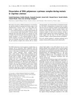
Figure 1 Nanoparticle preparation: Emulsion 1 (W/O) was obtained after an aqueous buff er contai ning Protamine Sulphat e (PS) +/-
DNA (blue) (solution I) was mixed with an organic buffer of PLGA with cationic lipids DOTAP (green) or DC-Chol (red) (solution II) and
sonicated. Then another aqueous buffer containing PVA (solution III) was added to form Emulsion 2 (W/O/W). The mixture was briefly sonicated
and NPs were formed by solvent evaporation. For DNA encapsulated NPs (in and both), pLuc DNA was added to solution I. For DNA adsorbed
NPs (out or both), pLuc DNA was added to the NPs as described in the methods. The nanoparticles are designated as: green for PLGA/DOTAP,
red for PLGA/DC-Chol and a blue plus inside the circle for encapsulated DNA. Blue on the outer circle designates adsorbed DNA.
Zhong et al. Journal of Nanobiotechnology 2010, 8:6
/>Page 3 of 10
potential module equipped with a 35 mW two laser
diode (658 nm). Scattered light was detected at a 90
angle and a temperature of 25°C. About 1.6 ml of a sus-
pension of charged particles in water was used for the
measurements. Zeta potential values (Tables 3 and 4)
were calculated from measured velocities using the
Smoluchowski equation.
The shape and surface morphology (smooth versus
porous structure) of the nanoparticles were investigated
using a scanning electron microscope (SEM) (S-4800N,
Tokyo, Japan). Nanoparticles suspended in deionized
water were freeze-dried. The dried nanoparticles were
mounted on metal stubs with double sided tape and
coated with a thin gold layer using an ion coater
(K550X, EMITECH, Kent, UK).
Quality Control for DNA Location on Nanoparticles
We used measurement of luciferase activity for transgene
expression, as the most sensitive assay to assign DNA
location (out, in or both) on the different NP/DNA com-
plexes. The six NPs were eac h suspended in water, trea-
ted with DNase I (Fermentas, Glen Burnie, MD) at 37°C
for 30 min, washed and delivered to 293 cells. Specifi-
cally, 16 μg NPs (with or without DNase I treatment)
were added to 10
5
cells in 48 well plates for 48 hours and
luciferase activity was measured as seen in Figure 2. We
had previously tried unsuccessfully, to measure residual
DNA by location on the NP/DNA complexes, using
DNA concentration (OD at 260 nm) or agarose gel elec-
trophoresis before and after DNase I digestion.
Evaluation of NP/DNA Complex Uptake in vitro by Cells
For dose responses assays, 293 cells were seeded onto 48
well plates at a density of 10
5
cells per well in 1 ml
DMEM (Invitrogen, Carisbad, CA) containing 10% FBS.
Incubation of cells was for 24 hr at 37°C in a 5% CO
2
incubator. Each of the six different NPs in 50 μlPBS
and co ntaining pLuc DNA was added at concentrations
of 164 ng to 100 μg (in 2 to 2.5 fold-stepwise intervals)
to separate wells. After 48 hrs incubation, luciferase was
assayed using a kit from Promega. DNA with Lip2 000
was the positive control (PC) and DNA only was the
negative control (NC).
Regression analysis and determination of the declina-
tion angles for DNA uptake of NPs by 293 cells was
performed using the trend line program from a Micro-
soft Excel 2007 software statistical package. Cells were
passaged at 10
5
cells per ml in a T25 flask containing 5
ml DMEM with 10% FBS. After 24 hr, each of the six
NPs containing pLuc DNA was added at 40 μgandcul-
turing was maintained for up to 4 weeks. At 3 or 4 day
intervals, cell density was adjusted to 10
5
cells per ml by
adding fresh medium. DNA activity was measured by
the luciferase assay.
Results and Discussion
Characterization of hybrid nanoparticle/DNA complexes
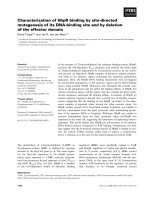
PLGA based NPs prepared by the solvent evaporation
method (Figure 1), with either DO TAP or DC-Chol
showed a similar particle size di stribution (Figure 3).
Fromtherepresentativesize distribution d iagrams, it
can be seen that in both formulations 70% of particles
were in the range of 100-400 nm. NPs formulated,
either with DOTAP or DC-Chol, exhibit a uniform
spherical shape with smooth surface as seen by scan-
ning electron microscopy. The particle size distribu-
tions and zeta potentials are described in Table 3.
Initially, PLGA NPs with PVA, a most commonly used
surfactant or stabilizer, have a negative surface charge
because of physical entrapment of liquid within the
surface layer of the polymer [38]. In our formulations,
after addition of cationic lipids (DOTAP and DC-
Chol) an overall positive charge is imparted to the NP
surface. The PLGA/DOTAP and PLGA/DC-Chol NPs
also were complexed with luciferase gene plasmid
DNA pLuc (pGL4.75), at the concentrations described
(Table 1, 2). Although the zeta potential is varied in
all formulations, it is still positive in all cases. The
lower positive zeta potentials of adsorbed NPs (out
and both) may possibly be due to the nullifying effects
of negative charge on DNA versus t he positive charge
of cationic lipid on the surface of these NPs, com-
pared to encapsulated NPs (in)(Table4).Previous
studies with such cationic lipid/DNA NP complexes
have shown that they are stable [34] and efficiently
taken up by tissue culture cells [35,39]. In this study
we have focused on delivery of such NPs to 293 and
other cells.
Table 3 Physical properties of PLGA cationic particles
Formulation Particle Size (nm) Zeta Potential (mv)
d (0.1) d (0.5) d (0.9)
A PLGA/DOTAP 95 218 425 52.64 ± 1.17
B PLGA/DC-Chol 86 210 523 41.67 ± 2.55
The mean size and distribution for different NPs are indicated; d(0.1), d(0.5), d
(0.9) means that less than 10%, 50%, 90% of the NPs respectively, are
distributed around the particle sizes indicated
Table 4 Zeta potential of nanoparticle DNA complexes
Formulation Zeta Potential (mv)
A1 DOTAP (out) 06.86 ± 0.72
B1 DC-Chol (out) 05.83 ± 0.24
C1 DOTAP (in) 31.95 ± 0.99
D1 DC-Chol (in) 14.84 ± 0.11
E1 DOTAP (both) 16.40 ± 0.27
F1 DC-Chol (both) 06.46 ± 0.07
DOTAP: PLGA/DOTAP DC-Chol: PLGA/DC-Chol
Zhong et al. Journal of Nanobiotechnology 2010, 8:6
/>Page 4 of 10
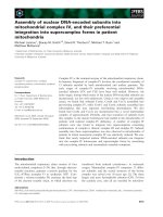
Figure 2 Quality control for pLuc DNA adsorbed to either surface NPs (out and both) or encapsulated NPs (in and both). The NP/DNA
complexes were treated with or without DNase I and delivered to 293 cells for 48 hours. Lipofectamine 2000 with pLuc DNA was a positive
control (Lip) and untreated 293 cells was the negative control (NC). The assay measures luciferase activity.
Figure 3 SEM photomic rograph of PLGA/DOTAP and PLGA/DC-Chol nanopart icles (top). The corresponding particle size distribution for
PLGA/DOTAP nanoparticles (green) and PLGA/DC-Chol nanoparticles (red) is on the bottom.
Zhong et al. Journal of Nanobiotechnology 2010, 8:6
/>Page 5 of 10
Optimization of NP DNA binding conditions
We determined the optimal conditions for binding the
maximal amount of DNA to the PLGA hybrid NPs. The
two types, DOTAP (A) or DC-Chol (B) hybr id NPs,
were complexed with luciferase gene plasmid DNA at a
w/w ratio of 10/1 and held at 4°C, room temperature
(22°C) or 50°C for 1, 2, 3, 4 hours, a s well as overnight.
Both types gave similar results, so we will describe spe-
cific findings for DOTAP/DNA NPs (out). After 3 hours
at 4°C or 22°C these NPs have a similar, high level of
DNA binding activity relative to those held at 50°C. 100
μg of such NP/DNA complexes formed at 4°C or room
temperature were then transferred for uptake to 10
5
293
cells in 1 ml and incubated for 1 day. About a 23%
increase in DNA binding was observed at 4°C. The max-
imal amount of DNA that could tightly bind to the NPs
at 4°C was then determined. For this, NP/DN A (w/w)
ratios of 10/1 to 50/1 were incubated overnight at 4°C.
Then the NPs were pelleted and the supernatant was
collected. DNA measurements were made both for the
NP/DNA complexes and free DNA using 1 mg of NP
complexed with 100 μg, 50 μg, 40 μgand20μgof
DNA. The amount of free DNA was highest at the 10/1
ratio and lowest at the 50/1 ratio; however all levels
showed that ≥ 95% of DNA was bound to the NP.
Based on these findings, our experiments utilized NPs at
a ratio of 20-30 μg DNA/1 mg NP, in order to avoid
competition with free DNA.
Localization of DNA in the nanoparticles/DNA complexes
The six NP/DNA complexes were suspended in water
at 10 mg/ml. In order to verify DNA location on the
outside or inside of the NP complexes respectively, we
used the following approach to determine sensitivity to
DNase I. NP/DNA complexes were treated with DNase
I and delivered to 293 cells. Expression of residual
DNA was assigned by measuring luciferase activity
after48hours.WenoteinFigure2thatthoseNP/
DNA complexes where DNA was adsorbed on outer
surfaces (out and both)wereabletobecleavedby
DNase I. Thus no expression was detected for out,but
about 50% expression was detected for both.As
expected, no difference was seen for NPs with encap-
sulated DNA (in)(Figure2).
Optimization of NP/DNA complex delivery conditions to
293 cells
We compared the efficiency of DNA delivery to 293
cells by the six NP/DNA complexes vs. a Lip2000/DNA
mixture. Lipofectamine 2000 is a cationic lipid widely
used to tr ansfect plasmid and other DNA into a variety
of mammalian cells. Invitrogen reports [40] that 293
cells transfected with pCMV-b gal DNA exhibited a
high transfection efficiency (99%) and 100% cell viability
at 24 hours post transfection. PLGA/DOTAP or PLGA/
DC-Chol NPs with the composition of pLuc DNA seen
in Tables 1 and 2 were formulated as in Figure 1, and
all six were used at a concentration of 25 μgDNA/1
mgNP.NPswereaddedto10
5
cells at 2 t o 2.5 fold
increasing concentrations starting at 164 ng and going
to 100 μg for 2 days (Figure 4 ). Based on the R
2
value
of the straight line seen in Figure 5 for the three
DOTAP NP/DNA complexes, the transfection efficiency
achieved is high and similar to that for Lip2000/DNA
complexes.
Although Lipofectamine 2000 appears effective at
lower concentrations of plasmid DNA (100 pg to 100
ng), it has the disadvantage of toxicit y, as n oted in the
introduction and thus would have limited applicability
in vivo. Specifically, high cytotoxicity in renal and arter-
ial tissue-based studies [41,42], as well as in animal
applications [43,44] have been reported. Hybrid NPs in
contrast, are safe in cell and animal studies [41,45].
Further, from Figures 4 and 5 we note that NPs are best
used at concentrations of 16-40 μg NPs/ml with 293
cells; NP levels ≥ 100 μg/ml are cytotoxic (data not
shown). The DNA binding experiment seen in Figure 5
was repeated with DC-Chol NPs and gave a similar
result. The relative transfection efficiency of pLuc DNA
calculated from these experiments show that DOTAP or
DC-Chol NPs are nearly as efficient as Lip2000 in deli-
vering DNA to 293 cells; however, when compared to
free DNA, NPs have a 500-600 fold higher transmission
efficiency. In conclusion, we find that after 2 days of
NP/DNA complex delivery to 293 cells (Figure 4), “Out”
NPs shows a higher luciferase expression than NPs with
only inside DNA (in) and luciferase expression is inter-
mediate for “ Both” NPs. This suggests that outside
DNA exhibits an initial high expression due to rapid
release of bound DNA. On the other hand, DNA encap-
sulated NPs (in) are slower to release DNA and are
probably affected by biodegradation of the NPs within
cells.
Study of gene delivery with hybrid nanoparticle/DNA
complexes using other cell lines
The optimal condition for DNA gene delivery to 293
cells was shown in Figures 4 and 5, and we found that
all six NP/DNA complexes showed a high efficiency of
gene transfection. We also were interested in checking
transfection with other cell lines and found that two
adherent prostate cell lin es (PC-3, LNcap) gave the
same high efficiency for the six different hybrid NP/
DNA complexes, again compared to Lip2000 (data not
shown). Interestingly, when non-adherent MOLT-4 cells
were used, only a high trans fection efficiency was found
with the NP/DNA complexes and not Lip2000 (data not
shown).
Zhong et al. Journal of Nanobiotechnology 2010, 8:6
/>Page 6 of 10
Figure 5 Dose/response bars and lines showing transfection efficiency. Luciferase activity was measured (blue bars) and the corresponding
straight lines generated (black lines). DOTAP NPs (25 μg DNA/mg NPs) were added at amounts of 410 ng to 16 μg NPs to 10
5
cells/ml (293
cells) for 48 hours. Top shows Out and In NP/DNA complexes. Bottom shows Both NP/DNA and Lip2000 (Lip) complexes; Lip/DNA complexes
were added at 100 pg to 100 ng DNA.
Figure 4 Dose/response bar graphs showing efficiency of DNA delivery to 293 cells after 48 hours incubation for three classes of NPs
made from two type of cationic lipid; DOTAP (top) and DC-Chol (bottom). NP/DNA complexes were added at concentrations from 164 ng
to 100 μg in 2.5 fold-stepwise intervals. Positive control (PC) is Lipofectamine 2000 with 100 ng DNA; DNA control (DC) uses 10 μg DNA alone;
Negative control (NC) is 293 cells only and no particles, lipofectamine or DNA.
Zhong et al. Journal of Nanobiotechnology 2010, 8:6
/>Page 7 of 10
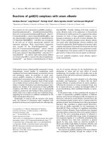
Degradation of NP/DNA complexes delivered to 293 cells
For these experiments, we freshly prepared the six NP/
DNA complexes, using a NP/DNA (w/w) ratio of 40/1
(Figure 1). Such complexes bound DNA at a level o f
96% to 99%. They were added to 293 cells for 3 days
and incubated at 37°C for about 4 weeks. Cell passages
were done at 3 to 4 day intervals. Samples were
removed at these times and the level of luciferase DNA
was measured. The results are shown in Figure 6 with a
positive control using Lipofectamine (Lip). The top fig-
ure presents the data in a graph format, while the
middle and bottom provide t he data as straight lines.
These results represent the release profile of DNA from
the NP/DNA complexes within 293 cells, o ver time.
Regression analysis was performed and lines were
drawn of the data points taken for the 4 week period.
DC-Chol NPs containing externally bound DNA (out)
(bottom graph) exhibited a large initial uptake followed
by a steep decay of pLuc DNA, similar to Lipofecta-
mine. However with DOTAP (middle graph), externally
bound DNA NPs (out) exhibited a diminished slope of
DNA dec ay relative to Lipofectamine. DOTAP NPs
Figure 6 Degradation analysis for DNA delivery to 293 cells by six different nanoparticle/DNA complexes over a four week period.
Two NP/cationic lipid mixtures (PLGA/DOTAP and PLGA/DC-Chol) and three classes of NP/DNA complexes (out, in and both) were used. Lip
(Lip2000/DNA mixture) was a positive control. Top columns show luciferase activity at 3 or 4 day intervals for 4 weeks. Middle graph is (DOTAP)
and bottom graph (DC-Chol) NPs. Regression analysis gave straight lines (blue for out, red for in and green for both) for nanoparticles and Lip
(purple).
Zhong et al. Journal of Nanobiotechnology 2010, 8:6
/>Page 8 of 10
(middle graph) and DC-Chol NPs (bottom graph) with
bound and encapsula ted DNA (both) also led to a large
initial uptake, but it was followed by sustained DNA
release over a longer time. This i s correlated with a
lower angle of decl ination of the regr ession line t han
Lip (average angle of 23.8° for DOTAP and 29.3° for
DC-Chol) (Table 5). NPs with only encapsulated DNA
(in) showed an intermediate level of DNA degradation.
Since all assays started with the same number of cells,
this different decline in luciferase activity with different
NPs is not likely to be a cell dilution problem. In sum-
mary, the “Lip ” and “ Out” NP complexes have similar
profiles (steep slope) because both have outside bound
DNA and the expression assay in 293 cells reflects the
rapid release of such bound DNA. On the other hand,
“In” and “Both” have longer retention profiles, indicat-
ing that this expression assay is affected by biodegrada-
tion in time, of encapsulated NP/DNA complexes
within cells. Howev er, our results show that the “Both”
NP/DNA complexes, which have DNA both outside and
inside show a higher level of luciferase activity after four
weeks than the “In” NP/DNA complexe s. This may be
because the former NPs with DNA on the outside can
stabilize the surface charge and allow for a longer reten-
tion time within 293 cells. These findings are important
for the future design of vaccines using NP/DNA com-
plexes. Thus, when an i nitial strong gene delivery
response over a short time is required, as in “priming”
for an a ntibody in animals, it appears that NP com-
plexes with adsorbed DNA (out) are best used. How-
ever, for a response where one wants a longer time of
gene delivery, as in a “ booster” inoculation, the
adsorbed/encapsulated DNA complexes (both)arebest
used. It should be noted with NPs that there is alw ays
the potential for an inflammatory response as with gene
delivery systems, but in both cases this is usually depen-
dent on immune response to the transgene product.
Conclusion
Nanoparticles provide a better vector than DNA alone
for luciferase gene delivery (500-600 times more effi-
cient).Adoseresponsecurveforgenedeliveryofsix
different NP/DNA complexes to 293 cells has been
generated; optimal delivery conditions occur for 10
5
cells over a range of 500 ng-10 μg of NPs containing
20-30 μg DNA per 1 mg of NPs. NPs with externally
bound DNA ( out) led to a steep slope on lines drawn
from regression analysis, while NPs with both adsorbed
and encapsu lated DNA (both) exhibited a l ong er reten-
tion time. This offers the potential of using hybrid NPs
with adsorbed DNA (out)for“ priming” in animal
immunization studie s, while DNA adsorbed/encapsu-
lated NPs (both) are optimal for “ booster”
immunization.
Acknowledgements
This work was supported, in part, by the Louisiana Vaccine Center and the
South Louisiana Institute for Infectious Disease Research sponsored by the
Louisiana Board of Regents and LEQSF(2007-12)-ENH-PKSFI-PRS-02.
Author details
1
Department of Microbiology Immunology and Parasitology, Louisiana State
University Health Sciences Center, New Orleans, Louisiana 70112, USA.
2
College of Pharmacy, Xavier University of Louisiana, New Orleans, Louisiana
70125, USA.
3
Guangdong Food and Drug Vocational College, Guang Zhou,
Guangdong 510520, PR China.
Authors’ contributions
QZ carried out design and performed study, data analysis and drafting of
the manuscript. TKM directed, while DMDC and SP carried out NP
formulation and characterization such as particle size, zeta potential and
morphology of nanoparticles. HW consulted and participated in the design
of the study. XY carried out the Luciferase assay in evaluation of NPs and
prepared cells. RBL was involved with the design, coordination, data analysis
and drafting of the manuscript through its many revisions. All authors read
and approved the final manuscript.
Competing interests
The authors declare that they have no competing interests.
Received: 24 November 2009
Accepted: 24 February 2010 Published: 24 February 2010
References
1. Maguire AM, Simonelli F, Pierce EA, Pugh EN Jr, Mingozzi F, Bennicelli J,
Banfi S, Marshall KA, Testa F, Surace EM, Rossi S, Lyubarsky A, Arruda VR,
Konkle B, Stone E, Sun J, Jacobs J, Dell’Osso L, Hertle R, Ma JX,
Redmond TM, Zhu X, Hauck B, Zelenaia O, Shindler KS, Maguire MG,
Wright JF, Volpe NJ, McDonnell JW, Auricchio A, et al: Safety and efficacy
of gene transfer for Leber’s congenital amaurosis. N Engl J Med 2008,
358:2240-2248.
2. Stewart DJ, Hilton JD, Arnold JM, Gregoire J, Rivard A, Archer SL,
Charbonneau F, Cohen E, Curtis M, Buller CE, Mendelsohn FO, Dib N,
Page P, Ducas J, Plante S, Sullivan J, Macko J, Rasmussen C, Kessler PD,
Rasmussen HS: Angiogenic gene therapy in patients with
nonrevascularizable ischemic heart disease: a phase 2 randomized,
controlled trial of AdVEGF(121) (AdVEGF121) versus maximum medical
treatment. Gene Ther 2006, 13:1503-1511.
3. Nikol S, Engelmann MG, Pelisek J, Fuchs A, Golda A, Shimizu M, Mekkaoui C,
Rolland PH: Local perivascular application of low amounts of a plasmid
encoding for vascular endothelial growth factor (VEGF165) is efficient
for therapeutic angiogenesis in pigs. Acta Physiol Scand 2002,
176:151-159.
4. Li C, Hirsch M, DiPrimio N, Asokan A, Goudy K, Tisch R, Samulski RJ:
Cytotoxic-T-lymphocyte-mediated elimination of target cells transduced
with engineered adeno-associated virus type 2 vector in vivo. J Virol
2009, 83:6817-6824.
5. Mulligan RC: The basic science of gene therapy. Science 1993,
260:926-932.
Table 5 Angle of regression line declination* over a four
week period for six nanoparticle preparations
Experiment DOTAP DC-Chol
out in both out in both
#1 35.5° 32.3° 25.3° 46.8° 35.9° 29.5°
#2 30.1° 28.1° 17.4° 39.2° 32.0° 23.7°
#3 42.3° 36.5° 28.7° 54.5° 36.5° 34.6°
Average 36.0° 32.3° 23.8° 46.8° 34.8° 29.3°
*Angle is in degrees and reflects pLuc DNA degradation over time in 293 cells
Zhong et al. Journal of Nanobiotechnology 2010, 8:6
/>Page 9 of 10
6. Miyake K, Suzuki N, Matsuoka H, Tohyama T, Shimada T: Stable integration
of human immunodeficiency virus-based retroviral vectors into the
chromosomes of nondividing cells. Hum Gene Ther 1998, 9:467-475.
7. Marshall E: Gene therapy. What to do when clear success comes with an
unclear risk?. Science 2002, 298:510-511.
8. Ott MG, Schmidt M, Schwarzwaelder K, Stein S, Siler U, Koehl U, Glimm H,
Kuhlcke K, Schilz A, Kunkel H, Naundorf S, Brinkmann A, Deichmann A,
Fischer M, Ball C, Pilz I, Dunbar C, Du Y, Jenkins NA, Copeland NG, Luthi U,
Hassan M, Thrasher AJ, Hoelzer D, von Kalle C, Seger R, Grez M: Correction
of X-linked chronic granulomatous disease by gene therapy, augmented
by insertional activation of MDS1-EVI1, PRDM16 or SETBP1. Nat Med
2006, 12:401-409.
9. von Kalle C, Fehse B, Layh-Schmitt G, Schmidt M, Kelly P, Baum C: Stem cell
clonality and genotoxicity in hematopoietic cells: gene activation side
effects should be avoidable. Semin Hematol 2004, 41:303-318.
10. Baum C, Dullmann J, Li Z, Fehse B, Meyer J, Williams DA, von Kalle C: Side
effects of retroviral gene transfer into hematopoietic stem cells. Blood
2003, 101:2099-2114.
11. Cohen J: AIDS research. Did Merck’s failed HIV vaccine cause harm?.
Science 2007, 318:1048-1049.
12. Tal J: Adeno-associated virus-based vectors in gene therapy. J Biomed Sci
2000, 7:279-291.
13. Campos SK, Barry MA: Current advances and future challenges in
Adenoviral vector biology and targeting. Curr Gene Ther 2007, 7:189-204.
14. Amalfitano A: Next-generation adenoviral vectors: new and improved.
Gene Ther 1999, 6:1643-1645.
15. Scallan CD, Liu T, Parker AE, Patarroyo-White SL, Chen H, Jiang H, Vargas J,
Nagy D, Powell SK, Wright JF, Sarkar R, Kazazian HH, McClelland A,
Couto LB: Phenotypic correction of a mouse model of hemophilia A
using AAV2 vectors encoding the heavy and light chains of FVIII. Blood
2003, 102:3919-3926.
16. Hartig PC, Hunter ES: Gene delivery to the neurulating embryo during
culture. Teratology 1998, 58:103-112.
17. Mintzer MA, Simanek EE: Nonviral vectors for gene delivery. Chem Rev
2009, 109:259-302.
18. Anwer K: Formulations for DNA delivery via electroporation in vivo.
Methods Mol Biol 2008, 423:77-89.
19. Kasuya T, Kuroda S: Nanoparticles for human liver-specific drug and gene
delivery systems: in vitro and in vivo advances. Expert Opin Drug Deliv
2009, 6:39-52.
20. Konig Merediz SA, Zhang EP, Wittig B, Hoffmann F: Ballistic transfer of
minimalistic immunologically defined expression constructs for IL4 and
CTLA4 into the corneal epithelium in mice after orthotopic corneal
allograft transplantation.
Graefes Arch Clin Exp Ophthalmol 2000,
238:701-707.
21. Reed SD, Li S: Electroporation Advances in Large Animals. Curr Gene Ther
2009, 9:316-326.
22. Mir LM: Nucleic Acids Electrotransfer-Based Gene Therapy
(Electrogenetherapy): Past, Current, and Future. Mol Biotechnol 2009,
43:167-176.
23. Wells DJ: Gene therapy progress and prospects: electroporation and
other physical methods. Gene Ther 2004, 11:1363-1369.
24. Liu Y, Liggitt D, Zhong W, Tu G, Gaensler K, Debs R: Cationic liposome-
mediated intravenous gene delivery. J Biol Chem 1995, 270:24864-24870.
25. Gao X, Huang L: Cationic liposome-mediated gene transfer. Gene Ther
1995, 2:710-722.
26. Canonico AE, Plitman JD, Conary JT, Meyrick BO, Brigham KL: No lung
toxicity after repeated aerosol or intravenous delivery of plasmid-
cationic liposome complexes. J Appl Physiol 1994, 77:415-419.
27. Porteous DJ, Dorin JR, McLachlan G, Davidson-Smith H, Davidson H,
Stevenson BJ, Carothers AD, Wallace WA, Moralee S, Hoenes C, Kallmeyer G,
Michaelis U, Naujoks K, Ho LP, Samways JM, Imrie M, Greening AP, Innes JA:
Evidence for safety and efficacy of DOTAP cationic liposome mediated
CFTR gene transfer to the nasal epithelium of patients with cystic
fibrosis. Gene Ther 1997, 4:210-218.
28. Nchinda G, Zschornig O, Uberla K: Increased non-viral gene transfer levels
in mice by concentration of cationic lipid DNA complexes formed under
optimized conditions. J Gene Med 2003, 5:712-722.
29. Cemazar M, Sersa G, Wilson J, Tozer GM, Hart SL, Grosel A, Dachs GU:
Effective gene transfer to solid tumors using different nonviral gene
delivery techniques: electroporation, liposomes, and integrin-targeted
vector. Cancer Gene Ther 2002, 9:399-406.
30. Huang YC, Riddle K, Rice KG, Mooney DJ: Long-term in vivo gene
expression via delivery of PEI-DNA condensates from porous polymer
scaffolds. Hum Gene Ther 2005, 16:609-617.
31. Nguyen DN, Raghavan SS, Tashima LM, Lin EC, Fredette SJ, Langer RS,
Wang C: Enhancement of poly(orthoester) microspheres for DNA vaccine
delivery by blending with poly(ethylenimine). Biomaterials 2008,
29:2783-2793.
32. Benita S, Benoit JP, Puisieux F, Thies C: Characterization of drug-loaded
poly(d, l-lactide) microspheres. J Pharm Sci 1984, 73:1721-1724.
33. Pamujula S, Graves RA, Moiseyev R, Bostanian LA, Kishore V, Mandal TK:
Preparation of polylactide-co-glycolide and chitosan hybrid
microcapsules of amifostine using coaxial ultrasonic atomizer with
solvent evaporation. J Pharm Pharmacol 2008, 60:283-289.
34. Singh M, Briones M, Ott G, O’Hagan D: Cationic microparticles: A potent
delivery system for DNA vaccines. Proc Natl Acad Sci USA
2000,
97:811-816.
35. Gvili K, Benny O, Danino D, Machluf M: Poly(D, L-lactide-co-glycolide acid)
nanoparticles for DNA delivery: waiving preparation complexity and
increasing efficiency. Biopolymers 2007, 85:379-391.
36. Diez S, Navarro G, de ICT: In vivo targeted gene delivery by cationic
nanoparticles for treatment of hepatocellular carcinoma. J Gene Med
2009, 11:38-45.
37. Shive MS, Anderson JM: Biodegradation and biocompatibility of PLA and
PLGA microspheres. Adv Drug Deliv Rev 1997, 28:5-24.
38. Evora C, Soriano I, Rogers RA, Shakesheff KN, Hanes J, Langer R: Relating
the phagocytosis of microparticles by alveolar macrophages to surface
chemistry: the effect of 1,2-dipalmitoylphosphatidylcholine. J Control
Release 1998, 51:143-152.
39. Oberl V, Zuhorn IS, Audouy S, Bakowsky U, Smisterova J, Engberts JBFN,
Hoekstra D: Targeting of Drugs. Gregoriadis G, McCormack B (Series Editor)
IOS PressGregoriadis G, McCormack B .
40. Ohki EC, Tilkins ML, Ciccarone VC, Price PJ: Improving the transfection
efficiency of post-mitotic neurons. J Neurosci Methods 2001, 112:95-99.
41. Bejjani RA, BenEzra D, Cohen H, Rieger J, Andrieu C, Jeanny JC, Gollomb G,
Behar-Cohen FF: Nanoparticles for gene delivery to retinal pigment
epithelial cells. Mol Vis 2005, 11:124-132.
42. Madry H, Reszka R, Bohlender J, Wagner J: Efficacy of cationic liposome-
mediated gene transfer to mesangial cells in vitro and in vivo. J Mol Med
2001, 79:184-189.
43. Armeanu S, Pelisek J, Krausz E, Fuchs A, Groth D, Curth R, Keil O, Quilici J,
Rolland PH, Reszka R, Nikol S: Optimization of nonviral gene transfer of
vascular smooth muscle cells in vitro and in vivo. Mol Ther 2000,
1:366-375.
44. Gebhart CL, Kabanov AV: Evaluation of polyplexes as gene transfer
agents. J Control Release 2001, 73:401-416.
45. Azarmi S, Lobenberg R, Roa WH, Tai S, Finlay WH: Formulation and in vivo
evaluation of effervescent inhalable carrier particles for pulmonary
delivery of nanoparticles. Drug Dev Ind Pharm 2008, 34:943-947.
doi:10.1186/1477-3155-8-6
Cite this article as: Zhong et al.: Optimization of DNA delivery by three
classes of hybrid nanoparticle/DNA complexes. Journal of
Nanobiotechnology 2010 8:6.
Submit your next manuscript to BioMed Central
and take full advantage of:
• Convenient online submission
• Thorough peer review
• No space constraints or color figure charges
• Immediate publication on acceptance
• Inclusion in PubMed, CAS, Scopus and Google Scholar
• Research which is freely available for redistribution
Submit your manuscript at
www.biomedcentral.com/submit
Zhong et al. Journal of Nanobiotechnology 2010, 8:6
/>Page 10 of 10