báo cáo khoa học: "Naturally occurring nanoparticles from English ivy: an alternative to metal-based nanoparticles for UV protection" doc
Bạn đang xem bản rút gọn của tài liệu. Xem và tải ngay bản đầy đủ của tài liệu tại đây (955.5 KB, 9 trang )
Xia et al. Journal of Nanobiotechnology 2010, 8:12
/>Open Access
RESEARCH
BioMed Central
© 2010 Xia et al; licensee BioMed Central Ltd. This is an Open Access article distributed under the terms of the Creative Commons At-
tribution License ( which permits unrestricted use, distribution, and reproduction in any
medium, provided the original work is properly cited.
Research
Naturally occurring nanoparticles from English ivy:
an alternative to metal-based nanoparticles for UV
protection
Lijin Xia
†
, Scott C Lenaghan
†
, Mingjun Zhang*, Zhili Zhang and Quanshui Li
Abstract
Background: Over the last decade safety concerns have arisen about the use of metal-based nanoparticles in the
cosmetics field. Metal-based nanoparticles have been linked to both environmental and animal toxicity in a variety of
studies. Perhaps the greatest concern involves the large amounts of TiO
2
nanoparticles that are used in commercial
sunscreens. As an alternative to using these potentially hazardous metal-based nanoparticles, we have isolated organic
nanoparticles from English ivy (Hedera helix). In this study, ivy nanoparticles were evaluated for their potential use in
sunscreens based on four criteria: 1) ability to absorb and scatter ultraviolet light, 2) toxicity to mammalian cells, 3)
biodegradability, and 4) potential for diffusion through skin.
Results: Purified ivy nanoparticles were first tested for their UV protective effects using a standard spectrophotometric
assay. Next the cell toxicity of the ivy nanoparticles was compared to TiO
2
nanoparticles using HeLa cells. The
biodegradability of these nanoparticles was also determined through several digestion techniques. Finally, a
mathematical model was developed to determine the potential for ivy nanoparticles to penetrate through human
skin. The results indicated that the ivy nanoparticles were more efficient in blocking UV light, less toxic to mammalian
cells, easily biodegradable, and had a limited potential to penetrate through human skin. When compared to TiO
2
nanoparticles, the ivy nanoparticles showed decreased cell toxicity, and were easily degradable, indicating that they
provided a safer alternative to these nanoparticles.
Conclusions: With the data collected from this study, we have demonstrated the great potential of ivy nanoparticles as
a sunscreen protective agent, and their increased safety over commonly used metal oxide nanoparticles.
Background
Ultraviolet (UV) radiation is highly energetic electromag-
netic radiation from light waves below that of the visible
light spectrum. The wavelengths for UV radiation range
from 100-400 nm, including UV-A (315-400 nm), UV-B
(280-315 nm), and UV-C (100-280 nm) [1]. While the
earth's ozone layer blocks 98.7% of UV radiation from
penetrating through the atmosphere, a small percentage
of UV, comprising UV-A and some UV-B, can still reach
the planet, which can cause harmful effects to humans
[2]. UV-C does not typically reach the surface of the
planet, but due to its ability to cause DNA damage it is
often used as a model for UV study in the laboratory.
Depending on the time of exposure to sunlight, the harm-
ful UV-A/UV-B effects include immediate distresses like
blistering sunburns, and long term problems like skin
cancer, melanoma, cataracts, and immune suppression
[3,4]. The underlying mechanism for UV-A damage
involves oxidative stress and protein denaturation, while
short wavelength UV-B radiation causes predominantly
DNA damage in the form of pyrimidine dimers and 6-4
photoproducts [5]. UV radiation induced DNA mutation
is one of the leading causes of skin cancer, with more than
one million cases diagnosed annually resulting in 11,590
deaths in the U.S. [6].
The demand for skin protection agents against the
harmful influence of UV solar radiation has become
increasingly important in light of the depletion of the
* Correspondence:
1
Department of Mechanical, Aerospace and Biomedical Engineering,
University of Tennessee, Knoxville, TN, 37996, USA
†
Contributed equally
Full list of author information is available at the end of the article
Xia et al. Journal of Nanobiotechnology 2010, 8:12
/>Page 2 of 9
ozone layer [7,8]. Sunscreens, which work by combining
organic and inorganic ingredients to reflect, scatter or
absorb UV radiation, provide significant protection
against the damage from solar UV. Early sunscreens
developed with inorganic UV filters, such as titanium
dioxide (TiO
2
) and zinc oxide (ZnO) particles, were often
opaque giving the skin a white tinge, which made them
unappealing to consumers [9]. With enhanced UV pro-
tection and low opacity, nanosize metal oxide particles
have been introduced into cosmetics products in recent
years and thousands of tons of nanomaterials are cur-
rently applied onto the faces and hands of hundreds of
millions of people every year [10]. With increased popu-
larity, the safety of these metal-based nanoparticles and
potential toxicity is under significant debate. Many stud-
ies indicated that when applied to skin for less than 8
hours, inorganic nanoparticle filters do not penetrate
through the stratum corneum (SC) layer of the skin [11-
14]. However, these studies typically examine the effects
of nanoparticles greater than 20 nm, and always use
healthy skin samples. Studies evaluating the penetration
of ultrafine nanoparticles found that TiO
2
, maghemite,
and iron nanoparticles less than 15 nm are capable of
penetrating through the SC [15,16]. Other studies have
also observed penetration of 4 nm and 60 nm TiO
2
parti-
cles through healthy skin in hairless mice after prolonged
exposure from 30-60 days [17]. This penetration leads to
increased aging of skin, pathological effects in the liver,
and particle accumulation in the brain. Studies like these
have raised significant concerns about the prolonged use
of these metal oxide nanoparticles for cosmetic applica-
tions which lead to investigation of alternative organic fil-
ters.
The properties of materials at the nanoscale differ sig-
nificantly from those at a larger scale, and safety claims by
cosmetics manufacturers based on their bulk properties
pose great risk without proper federal regulation of their
applications [18]. When decreasing size to the nanoscale,
materials alter many of their physical and chemical prop-
erties, including but not limited to color, solubility, mate-
rial strength, electrical conductivity, magnetic behavior,
mobility (within the environment and within the human
body), chemical and biological activities [19]. The
increased surface to volume ratio also enhances chemical
activity, which can result in the increased production of
reactive oxygen species (ROS) [20]. ROS production,
which has been found in metal oxide nanoparticles, car-
bon nanotubes, and fullerenes, is the leading force of oxi-
dative stress, inflammation, and consequent damage to
DNA, proteins and membranes [20]. Further concern for
these nanomaterials in applications is their photoactivity
when exposed to UV light, which results in greater ROS
and free radical production [21]. TiO
2
nanoparticles have
been shown to cause far greater damage to DNA than
does TiO
2
of larger particle size [22]. While 500 nm TiO
2
particles have some ability to cause DNA strand break-
age, 20 nm TiO
2
nanoparticles are capable of causing
complete destruction of super-coiled DNA, as demon-
strated in a plasmid DNA assay, even at lower doses and
without exposure to UV. In addition to the increased
potential for DNA damage from engineered metal oxide
nanoparticles, another concern for their application in
cosmetics is the potential for inhalation, ingestion, and
penetration through the skin. Once in the blood stream,
nanomaterials can be circulated inside the body and are
taken up by organs and tissues such as the brain, liver,
spleen, kidney, heart, bone marrow, and nervous system
[19]. With their stability, the damage of these nanoparti-
cles to human tissues and organs can occur through a tra-
ditional ROS pathway, or through accumulation that can
impair their normal functions. In vitro studies on BRL 3A
rat liver cells exposed to 100-250 μg/ml of Fe
3
O
4
, Al,
MoO
3
and TiO
2
nanoparticles revealed significant dam-
age from ROS in these cells [23]. Carbon nanotubes have
also been shown to be toxic to kidney cells and inhibit cell
growth [24]. The stability of nanomaterials in the envi-
ronment has also been linked to brain damage and mor-
tality in several aquatic species [25,26].
Due to the potential toxicity associated with prolonged
use of metal oxide nanoparticle sunscreens, it is crucial to
search for alternative ingredients that are non-toxic and
effective at blocking UV. It is highly expected that these
ingredients should be biodegradable, and less toxic to
mammalian cells than metal oxide nanoparticles. The
recent discovery of ivy and other naturally occurring
nanoparticles provides a promising alternative to engi-
neered metal oxide nanomaterials for cosmetics applica-
tions [27]. To explore the possibility of using ivy
nanoparticles for sunscreen, the UV protection proper-
ties of ivy nanoparticles were investigated in this study. In
addition, skin penetration, cytotoxicity, and environmen-
tal risks of ivy nanoparticles have also been investigated
in this study.
Results and Discussion
Ivy nanoparticle isolation and topographic
characterization
The first stage in assessing the UV protective abilities of
the nanoparticles required isolation and purification of
the nanoparticles from the aerial rootlets. In order to
facilitate easier collection of rootlets, we developed a tis-
sue culture method for the aerial rootlets that allowed
them to grow on culture plates in a nutrient agar. Another
advantage of this technique is that the aerial rootlets
grown in this culture system are sterile, and free from any
environmental contaminants. Atomic force microscopy
Xia et al. Journal of Nanobiotechnology 2010, 8:12
/>Page 3 of 9
(AFM) studies were conducted on the cultured rootlets,
and their wild counterparts. Similar nanoparticles were
observed as originally discovered in wild ivy [27]. In order
to generate bulk preparations of nanoparticles, we har-
vested the aerial rootlets from culture plates and homog-
enized them in a microfuge tube with heat sterilized
forceps. To remove the large debris, the homogenate was
centrifuged at 9,000 × g for 10 minutes. This separated
large cellular debris from the much smaller nanoparticles.
The supernatant was then filtered through a 200 nm filter
and dialyzed to remove compounds with a molecular
weight less than 12,000 daltons. The dialyzed solution
was then loaded to a BioSep-SEC-S 4000 column devel-
oped by Phenomenex (Phoenix, AZ), with a flow rate of
0.5 ml/min, and all fractions were collected. These frac-
tions were then analyzed by AFM for the existence of
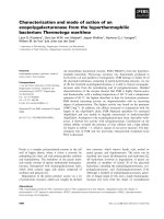
expected ivy nanoparticles. From Figure 1, we could
observe that the isolated ivy nanoparticles had a diameter
of 65.3 ± 8.04 nm, based on measurements of 30 ran-
domly counted nanoparticles that were not dominated by
other particles. Large agglomerates of nanoparticles
ranging from 100-200 nm were ignored from the particle
size determination.
UV extinction of ivy nanoparticles
To demonstrate the ability of ivy nanoparticles for UV
protection, we measured the optical extinction spectra of
these nanoparticles using an ultraviolet and visible wave-
length (UV-Vis) spectrophotometer. From the experi-
mental data, we observed that the ivy nanoparticles, at a
concentration of 4.92 μg/ml, had significant extinction in
the ultraviolet region, while having little extinction at the
visible and near infrared regions. This indicated that ivy
nanoparticles would effectively block UV radiation with-
out the opacity observed in other metal-based nanoparti-
cles.
Comparison of the UV blockage with TiO
2
nanoparti-
cles at the same concentration indicated that the total
extinction of the ivy nanoparticles from 280 nm to 400
nm was much better than that of the TiO
2
nanoparticles
(Figure 2). The extinction of the ivy nanoparticles
decreased sharply after the UV region, which makes ivy
nanoparticles more effective in the UV-A/UV-B region
and gives them high transmittance in the visible region
making them virtually "invisible".
Our previous studies have confirmed that ivy nanopar-
ticles are organic and have unique adhesive properties
[27]. The adhesive effect of these nanoparticles will allow
the ivy nanoparticles to remain on the skin for a longer
period of time, and thus enhance their UV protective
effect. The combination of these unique factors makes
the ivy nanoparticles an appealing candidate for the
development of a novel sunscreen product.
Cytotoxicity
Although nanoparticles greater than 20 nm in diameter
have not been reported to permeate through human skin,
this data was obtained using healthy individuals in an
optimal setting [15]. In specific cases, the skin structure
can be changed to allow the penetration of large particles
into the blood system, which has been demonstrated by
the ability of 1,000 nm particles to access the dermis
when intact skin is flexed [28]. More frequently, however,
Figure 1 AFM characterization of ivy nanoparticles. The image
shows a 2.05 × 2.05 um AFM view of isolated nanoparticles from cul-
tured ivy rootlets, which indicated a high abundance of nanoparticles
with an average diameter of 65.3 ± 8.04 nm.
Figure 2 UV extinction spectra. Spectral profiles represent the UV
extinction of the 4.92 μg/ml ivy and TiO
2
nanoparticles. The green line
corresponds to the ivy nanoparticles, while the blue line corresponds
to the TiO
2
nanoparticles.
Xia et al. Journal of Nanobiotechnology 2010, 8:12
/>Page 4 of 9
when skin is damaged, as in the case of people with sun-
burn, blemished skin, frequent shaving, or massages,
there will be an increased risk of penetration [29-31]. A
recent report by the US-based Environmental Working
Group on the health risks of commercially available cos-
metics and personal care products found that more than
half of all cosmetics contained ingredients that act as
"penetration enhancers" [32]. This raises further concerns
for the safety of applied nanoparticles for personal care
and cosmetics, since these agents will presumably
increase the penetration potential of nanoparticles. As
such, the cytotoxicity of nanoparticles should be thor-
oughly tested before their application in sunscreens.
Due to the increased toxicity associated with internal-
ized nanoparticles, as mentioned earlier, we have chosen
to examine the toxicity of a mammalian endothelial cell
line, HeLa cells. HeLa cells are commonly used for testing
the toxicity and trafficking of nanoparticles [33-35]. In
the experimental study, we incubated 1 μg/ml ivy nano-
particles with HeLa cells for 24 hours to test the cytotox-
icity of these ivy nanoparticles. The toxicity was
determined using propidium iodide staining and was
examined by flow cytometry. We observed no toxicity
compared to the control cells upon incubation with the
ivy nanoparticles. However, in the same study, the same
concentration of TiO
2
nanoparticles exhibited significant
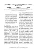
toxicity to HeLa cells. In a standard flow cytometry
experiment (Figure 3A), Gate C in each plot was defined
for the cell population with less DNA, and thus repre-
sented the cells experiencing apoptosis. Gate B from the
plots was the HeLa cells with more DNA, which indicated
replicating cells at differing growth stages. Statistical
analysis concluded that there was no significant differ-
ence in the percentage of cells experiencing apoptosis
between the control cell population (13.5% ± 1.27%) and
cells incubated with ivy nanoparticles (11.5% ± 1.06%)
(Figure 3B). However, in the cells incubated with TiO
2
,
24.3% ± 0.7% of cells were experiencing apoptosis, which
was significantly higher than the control cell population
(p = 0.011) and the cells incubated with ivy nanoparticles
(p = 0.007).
Degradation
Although we addressed the cytotoxicity of ivy nanoparti-
cles in the HeLa cell line, the possibility of these ivy nano-
particles exhibiting toxicity in the body may still exist.
There have been observations with gold-dendrimer
nanoparticles accumulating in the liver that might dam-
age normal liver function [36]. To address this concern
for ivy nanoparticles, we tested the ability of the nanopar-
ticles to be degraded should they pass through the skin or
mucous membranes. If the ivy nanoparticles were
degradable, then they would be digested after their pene-
tration through the skin and lose their normal nano-
structure and thus any toxicity based on the nano-mor-
phology of the particles. The degradability of nanomate-
rial is also beneficial to the environment when
considering reports that nanomaterials have been linked
to damage in fish, mortality in water fleas, and have bac-
tericidal properties that can impact ecosystems [14,19].
Thus, the biodegradability of these ivy nanoparticles was
also investigated in this study.
Our experimental studies indicated that at tempera-
tures from 4-37°C, the ivy nanoparticles were stable and
could be readily imaged by AFM. In addition, sonication
from 5-9 W was not effective at destroying the particle
structure, but did serve to disperse the particles and pre-
vent the formation of large agglomerates. Incubation of
the ivy nanoparticles in RPMI, a common cell culture
media, at 37°C for up to 24 hours did not result in diges-
tion of the nanoparticles as assessed by AFM. To test the
ability for the particles to be broken down by enzymatic
digestion, Proteinase K was used in an attempt to digest
the nanoparticles. Digestion was carried out from 15
minutes to 4 hours, to determine if the extent of incuba-
tion affected the digestion of the particles. After incuba-
tion with Proteinase K for 30 min, it was no longer
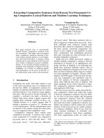
possible to image the nanoparticles with AFM. As shown
in Figure 4, after enzymatic digestion, the ivy nanoparti-
cles were degraded and lost their normal structure. This
enzymatic digestion by a common proteinase could fur-
ther reduce the risk to the environment and human tis-
sues and organs. This gives organic nanoparticles a
definitive advantage over metal oxide nanoparticles, since
these particles resist breakdown by biological organisms
and remain in the body or environment for prolonged
periods of time. We would like to point out that the above
nanoparticles used for the Proteinase K digestion were
collected based on size which also matched well with the
UV 280 nm detection. As expected, they have been totally
degraded. However, we could not eliminate the possibility
that there might be other type/size of nanoparticles that
had not been detected by UV detector, and as a result,
were not collected for the Proteinase K digestion. The
long-term objective of this study is to propose protein-
based ivy nanoparticles for UV protection.
Skin penetration
Another major concern with cosmetic nanoparticles is
their probability to penetrate through the skin into the
circulatory system [37,38]. The development of proper
markers for the detection of ivy nanoparticles in the skin
takes considerable time, however, a simple mathematical
modeling and computational approach may allow rapid
analysis for the potential of ivy nanoparticles to penetrate
though skin.
Skin structure, is composed of the protective outer SC
layer and a viable epidermis and dermis layer with other
Xia et al. Journal of Nanobiotechnology 2010, 8:12
/>Page 5 of 9
accessory glands [39]. The penetration of particles in the
skin can occur through pilosebaceous pores (diameter:
10-70 μm), sweat gland pores (diameter: 60-80 μm) and
lipid matrix that fills a gap of 75 nm between dead cor-
neocytes in the SC [39-41]. As the intact skin has more
than one layer in humans, it is expected that ivy nanopar-
ticles will have different diffusion activities in different
layers. Ex vivo and in vivo experimental data supported
that the SC has the most packaged properties and is not
permeable to many chemicals and drugs [42]. A skin dif-
fusion study indicated that the SC has a diffusion coeffi-
cient 10
3
times lower than the deeper viable layer for the
same chemical [43]. Another study for nanoparticles in
human skin also indicated that nanoparticles with a size
of more than 20 nm rarely have a chance to penetrate
through the SC layer [44]. Therefore, to understand ivy
nanoparticle diffusion and penetration in human skin, it
is essential to understand the diffusion process of these
nanoparticles in the SC.
There are many papers dealing with the transport of
nanoparticles through the SC layer of the skin in the cur-
rent literature [15,31,45-48]. While the data vary depend-
ing on the experimental setup, it is generally agreed that
the depth of penetration varies with the material proper-
ties of the nanoparticles, the size of individual particles,
their shape, and other physicochemical factors [45]. Stud-
ies have suggested that sunscreens composed of TiO
2
and
ZnO nanoparticles do not pass into the upper layers of
the SC. However, as mentioned earlier, these studies have
only examined healthy adult skin models [11-14,49].
More realistically the skin to which the sunscreen will be
applied has been damaged, either by prior sun exposure,
or by a variety of other factors that damage the skin. We
have previously discussed how damage to the skin and
small particle size increase the depth to which nanoparti-
cles will penetrate [15,16,29-31].
Despite the heterogeneous structure of the SC layer, in
cases where penetration is concerned, the skin behaves as
a homogeneous membrane and the diffusion law still
holds [50-53]. To understand the dynamic activities of the
ivy nanoparticles applied to the skin, Fick's Second Law is
Figure 3 Cytotoxicity analysis. HeLa cells were incubated with or without nanoparticles for 24 hours and stained with propidium iodide. The cell
apoptosis was then determined by detection of fluorescence using flow cytometry. A) Representative flow cytometry plots for each of the three sam-
ples: negative control (Neg control), ivy nanoparticle (Ivy), TiO
2
nanoparticle (TiO
2
). B) Cells experiencing apoptosis in the three samples. Each point
represents an average of 3 samples from one of three experiments. * denotes significant difference based on Student's t test (p < 0.05).
Xia et al. Journal of Nanobiotechnology 2010, 8:12
/>Page 6 of 9
applied and described as the follows: (∂ϕ;/∂t) = D(∂
2
ϕ/
∂χ
2
). The determination factor is the diffusion coefficient
(D). This D is normally defined to be: D = k
B
T/6πηR. In
the case of ivy nanoparticles, R is radius of ivy nanoparti-
cles and is set at the value of 32.65 nm, η is the viscosity of
the SC lipid matrix (0.02 kg/m s), k is the Boltzmann con-
stant (1.38 × 10
-23
m
2
kg s
-2
K
-1
), and T is the absolute
temperature for human skin (310.15 K). In this simplified
model the surface properties of varying nanoparticles are
ignored and only the radius of the nanoparticles effects
diffusion. The SC layer, which is 20 μm deep, is lipophilic
and acidic with variations in gender, anatomical sites, and
environmental settings [54].
Based on the model and obtained parameters, the
dynamics of nanoparticle diffusion in the SC layer of the
skin was simulated. In Figure 5, the predicted distribution
of different-sizes of nanoparticles after 8 and 20 hours of
application is shown. While nanoparticles with a diame-
ter less than 10 nm have a chance to reach into the bot-
tom of the SC layer (Figure 5), nanoparticles over 40 nm
can only reach 5-8 μm into the SC layer after 8 hours of
application and 8-13 μm after 20 hours, as displayed in
Figure 5. This agrees well with previous experimental
studies about other nanoparticles within the same size
range [55]. Considering that the normal period of expo-
sure to sunlight in humans is less than 8 hours, and the
diameter of the ivy nanoparticles is 65.3 nm, we expect
that ivy nanoparticles can be used in cosmetics applica-
tions without a risk of penetration.
Conclusions
The concern for the biosafety and health risk for the
metal-based and engineered nanoparticles in sunscreens
has led to the search for alternative replacement nanopar-
ticles. In this study, naturally occurring ivy nanoparticles
were investigated to replace TiO
2
and ZnO that are cur-
rently widely used in sunscreen products. Based on
experimental data, we have demonstrated that ivy nano-
particles have the potential levels of UV protection neces-
sary to warrant further investigation for uses in
cosmetics. The cell toxicity of ivy nanoparticles was next
tested and it was determined that ivy nanoparticles
exhibited much less toxicity than widely used TiO
2
nano-
particles. Without obtaining the proper marker for exper-
imental determination, a mathematical model was used
to analyze the diffusion dynamics in the human skin,
especially in the SC layer. Through this analysis, we found
ivy nanoparticles with a diameter of 65.3 nm will not
reach the bottom of SC layer in normal conditions for
short periods of time after application. The biodegrad-
ability of these ivy nanoparticles further eliminates con-
cerns regarding environmental contamination and in the
case of entry into the body. All of the above studies dem-
onstrated that naturally occurring ivy nanoparticles could
be a promising alternative for UV protection in cosmet-
ics, especially with concerns regarding the safety of
metal-based nanoparticles. With increased dangers asso-
ciated with more UV passing through the atmosphere
[56], the need to protect human from skin cancer elicits
the need for safe and effective UV protective agents. The
Figure 4 Biodegradability of ivy nanoparticles. The isolated ivy nanoparticles were incubated without (A) or with (B) Proteinase K at 37°C for 30
min. The structure of the samples was then analysed with AFM using tapping mode.
Xia et al. Journal of Nanobiotechnology 2010, 8:12
/>Page 7 of 9
promising application of these ivy nanoparticles thus pro-
vides a better chance to help protect people from UV
radiation.
Methods
Ivy nanoparticle isolation
Juvenile Hedera helix shoots were grown in a greenhouse.
The plant was originally taken naturally on the University
of Tennessee, Knoxville campus that was transplanted to
a pot in a greenhouse. Leaves were removed from the
shoots and shoots were trimmed to 6 cm in length.
Shoots were sterilized using 1.23% sodium hypochlorite
(20% [v/v] commercial bleach) plus 0.05% Tween 20,
shaken at 200 rpm for 20 min, and followed by washing
three times with sterile water. Sterile shoots were then
placed upright into Magenta GA7 boxes containing
Murashige and Skoog (MS) medium and were grown at
24°C at 16:8 h photoperiod under 82 μmol m
-2
s
-
1
irradiance. Aerial roots (rootlets) were produced after
ca. 2 d, which were allowed to grow for an additional 2
days in MS medium to reach approximately 3 cm in
length. Aerial roots were then excised from source plants
and were transferred to Petri dishes containing MS
medium until analysis was performed (between 3 days
and 2 weeks). To isolate ivy nanoparticles, the tips of
rootlets were homogenized in double-distilled water.
After centrifugation to remove tissue residuals, the solu-
tion was dialysed overnight, then was loaded the BioSep-
SEC-S 4000 size exclusion chromatography column and
the elution fractions were collected for further analysis.
Atomic force microscopy
The isolated ivy nanoparticle fractions were first air-dried
overnight. The air-dried ivy samples were scanned for the
existence of nanoparticles using an Agilent 5500 atomic
force microscope (Agilent Technologies, Santa Clara,
CA). The samples were imaged at room temperature
(20°C) using Picoview™ in tapping mode, which mini-
mizes sample distortion due to mechanical interactions
between AFM tip and the surface. To further optimize
imaging, the set point amplitude and the amplitude of the
oscillating cantilever were adjusted to avoid excessive
loading force applied to the samples. In this way, three-
dimensional imaging of the surface morphology with very
high lateral and vertical resolution has been obtained.
The used tips are commercially available silicon probes
TAP300Al
®
(Budget Sensors, Sofia, Bulgaria) with a spring
constant of 20-75 N/m, a resonant frequency of 300 ± 100
kHz, and a radius of curvature in less than 10 nm.
Sample preparation
The calculation of isolated ivy nanoparticles was based
on the number of the particles on the AFM images and
the used volume to obtain these nanoparticles. To pre-
pare the same concentration of TiO
2
suspension, TiO
2
particles with a diameter of 50 nm (99% purity) were pur-
chased from Nanostructured & Amorphous Materials
Inc. (Houston, TX). The particles were first ultrasonically
dispersed in water at 5 W for 30 min, then were used for
later UV-Vis study and cytotoxicity study.
UV-Vis extinction
The UV-Vis extinction (absorption and scattering) spec-
tra were measured using a Thermo Scientific Evolution
600 UV-Visible spectrophotometer (Thermo Fisher Sci-
entific, Waltham, MA). The optical length of the quartz
cuvette was 10 mm. The wavelength of the light started at
250 nm and stopped at 800 nm. Ivy nanoparticles at 4.92
μg/ml were measured for UV-Vis. The same concentra-
tion of TiO
2
nanoparticles was measured for UV-Vis
under the same conditions.
Cytotoxicity study
HeLa cells were cultured in a DMEM (Mediatech Inc,
Manassas, VA) solution supplemented with 10% heat-
inactivated fetal bovine serum in a humidified incubator
Figure 5 Distribution of nanoparticles in SC layer of skin. Compu-
tational simulation results for the distribution of nanoparticles in the SC
layer of human skin 8 hours (A) and 20 hours (B) after application to the
surface of the skin.
Xia et al. Journal of Nanobiotechnology 2010, 8:12
/>Page 8 of 9
with an atmosphere of 5% CO
2
in air at 37°C. The TiO
2
or
ivy nanoparticle aqueous suspension was added to
DMEM solution supplemented with 10% fetal bovine
serum to prepare a DMEM solution containing nanopar-
ticles, which was used to investigate the cytotoxicity
against HeLa cells. Negative controls consisted of DMEM
with 10% fetal bovine serum, without the presence of
nanoparticles. For apoptosis analysis, the cells were har-
vested 24 hours after addition of nanoparticles, fixed and
stained with propidium iodide, then were analyzed using
Bechman Coulter Episc XL (Bechman Coulter, Brea, CA)
with a 488 nm argon laser.
Nanoparticle degradation
Purified nanoparticles were sonicated at 5 W for 20 min-
utes to physically disperse the nanoparticles so that indi-
vidual ones that could be analyzed. SDS was then added
to the nanoparticle solution to make a final concentration
of 0.5%. To the following, 50 μg/ml of Proteinase K was
added. The solution was then incubated at 37°C from 15
minutes to 4 hours. Upon completion of the digestion,
the sample was air-dried and imaged using AFM to deter-
mine if the nanoparticles were degraded. The control
sample was prepared in the same way except that the Pro-
teinase K was not added before incubation. In addition to
examining enzymatic digestion with Proteinase K, the
effects of varying temperatures were examined by incu-
bation of the nanoparticles from 4-37°C. Similarly, the
nanoparticles were added to RPMI for 24 hours to deter-
mine their stability in a typical cell culture media. All
samples were then air-dried and imaged by AFM.
Statistical analysis
To determine if there were significant differences in cyto-
toxicity among HeLa cells incubated without or with dif-
ferent nanoparticles, a Student's t test for comparisons of
each pair with 95% confidence was carried out using JMP
8 statistical software.
Competing interests
The authors declare that they have no competing interests.
Authors' contributions
LX performed the majority of the experiments and wrote the manuscript with
SCL and MZ. QL contributed with the characterization by spectrophotometry
and helped with data analysis. MZ, ZZ, SCL and LX designed the overall project.
MZ and SCL helped with the interpretation of data and revised the manuscript.
All authors read and approved the manuscript.
Acknowledgements
The authors would like to extend special thanks to Dr. Yu Wu for helpful discus-
sion concerning mathematical modelling and simulation, and Ms Dianne Trent
for her support with flow cytometry data collection and analysis. The cultured
ivy rootlets were generously supplied by Jason B Burris and Dr. Stewart's group
from the Department of Plant Sciences, University of Tennessee, Knoxville. We
would also like to thank the partial support for this study by the US Army
Research Office, Life Sciences Division, Biochemistry Program under the con-
tract W911NF-10-1-0114.
Author Details
Department of Mechanical, Aerospace and Biomedical Engineering, University
of Tennessee, Knoxville, TN, 37996, USA
References
1. Diffey BL: Solar ultraviolet radiation effects on biological systems. Phys
Med Biol 1991, 36:299-328.
2. Pathak SK, Mason NJ: Our shrinking ozone layer. Resonance 2002,
7:71-80.
3. Hockberger PE: A history of ultraviolet photobiology for humans,
animals and microorganisms. Photochem Photobiol 2002, 76:561-579.
4. Longstreth J, de Gruijl FR, Kripke ML, Abseck S, Arnold F, Slaper HI, Velders
G, Takizawa Y, van der Leun JC: Health risks. J Photochem Photobiol B
1998, 46:20-39.
5. Friedberg EC: DNA damage and repair. Nature 2003, 421:436-440.
6. American Cancer Society: [ />ped_7_1_what_you_need_to_know_about_skin_cancer.asp].
7. van der Leun JC, de Gruijl FR: UV-B radiation and ozone depletion:
effects on humans, animals, plants, microorganisms, and materials. In
Influences of ozone depletion on human and aniaml health Edited by: Tevini
M. Ann Arbor: Lewis Publishers; 1993:95-123.
8. Smith RC, Prézelin BB, Baker KS, Bidigare RR, Boucher NP, Coley T, Karentz
D, MacIntyre S, Matlick HA, Menzies D, et al.: Ozone depletion: ultraviolet
radiation and phytoplankton biology in antarctic waters. Science 1992,
255:952-959.
9. Wolf R, Wolf D, Morganti P, Ruocco V: Sunscreens. Clin Dermatol 2001,
19:452-459.
10. Nohynek GJ, Lademann J, Ribaud C, Roberts MS: Grey goo on the skin?
Nanotechnology, cosmetic and sunscreen safety. Crit Rev Toxicol 2007,
37:251-277.
11. Durand L, Habran N, Henschel V, Amighi K: In vitro evaluation of the
cutaneous penetration of sprayable sunscreen emulsions with high
concentrations of UV filters. Int J Cosmet Sci 2009, 31:279-292.
12. Gontier E, Ynsa M-D, Bíró T, Hunyadi J, Kiss B, Gáspár K, Pinheiro T, Silva J-N,
Filipe P, Stachura J, et al.: Is there penetration of titania nanoparticles in
sunscreens through skin? A comparative electron and ion microscopy
study. Nanotoxicology 2009, 2:218-231.
13. Newman MD, Stotland M, Ellis JI: The safety of nanosized particles in
titanium dioxide- and zinc oxide-based sunscreens. J Am Acad
Dermatol 2009, 61:685-692.
14. Oberdörster E: Manufactured nanomaterials (fullerenes, C
60
) induce
oxidative stress in the brain of juvenile largemouth bass. Environ Health
Perspect 2004, 112:1058-1062.
15. Baroli B, Ennas MG, Loffredo F, Isola M, Pinna R, López-Quintela MA:
Penetration of metallic nanoparticles in human full-thickness skin. J
Invest Dermatol 2007, 127:1701-1712.
16. Menzel F, Reinert T, Vogt J, Butz T: Investigations of percutaneous
uptake of ultrafine TiO
2
particles at the high energy ion nanoprobe
LIPSION. Nucl Instrum Methods Phys Res Sect B 2004, 219-220:82-86.
17. Wu J, Liu W, Xue C, Zhou S, Lan F, Bi L, Xu H, Yang X, Zeng F-D: Toxicity
and penetration of TiO
2
nanoparticles in hairless mice and porcine skin
after subchronic dermal exposure. Toxicol Lett 2009, 191:1-8.
18. Buzea C, Pacheco II, Robbie K: Nanomaterials and nanoparticles: sources
and toxicity. Biointerphases 2007, 2:MR17-MR71.
19. Oberdörster G, Oberdörster E, Oberdörster J: Nanotoxicology: an
emerging discipline evolving from studies of ultrafine particles.
Environ Health Perspect 2005, 113:823-839.
20. Nel A, Xia T, Mädler L, Li N: Toxic potential of materials at the nanolevel.
Science 2006, 311:622-627.
21. Dunford R, Salinaro A, Cai L, Serpone N, Horikoshi S, Hidaka H, Knowland J:
Chemical oxidation and DNA damage catalysed by inorganic
sunscreen ingredients. FEBS Lett 1997, 418:87-90.
22. Donaldson K, Beswick PH, Gilmour PS: Free radical activity associated
with the surface of particles: a unifying factor in determining biological
activity? Toxicol Lett 88:293-298.
23. Hussain SM, Hess KL, Gearhart JM, Geiss KT, Schlager JJ: In vitro toxicity of
nanoparticles in BRL 3A rat liver cells. Toxicol In Vitro 2005, 19:975-983.
Received: 17 March 2010 Accepted: 9 June 2010
Published: 9 June 2010
This article is available from: 2010 Xia et al; licensee BioMed Central Ltd. This is an Open Access article distributed under the terms of the Creative Commons Attribution License ( .0), which permits unrestricted use, distribution, and reproduction in any medium, provided the original work is properly cited.Journal of Na nobiotechnolog y 2010, 8:12
Xia et al. Journal of Nanobiotechnology 2010, 8:12
/>Page 9 of 9
24. Oberdörster G, Maynard A, Donaldson K, Castranova V, Fitzpatrick J,
Ausman K, Carter J, Karn B, Kreyling W, Lai D, et al.: Principles for
characterizing the potential human health effects from exposure to
nanomaterials: elements of a screening strategy. Part Fibre Toxicol 2005,
2:8.
25. Luo J: Toxicity and bioaccumulation of nanomaterial in aquatic species.
Journal of the US SJWP 2007, 2:1-16.
26. Zhu S, Oberdörster E, Haasch ML: Toxicity of an engineered nanoparticle
(fullerene, C
60
) in two aquatic species, Daphnia and fathead minnow.
Mar Environ Res 2006, 62(Suppl):S5-S9.
27. Zhang M, Liu M, Prest H, Fischer S: Nanoparticles secreted from ivy
rootlets for surface climbing. Nano Lett 2008, 8:1277-1280.
28. Tinkle SS, Antonini JM, Rich BA, Roberts JR, Salmen R, DePree K, Adkins EJ:
Skin as a route of exposure and sensitization in chronic beryllium
disease. Environ Health Perspect 2003, 111:1202-1208.
29. Larese FF, D'Agostin F, Crosera M, Adami G, Renzi N, Bovenzi M, Maina G:
Human skin penetration of silver nanoparticles through intact and
damaged skin. Toxicology 2009, 255:33-37.
30. Mortensen LJ, Oberdörster G, Pentland AP, DeLouise LA: In vivo skin
penetration of quantum dot nanoparticles in the murine model: the
effect of UVR. Nano Lett 2008, 8:2779-2787.
31. Zhang LW, Yu WW, Colvin VL, Monteiro-Riviere NA: Biological
interactions of quantum dot nanoparticles in skin and in human
epidermal keratinocytes. Toxicol Appl Pharmacol 2008, 228:200-211.
32. Hoet PH, Brüske-Hohlfeld I, Salata OV: Nanoparticles - known and
unknown health risks. J Nanobiotechnology 2004, 2:12.
33. Park IY, Kim IY, Yoo MK, Choi YJ, Cho MH, Cho CS: Mannosylated
polyethylenimine coupled mesoporous silica nanoparticles for
receptor-mediated gene delivery. Int J Pharm 2008, 359:280-287.
34. Chithrani BD, Stewart J, Allen C, Jaffray DA: Intracellular uptake,
transport, and processing of nanostructures in cancer cells.
Nanomedicine 2009, 5:118-127.
35. Harush-Frenkel O, Bivas-Benita M, Nassar T, Springer C, Sherman Y, Avital
A, Altschuler Y, Borlak J, Benita S: A safety and tolerability study of
differently-charged nanoparticles for local pulmonary drug delivery.
Toxicol Appl Pharmacol in press.
36. Minchin R: Nanomedicine: sizing up targets with nanoparticles. Nat
Nanotechnol 2008, 3:12-13.
37. Ryman-Rasmussen JP, Riviere JE, Monteiro-Riviere NA: Penetration of
intact skin by quantum dots with diverse physicochemical properties.
Toxicol Sci 2006, 91:159-165.
38. Benson HA: Transdermal drug delivery: penetration enhancement
techniques. Curr Drug Deliv 2005, 2:23-33.
39. Elias PM: Stratum corneum defensive functions: an integrated view. J
Invest Dermatol 2005, 125:183-200.
40. Lauer AC, Ramachandran C, Lieb LM, Niemiec S, Weiner ND: Targeted
delivery to the pilosebaceous unit via liposomes. Adv Drug Delivery Rev
1996, 18:311-324.
41. Johnson ME, Blankschtein D, Langer R: Evaluation of solute permeation
through the stratum corneum: lateral bilayer diffusion as the primary
transport mechanism. J Pharm Sci 1997, 86:1162-1172.
42. Wartewig S, Neubert RH: Properties of ceramides and their impact on
the stratum corneum structure: a review. Part 1: ceramides. Skin
Pharmacol Physiol 2007, 20:220-229.
43. Sugibayashi K, Hayashi T, Morimoto Y: Simultaneous transport and
metabolism of ethyl nicotinate in hairless rat skin after its topical
application: the effect of enzyme distribution in skin. J Control Release
1999, 62:201-208.
44. Alvarez-Román R, Naik A, Kalia YN, Guy RH, Fessi H: Skin penetration and
distribution of polymeric nanoparticles. J Control Release 2004,
99:53-62.
45. Kohli AK, Alpar HO: Potential use of nanoparticles for transcutaneous
vaccine delivery: effect of particle size and charge. Int J Pharm 2004,
275:13-17.
46. Díaz-Torres R, Sandoval SJJ, Ibañez-Orozco O, Rodríguez-Romo S:
Polyethylcyanoacrylate nanoparticle transport through the stratum
corneum. Appl Phys Lett 2009, 95:043702.
47. Kuntsche J, Bunjes H, Fahr A, Pappinen S, Rönkkö S, Suhonen M, Urtti A:
Interaction of lipid nanoparticles with human epidermis and an
organotypic cell culture model. Int J Pharm 2008, 354:180-195.
48. Coulman SA, Anstey A, Gateley C, Morrissey A, McLoughlin P, Allender C,
Birchall JC: Microneedle mediated delivery of nanoparticles into human
skin. Int J Pharm 2009, 366:190-200.
49. Zvyagin AV, Zhao X, Gierden A, Sanchez W, Ross JA, Roberts MS: Imaging
of zinc oxide nanoparticle penetration in human skin in vitro and in
vivo. J Biomed Opt 2008, 13:064031.
50. Kalia YN, Pirot F, Guy RH: Homogeneous transport in a heterogeneous
membrane: water diffusion across human stratum corneum in vivo.
Biophys J 1996, 71:2692-2700.
51. Kalia YN, Alberti I, Sekkat N, Curdy C, Naik A, Guy RH: Normalization of
stratum corneum barrier function and transepidermal water loss in
vivo. Pharm Res 2000, 17:1148-1150.
52. Alberti I, Kalia YN, Naik A, Bonny JD, Guy RH: In vivo assessment of
enhanced topical delivery of terbinafine to human stratum corneum. J
Control Release 2001, 71:319-327.
53. Schwindt DA, Wilhelm KP, Maibach HI: Water diffusion characteristics of
human stratum corneum at different anatomical sites in vivo. J Invest
Dermatol 1998, 111:385-389.
54. Williams AC: Transdermal drug delivery: second edition (revised and
expanded). Int J Pharm 2003, 261:171. ISBN: 0-8247-0861-x.
55. Popov AP, Lademann J, Priezzhev AV, Myllylä R: Effect of size of TiO
2
nanoparticles embedded into stratum corneum on ultraviolet-A and
ultraviolet-B sun-blocking properties of the skin. J Biomed Opt 2005,
10:064037.
56. Pitcher HM, Longstreth JD: Melanoma mortality and exposure to
ultraviolet radiation: an empirical relationship. Environ Int 1991,
17:7-21.
doi: 10.1186/1477-3155-8-12
Cite this article as: Xia et al., Naturally occurring nanoparticles from English
ivy: an alternative to metal-based nanoparticles for UV protection Journal of
Nanobiotechnology 2010, 8:12