báo cáo khoa học: "Anti-oxidant effect of gold nanoparticles restrains hyperglycemic conditions in diabetic mice" ppt
Bạn đang xem bản rút gọn của tài liệu. Xem và tải ngay bản đầy đủ của tài liệu tại đây (2.01 MB, 15 trang )
BarathManiKanth et al. Journal of Nanobiotechnology 2010, 8:16
/>Open Access
RESEARCH
© 2010 BarathManiKanth et al; licensee BioMed Central Ltd. This is an Open Access article distributed under the terms of the Creative
Commons Attribution License ( which permits unrestricted use, distribution, and repro-
duction in any medium, provided the original work is properly cited.
Research
Anti-oxidant effect of gold nanoparticles restrains
hyperglycemic conditions in diabetic mice
Selvaraj BarathManiKanth
†1
, Kalimuthu Kalishwaralal
†1
, Muthuirulappan Sriram
1
, SureshBabu Ram Kumar Pandian
1
,
Hyung-seop Youn
2
, SooHyun Eom
2
and Sangiliyandi Gurunathan*
1
Abstract
Background: Oxidative stress is imperative for its morbidity towards diabetic complications, where abnormal
metabolic milieu as a result of hyperglycemia, leads to the onset of several complications. A biological antioxidant
capable of inhibiting oxidative stress mediated diabetic progressions; during hyperglycemia is still the need of the era.
The current study was performed to study the effect of biologically synthesized gold nanoparticles (AuNPs) to control
the hyperglycemic conditions in streptozotocin induced diabetic mice.
Results: The profound control of AuNPs over the anti oxidant enzymes such as GSH, SOD, Catalase and GPx in diabetic
mice to normal, by inhibition of lipid peroxidation and ROS generation during hyperglycemia evidence their anti-
oxidant effect during hyperglycemia. The AuNPs exhibited an insistent control over the blood glucose level, lipids and
serum biochemical profiles in diabetic mice near to the control mice provokes their effective role in controlling and
increasing the organ functions for better utilization of blood glucose. Histopathological and hematological studies
revealed the non-toxic and protective effect of the gold nanoparticles over the vital organs when administered at
dosage of 2.5 mg/kilogram.body.weight/day. ICP-MS analysis revealed the biodistribution of gold nanoparticles in the
vital organs showing accumulation of AuNPs in the spleen comparatively greater than other organs.
Conclusion: The results obtained disclose the effectual role of AuNPs as an anti-oxidative agent, by inhibiting the
formation of ROS, scavenging free radicals; thus increasing the anti-oxidant defense enzymes and creating a sustained
control over hyperglycemic conditions which consequently evoke the potential of AuNPs as an economic therapeutic
remedy in diabetic treatments and its complications.
Background
Diabetes mellitus a lifelong progressive disease is a
chronic metabolic disorder due to the relative deficiency
of insulin secretion and varying degrees of insulin resis-
tance and is characterized by high circulating glucose [1].
This disease has reached epidemic proportion among the
challenging unresolved health problems of the 21st cen-
tury. Around 230 million people worldwide have been
affected by diabetes and around 366 million people are
expected to get affected by 2030 [2]. Several pathogenic
pathways are activated in diabetes among which reactive
oxygen species (ROS) generated by high glucose levels is
responsible for metabolic abnormalities and chronic
complications [3]. A counteractive defense system that
eliminates the ROS produced during normal oxidative
metabolism is being maintained and any imbalance in the
production and scavenging of ROS leads to excessive lev-
els of either molecular oxygen or ROS, thus resulting in
increased 'oxidative stress' [4]. Since numerous studies
have demonstrated that oxidative stress, mediated mainly
by hyperglycemia-induced generation of free radicals,
contributes to the development and progression of diabe-
tes and its complications, it will be an effective strategy to
use antioxidants to ameliorate treatments for oxidative
stress. The management of diabetic conditions by insulin
therapy has several drawbacks like insulin resistance and
in chronic treatment causes anaeroxia nervosa, brain
atrophy and fatty liver. Thus an effective and economic
therapeutic molecule capable of up drifting the treat-
ments for diabetes mellitus, by controlling the oxidative
stress induced by hyperglycemia, disquieting various
* Correspondence:
1
Department of Biotechnology, Division of Molecular and Cellular Biology,
Kalasalingam University, Anand Nagar, Krishnankoil-626190, Tamilnadu, India
†
Contributed equally
Full list of author information is available at the end of the article
BarathManiKanth et al. Journal of Nanobiotechnology 2010, 8:16
/>Page 2 of 15
metabolic pathways and thereby preventing the onset of
complications is still the need of the era.
Discovery of new molecules and manipulating those
available naturally in nanosize could be appealing for
their greater potential to improve health care [5]. Several
pharmacological companies have won approval from the
Food and Drug Administration (FDA) for the use and
development of nanotechnology-based drugs in the last
few years.
Gold compounds have received great attention as an
anti-inflammatory agents through their ability to inhibit
expression of NF-kappa B and subsequent inflammatory
reactions [6-8]. The immunomodulatory, antioxidative
and restorative activity of Swarna Bhasma in cerebral
ischaemic rats has revealed their perceptive application
in the treatment of ischaemia and cerebral damages [9].
The major drawback of ionic gold lies on the fact that
they are easily inactivated by complexation and precipita-
tion thus limiting their desired functions in human sys-
tem. Here zerovalent gold nanoparticles can be a valuable
alternative replacing the potential of metallic gold [10].
Gold nanoparticles (AuNPs), an emerging nanomedicine
is renowned for its promising therapeutic possibilities,
due to its significant properties such as biocompatibility,
high surface reactivity, resistance to oxidation and plas-
mon resonance[11]. The inhibitory activity of gold nano-
particles against VPF/VEGF165 induced proliferation of
endothelial cells provides clear evidence over their thera-
peutic potential in the treatment of diseases like chronic
infiammation, pathological neo-vascularization, rheuma-
toid arthritis, and neoplastic disorders [12]. The role of
gold nanoparticles invading the treatment for various
inflammatory diseases and other relative disorders that
are context dependent, in orientation with the evidences
towards the anti-oxidative effect of traditional gold in
treatment of diseases, have affirmed the urge for the need
of study over restorative effect of gold nanoparticles at
conditions of, hyperglycemia leading to, oxidative stress
which has not been revealed yet.
Hence the effect of biologically synthesized gold nano-
particles on streptozotocin induced diabetic mice at
hyperglycemic conditions leading to oxidative stress,
have been investigated in this study.
Results
Characterization of Au-NPs
Characterization of the synthesized gold nanoparticles
was carried out before testing for their potent anti-oxida-
tive effect in hyperglycemic conditions. The morphology
and size of the biologically synthesized gold nanoparticles
was determined using Transmission electron microscopy
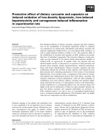
(TEM). The images clearly show that the average size of
the particles was found to be in the order of 50 nm and
depicts that they are relatively uniform in diameter and
spherical in shape. (Figure 1A) The XRD pattern obtained
showed four intense peaks in the whole spectrum of 2θ
values ranging from 20 to 80. The presence of intense
peaks of nanoparticles (111), (200), (220) and (311)
appeared which are indexed as crystalline gold face cen-
tered cubic phase. The standard XRD patterns for Au are
found to be almost similar [Joint Committee on Powder
Diffraction Standards (JCPDS) file no: 01-1174 for Au].
The XRD pattern thus clearly shows that the gold nano-
particles formed by the reduction of AuCl
4
-
ions by Bacil-
lus licheniformis are crystalline in nature (Figure 1B). The
Lal test revealed that the synthesized nanoparticles were
endotoxin free based on the qualitative analysis which did
not show any formation of gel clot.
Figure 1 A. TEM micrograph of the 1 mM AuCl
4
-
ions-treated son-
icated sample of B. licheniformis showing synthesized AuNPs. Pu-
rified nanoparticles from B. licheniformis were examined by electron
microscopy. Several fields were photographed and were used to de-
termine the diameter of nanoparticles. The range of observed diame-
ter of the synthesized gold nanoparticles was about 50 nm. B.
Representative XRD pattern of gold nanoparticles synthesized
after θ24 h. The XRD pattern shows four intense peaks in the whole
spectrum of 2 θ values ranging from 20 to 80. Note 2 θ peak values of
39.01°, 46.48°, 64.69° and 77.62°, corresponding to 111, 200, 220, 311
planes, respectively, for gold.
BarathManiKanth et al. Journal of Nanobiotechnology 2010, 8:16
/>Page 3 of 15
Toxicity studies
In vivo nanoparticles toxicity studies are focused mainly
on examining changes in blood serum chemistry and cell
population; changes in tissue morphology through histo-
logical analysis, along with nanoparticles biodistribution.
These in vivo studies not only provide the toxicity infor-
mation unavailable through in vitro studies but also
inform the choice of relevant model system for carrying
out further in vitro studies [13]. Thus the mice were
injected with AuNPs at a dosage of 2.5 mg/kg.b.wt/day
for 15 days and daily examined for any changes in the
morphology and behavior. All the mice survived through-
out the experimental period without exhibiting any
abnormalities. The mice did not show any symptoms of
toxicity such as fatigue, loss of appetite, change in fur
color, weight loss, etc. Comparative analysis of various
hematological parameters in the gold treated and control
animals, clearly showed that there was no significant
alteration except marginal variations in certain parame-
ters (Table 1). Histological studies are well thought-out to
be a reliable method to detect morphological changes due
to toxicities. These histological/histopathological assays
provide evidences over the morphological changes, evi-
dencing that toxicity correlates with changes in tissue and
cell morphology of a scale that can be visualized using
light microscopy [14]. Thus the pathological effect of the
nanoparticles over the morphological characteristics of
the organs was examined through the histological obser-
vations using light microscope. The histopathological
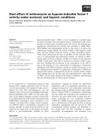
findings of the non-toxic effect of the gold nanoparticles
over liver, kidney, spleen and lung that were observed are
presented in (Figure 2). The examined reports obtained
from the senior pathologist confirmed that the gold
nanoparticles treated organs did not show any significant
morphological changes in comparison to control. In the
lung histopathology the sections from control animals
was showing normal alveolar geometry and normal
appearing alveolar septum (Figure 2A). The same histo-
pathological finding was seen after the treatment of gold
nanoparticles at a concn of 500 nm day
-1
(Figure 2B)
showing normal alveolar membranes with normal paren-
chyma blood vessels. The kidney histological studies
showed the control kidney with normal renal cortex and
glomerular tufts (Figure 2C) and the treatment of gold
nanoparticles at a dosage of 2.5 mg/kg.b.wt/day did not
lead to any disruptions in the histology. They showed
normal glomerular tubules and renal cortex (Figure 2D).
In the Liver histopathology sections from the control ani-
mals are showing normal hepatic portal triad and central
vein (Figure 2E). The gold nanoparticles treated liver also
showed normal hepatocytes with clear central vein show-
ing no morphological changes significant in comparison
to control (Figure 2F). The study over the spleen histology
also revealed that there were no any disruptions due to
the treatment of gold nanoparticles at a dosage of 2.5 mg/
kg.b.wt/day. The control and gold treated spleens showed
normal lymphoid follicles and sinuses (Figure 2G-H).
Blood parameters
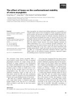
The control effect of gold nanoparticles over the blood
glucose and blood urea level obtained is represented in
Figure 3. The blood glucose level increased two fold and
blood urea level were observed to be elevated signifi-
cantly in the diabetic control mice in comparison to con-
trol group. The diabetic treated group showed a
controlled effect over the induced hyperglycemic condi-
tion by significantly decreasing the blood glucose by 75%
in comparison to the diabetic control. The blood glucose
and Urea level of gold treated group also did not show
Table 1: Hematological analysis revealing the nontoxic effect of AuNPs in mice
Parameters Control Gold treated
Hb (g/dl) 10.8 (± 1.19) 11.10 (± 1.3)
RBC Distrib Width (%) 17.4 (± 3.1) 17.95 (± 2.5)
MCV (fL) 48.30 (± 0.8) 44.0 (± 0.66)
MCH (pg) 24.75(± 5.33) 26.3(± 4.89)
MCHC (g/dl) 32.10(± 0.43) 33.8(± 0.28)
Platelet count [× 10
(9)
/L]
289(± 39.72) 298(± 44.69)
RBC [× 10
(12)
/L]
4.22((± 0.15) 4.74((± 0.3)
Leukocytes[× 10
(9)
/L]
2.74(± 0.6) 3.91(± 0.8)
HCT (%) 31.32((± 2.4) 33.0((± 1.55)
Each value represents the mean ± S.D of n = 6 Hb, hemoglobin; cells; RBC, red blood cells; MCV, mean corpuscular volume; MCH, mean
corpuscular hemoglobin; MCHC, mean corpuscular hemoglobin content; HCT, hematocrit. Numerical values (±) in the parenthesis are
considered as 'Standard Deviation (SD)'. P values were calculated using one way ANOVA followed by Students-'t' test by comparing between
different groups (control vs. treatment) and values are considered to be non-significant (P > 0.05).
BarathManiKanth et al. Journal of Nanobiotechnology 2010, 8:16
/>Page 4 of 15
any significant changes in comparison to the control
group (p < 0.05).
Various parameters of blood lipid profile were tested in
streptozotocin-induced diabetic mice before and after the
treatment with the gold nanoparticles. Treatment with
gold nanoparticles lowered the levels of TC, LDL, VLDL
and TG in diabetic mice near to normal. The level of TC,
LDL cholesterol and TG, were significantly decreased at
about 55%, 65% and 45% respectively in diabetic mice
treated with gold nanoparticles as compared to diabetic
control. Similarly, HDL levels were found to be increased
partially in diabetic mice after the treatment with gold
nanoparticles as compared to diabetic control. (p < 0.05)
(Table 2). The gold nanoparticles treated mice group IV
did not show any significant changes in comparison to
control group I.
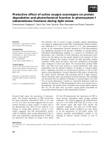
OGTT
The control effect of the gold nanoparticles over high glu-
cose conditions was studied by Oral Glucose Tolerance
Test (OGTT). The blood glucose level at fasting condi-
tions (FBG) and after the oral administration of glucose
in control and experimental animals are represented in
(Figure 4). Blood glucose levels, estimated in overnight
fasting diabetic mice (FBG), were significantly elevated.
However, this level was reduced significantly upon treat-
ment with gold nanoparticles at a dosage of 2.5 mg/
kg.b.wt/day (Figure.4, FBG data). For GTT, 1 g/kg.b.wt of
glucose dissolved in water were fed to the overnight-
fasted mice and the blood glucose level was determined
up to 120 min. The blood glucose level had decreased sig-
nificantly by 90 min in comparison with the elevation by
30 min and this was maintained until 120 min with an
effective dose of gold nanoparticles (p < 0.05).
Serum analysis
The enzymes such as ALT, AST, ACP and ALP are
responsible for the proper functioning of the liver and
any damages induced in the liver due to the hyperglyce-
mic conditions may lead to excessive leakage of these
enzymes in the blood stream. Thus the effect of gold
nanoparticles over the level of different metabolic
enzymes shaping the effective functioning of the liver
through the serum analysis was analyzed and their pro-
tective effect of gold nanoparticles over the liver damage
is shown in Table 3. The enzymes ALT, AST, ACP and
ALP showed significant elevated levels in the diabetic
control group (G2) in comparison to control group. Fol-
lowing treatment of gold nanoparticles at a dosage of 2.5
mg/kg. b. wt, the diabetic treated group (G3) presented a
partial decrease significantly in comparison to the dia-
Figure 2 Toxicity studies of gold nanoparticles in mouse organs.
Histological specimens of mice tissues (lung, kidney, liver and spleen)
collected from mice euthanized on day 15, stained with hematoxylin
and eosin (H and E) showed normal histology. 100% long-term survival
of mice was also observed in the mice treated with gold nanoparticles
at a concn of 500 nm for 15 days. A. Control animal lung section show-
ing normal alveolar geometry and normal appearing alveolar septum.
B. gold treated animal lung section showing normal alveolar mem-
branes with normal parenchyma blood vessels. C. Control kidney sec-
tion showing normal renal cortex and glomerular tufts D. gold treated
kidney section showing normal glomerular tubules and renal cortex E.
Control animal liver section showing normal, central vein and hepato-
cytic architecture F. Gold nanoparticles treated liver also showed nor-
mal hepatocytes with clear central vein G. Spleen sections of control
animals showing normal splenic architecture with normal lymphoid
follicles and sinuses H. gold treated spleen showing no pathological
changes.
Figure 3 Control effect of gold nanoparticles over blood glucose
and urea in experimental mice. The treatment of gold nanoparticles
significantly restrained the blood glucose and urea level to normal
near to control in comparison to diabetic control. Datas are given as
mean ± S.D for n = 6. Values are statistically significant at * p < 0.05.
a
Diabetic control compared with control group.
b
Gold treated diabetic
group compared with diabetic control group.
c
Gold treated group
compared with control group.
BarathManiKanth et al. Journal of Nanobiotechnology 2010, 8:16
/>Page 5 of 15
betic control group (G2), which directly reveals the pro-
tective/regenerative effect over the exaggerated activity of
liver. The level of creatinine symptomatic of the renal
functions was also decreased significantly near to normal
in the diabetic treated groups in comparison to the dia-
betic control group. The gold nanoparticles treated mice
did not show any significant changes of creatinine level in
comparison to the control (p < 0.05). These results
obtained over the restorative effect of gold nanoparticles
over the metabolic enzymes confirm the ability of gold
nanoparticles to protect the organs from damage due to
hyperglycemia induced oxidative stress.
ROS generation and lipid peroxidation
ROS generated by high glucose levels play a vital role in
the development of diabetic complications [15]. It is the
resultant of the oxidative stress developed due to the
release of free radicals, thereby decreasing the level of
antioxidant enzymes. Estimation of ROS generation in
the liver revealed that gold nanoparticles blocked the
high glucose-induced increase in ROS generation to a
maximum extent in the liver which is shown in Figure 5.
Induction of diabetes in the group II mice results in a
twofold level of increase in ROS generation relative to the
control mice. The diabetic mice treated with gold nano-
particles significantly decreased the high glucose-induced
rise in ROS generation in the liver in comparison to dia-
betic control mice. This makes clear the inhibitory effect
of gold nanoparticles over ROS generation during hyper-
glycemia induced oxidative stress.
Functional damage to cells under oxidative stress is not
only by oxygen free radicals and unbalanced redox poten-
tial but also due to enhanced lipid peroxidation [16]. The
inhibitory effect of gold nanoparticles over the occur-
rence of lipid peroxidation in the enzyme source is con-
firmed which is shown in Figure 5. A potent control effect
of gold nanoparticles (500 nM) treated to the diabetic
treated group showed a significant decrease in lipid per-
oxidation compared with diabetic control group mice.
The gold nanoparticles treated normal mice did not show
any significant elevation of the peroxidation in compari-
son to control (p < 0.05).
Effect of gold nanoparticles over the Antioxidant system
Glutathione (GSH) is a tripeptide with a free reductive
thiol functional group, responsible for the detoxification
of peroxides such as hydrogen peroxide or lipid perox-
ides, and acting as an important anti-oxidant in cells.
During the detoxification process GSH (reduced form)
becomes oxidized glutathione (GSSG) which is then recy-
cled to GSH by the enzyme glutathione reductase present
in cells. The increased ROS levels in diabetes could be
due to their increased production and/or decreased
destruction by antioxidants such as GSH, SOD, catalase
and glutathione peroxidase [17-21].
To define the molecular mechanisms of the anti-oxida-
tive effect of gold nanoparticles due to high glucose-
induced oxidative stress in the mice, the effects of gold
nanoparticles on GSH levels in the diabetic treated mice
were investigated. GSH levels were measured, and shown
in Figure 6 stating that GSH levels increased significantly
in the diabetic control group relative to the control group
mice treated with citrate buffer alone. The GSH levels
reached a plateau when treated with AuNPs at dosage of
2.5 mg/kg.b.wt/day in comparison with diabetic control.
These results suggest that gold nanoparticles could exert
Table 2: Control Effect of gold nanoparticles over the Lipid profile
Group TC HDLC LDLC TG VLDLC
Control 93 ± 5.8 20 ± 2.0 55 ± 4.7 82 ± 3.6 16 ± 2.0
Diabetic control
84 ± 10
a
* 15.4 ± 1.9
a
* 134 ± 6.4
a
*121 ± 9.6
a
* 22.4 ± 3.1
a
*
Diabetic treated
98 ± 4.3
b
* 25 ± 3.2
b
* 41.8 ± 5.1
b
* 82.6 ± 3.5
b
* 16.9 ± 1.7
b
*
Gold treated
99 ± 8.2
c
* 28 ± 1.7
c
* 56.8 ± 2.7
c
* 89.6 ± 6.0
c
* 17.9 ± 1.3
c
*
Datas are given as mean ± S.D for n = 6. Values are statistically significant at * p < 0.05.
a
Diabetic control compared with control group.
b
Gold
treated diabetic group compared with diabetic control group.
c
Gold treated group compared with control group.
Figure 4 Oral glucose tolerance test (OGTT). The glucose tolerance
of streptozotocin-induced diabetic mice in response to gold nanopar-
ticles treatment. The ability of gold nanoparticles to maintain the
blood glucose of the diabetic treated mice near to the control in vari-
ous time intervals is shown. Results are means ± S.D of n = 6. FBG, fast-
ing blood glucose.
BarathManiKanth et al. Journal of Nanobiotechnology 2010, 8:16
/>Page 6 of 15
cytoprotective effects on diabetic mice through the stim-
ulation of GSH activity.
SOD is responsible for the catalysis of the dismutation
of the superoxide anion into hydrogen peroxide and
molecular oxygen. The cellular levels of SOD were signif-
icantly turned down in the diabetic group mice as com-
pared with the control group. Compared with the
diabetic control group, diabetic treated group, treated
with AuNPs showed the significant increase in the SOD
activity to 80% that was near to normal (p < 0.05) (Figure
6).
The catalase and Glutathione peroxidase that are con-
sidered as primary anti-oxidants responsible for the
direct elimination of ROS generated. A significant decline
in the level of the enzymes respectively in the diabetic
group mice as shown in Figure 6, were restored near to
control through a significant increase in the diabetic
treated mice with gold nanoparticles (p < 0.05).
Histopathological studies
Histological analysis over the liver and pancreas was car-
ried out in order to examine the potency of gold nanopar-
ticles to prevent the organs from damage. The results
obtained as shown in Figure 7 and 8, revealed the inhibi-
tory and protective effect of gold nanoparticles over the
organ damages at hyperglycemic conditions. The liver of
the control mice showed normal hepatic architecture,
portal traid and central vein (Figure 7A). The diabetic
control mice showed ground glass nuclei and lympho-
cytic infiltrations along with lobular inflammation with
high fatty cells (Figure 7B). The diabetic induced mice
treated with gold nanoparticles showed a significant
reduction in fatty cells, normal central vein with no
ground glass nuclei with, stating the restorative effect of
AuNPs over the organ damage. (Figure 7C). The gold
nanoparticles treated mice also showed normal whole
nuclei with central vein without any significant morpho-
logical disruptions in comparison to normal (Figure 7D).
Sections of pancreas from the control group showed nor-
mal islets (Figure 8A). The diabetic control mice showed
degeneration of pancreatic cells along with lymphocytic
Table 3: Effect of gold nanoparticles over the metabolic enzymes
Group AST ALT ALP ACP creatinine
Control 14.3 ± 0.64 12.17 ± 1.11 43.6 ± 1.78 5.18 ± 0.13 0.1 ± 0.02
Diabetic control
34.72 ± 1.12
a
* 22.67 ± 2.96
a
* 76.92 ± 2.06
a
* 9.76 ± 0.37
a
* 3.82 ± 0.24
a
*
Diabetic treated
15.8 ± 0.89
b
* 13.4 ± 1.34
b
* 53.4 ± 0.71
b
* 7.32 ± 0.19
b
* 0.49 ± 0.01
b
*
Gold treated
18.91 ± 5.01
c
* 15.13 ± 0.62
c
* 46.02 ± 0.61
c
* 5.92 ± 0.14
c
* 0.77 ± 0.15
c
*
Results are given as mean ± S.D (n = 6). Values are statistically significant at *p < 0.05.
a
Diabetic control compared with control group.
b
Gold
treated diabetic group compared with diabetic control group.
c
Gold treated group compared with control group. All the values of AST, ALT,
ALP and ACP are expressed as IU/L and creatinine in mg/dL.
Figure 5 Influence of gold nanoparticles over the anti-oxidant
system in experimental mice. The gold nanoparticles restored the el-
evated level of antioxidant enzymes such as GSH, SOD, GPx and cata-
lase to normal. Values are expressed as mean ± S.D (n = 6). Values are
statistically significant at *p < 0.05.
Figure 6 Effect of gold nanoparticles over the ROS generation
and Lipid peroxidation in experimental mice. The gold nanoparti-
cles inhibited increased ROS generation and Lipid peroxidation there-
by restoring the anti-oxidant system to normal. Datas are given as
mean ± S.D for n = 6. Values are statistically significant at * p < 0.05.
a
Diabetic control compared with control group.
b
Gold treated diabetic
group compared with diabetic control group.
c
Gold treated group
compared with control group.
BarathManiKanth et al. Journal of Nanobiotechnology 2010, 8:16
/>Page 7 of 15
infiltration (Figure 8B) and the diabetic treated mice had
clearly shown the protective effect of AuNPs with the
clear area occupied by the β cells stating their regenera-
tion (Figure 8C). The gold treated pancreas also did not
exhibit any degenerative effects in the cells as shown in
Figure 8D.
Biodistribution of gold nanoparticles
The distribution of gold element was detected in diverse
organs such as liver, kidney, spleen and lungs using the
ICP-MS. The gold nanoparticles were distributed in all
organs, and the distribution pattern obtained is shown in
Figure 9. The concentration of gold element in different
organs was analyzed by inductively coupled plasma mass
spectrometry (ICP-MS). The biodistribution of gold ele-
ment (per gram of tissue) in different organs of control
and gold treated mice after intra-peritoneal injection dur-
ing the treatment are shown in Figure 9. The accumulated
gold concentration in spleen, lungs, kidney and liver was
found to be 10.19, 0.32, 1.21, 1.74 ppm of the tissue by
volume respectively.
Discussion
The promising potential of gold nanoparticles in treating
inflammatory and auto immune diseases [22] have aug-
mented greater interest to investigate the anti-oxidative
and anti-hyperglycemic activity of the gold nanoparticles
in the diabetic system.
In this study the gold nanoparticles were biologically
synthesized by slight modification in the method
described earlier [23]. In the previous method biological
gold nanocubes are synthesized using nitrate media as a
Figure 7 Protective effect of gold nanoparticles over hyperglyce-
mia induced liver damage in diabetic mice. Histological specimens
of mice liver after treatment of gold nanoparticles for 45 days in exper-
imental group of mice revealing the preventive effect of gold nanopar-
ticles over oxidative stress induced organ damage in the liver. A.
control liver showing normal hepatic architecture, portal traid and cen-
tral vein B. diabetic control liver showing ground glass nuclei, lympho-
cytic infiltrations along with lobular inflammation and high fatty cells
C. diabetic treated liver showing a significant reduction in the fatty
cells near to normal along with a clear central vein. D. gold treated liver
for 45 days showing whole nuclei with central vein without any signif-
icant morphological disruptions.
Figure 8 Protective effect of gold nanoparticles over hyperglyce-
mia induced damage in pancreas of diabetic mice. Histological sec-
tions of pancreas of experimental group of mice after treatment with
gold nanoparticles for 45 days revealing the preventive effect of gold
nanoparticles over oxidative stress induced organ damage in the pan-
creas. A. normal islets with clusters of purple stained β-cells B. the
greater atrophy of β-cells and vascular degeneration C. increased size
of β-cells and clear islets near to normal D. normal atrophy of pancre-
atic cells similar to normal without any degenerative effects.
Figure 9 Biodistribution of gold nanoparticles in mice. ICP MS data
shows the biodistribution of gold nanoparticles in different organs
(lungs, kidney, spleen, liver) of mice euthanized (toxicity study) after
treatment with gold nanoparticles suspended in deionized water for
fifteen days through intra-peritoneal injection which reveals the great-
er accumulation of gold nanoparticles in the spleen comparatively
higher than in other vital organs. Values are statistically significant at p
< 0.05.
BarathManiKanth et al. Journal of Nanobiotechnology 2010, 8:16
/>Page 8 of 15
prime source at optimum alkaline pH whereas in the
present study the use of nutrient media replacing the
nitrate media, at working pH 7 is responsible for the syn-
thesis of spherical gold nanoparticles. The results
obtained in the synthesis and characterization of the syn-
thesized nanoparticles is strongly supported by previ-
ously published reports on synthesis of silver
nanoparticles using the same biological method and
strain [24].
The preliminary objective of the study was to confirm
the nontoxic nature of the biologically synthesized gold
nanoparticles of size 50 nm in the in vivo system. Cells are
capable of taking up gold nanoparticles without any cyto-
toxic effects [25] and in case PEG modified gold nanorods
removing the stabilizer CTAB did not show any cytotox-
icity [26]. The nontoxic effect of the gold nanoparticles in
the present study was confirmed for no sub clinical toxi-
cology through hematological analysis and histological
studies over the vital organs (liver, kidney, spleen and
lung) after the administration of gold nanoparticles for 15
days, which is supported by the evidence over the size
dependent toxicity of gold nanoparticles in experimental
mice that revealed the acute toxic effects of gold nanopar-
ticles of size range about 8, 17, 12 and 37 nm over the
mice, whereas gold nanoparticles of size ranging about 3,
5, 50 and 100 nm did not show signs of any toxic effects
[27]. Our results corroborate with the previous
researches made by Hainfeld et al [28] in using gold nano-
particles as advantageous X-Ray contrast agent than
other existing chemical contrasts in which the gold nano-
particles exhibited a non-toxic effect over the blood
chemistry and vital organs. Recently the anti-glycation
activity of gold nanoparticles in addition to their biocom-
patibility has made them preferable for ophthalmological
implications [29]. Therefore in the present study, after
confirmation of the non toxic nature of the AuNPs of size
50 nm, the effect of the gold nanoparticles over the oxida-
tive stress induced at hyperglycemic conditions was
investigated, which auspiciously showed the significant
reduction of peak levels of sugar within two hours during
GTT that strengthens the anti-diabetogenic potential of
the gold nanoparticles in the mice model. Further, the
AuNPs at a dosage of 2.5 mg/kg.b.wt significantly
decreased the blood glucose level and the blood urea level
at a range compared to the diabetic control groups when
analyzed for the blood parameters in consistent to the
previous researches made.
Hypertriglyceridemia which is a widespread finding in
patients with diabetes mellitus and plays a leading role in
vascular complications [30]. The treatment of gold nano-
particles in the diabetic mice for a period of 45 days have
restored the total cholesterol and the triglycerides levels
near to the normal thus resuming lipid functioning simi-
lar to that of non diabetic control group. The enzymes
ALT (SGPT), AST (SGOT), ALP and ACP are the meta-
bolic enzymes which leak into the blood stream during
liver damage due to oxidative stress and the potential of
AuNPs to control these enzymes to normal affirm their
ability to prevent the organs from damage. ALP is also
called cholestatic liver enzymes. Chloestasis is a condi-
tion that causes partial or full blockage of the bile ducts
[31]. Bile ducts bring bile from the liver into the gall blad-
der and the intestines. Bile the green fluid produced in
liver cells helps the body to break down fat, process cho-
lesterol and get rid of toxins. If the bile duct is inflamed or
damaged, ALP can get backed up and spill out from the
liver into the bloodstream. This restorative activity of
gold nanoparticles to normalize the bile action confirms
the ability of gold nanoparticles to bring the lipid profile
in the diabetic mice to normal which is consistent with its
potential activities against inflammatory responses [22].
The level of creatinine which shows the normal function-
ing of renal activities was also restored near to normal in
the diabetic treated mice that state the role of AuNPs in
preventing the kidney from damage. These restorative
and nontoxic effects of gold nanoparticles over the serum
clinical chemistry correlate with previous evidences of
researches made using gold nanoparticles in enhance-
ment of radiotherapies in mice in which the mice treated
with gold nanoparticles for 11 and 28 days did not exhibit
any significant changes in comparison to the control [32].
The activities of antioxidant defense enzymes in charge
for scavenging free radicals and maintaining redox
homeostasis such as SOD and GSH are diminished dur-
ing hyperglycemia. Increased glucose flux both enhances
oxidant production and impairs antioxidant defenses by
multiple interacting pathways [33]. In the present study a
statistically significant increase in the levels of GSH,
SOD, catalase and GPx in the diabetic treated mice with
AuNPs in comparison to diabetic control is being proved
which is due to the significant decrease in lipid peroxida-
tion and ROS generation that was accomplished in dia-
betic treated mice with AuNPs, relative to diabetic
control suggesting that AuNPs prevents disruption of
organs by protecting lipids from peroxidation by ROS
under hyperglycemic conditions.
Oxidative stress plays a foremost role in etiology of sev-
eral diabetic complications [34-36]. The ability of gold
nanoparticles in inhibiting the lipid from peroxidation
thereby preventing the ROS generation has restored the
imbalances in the antioxidants and liver enzymes respon-
sible for the cell dysfunction and destruction, leading to
tissue injury in the diabetic control group at hyperglyce-
mic conditions. Our result suggesting gold nanoparticles'
potential as antioxidant is shored up with previous
reports delivering the control effects of gold nanoparti-
cles as an antioxidant [37] and potential of other rare
earth metals like cerium oxide to scavenge free radicals
BarathManiKanth et al. Journal of Nanobiotechnology 2010, 8:16
/>Page 9 of 15
ROIs in retinal neurons [38]. These results are also sup-
ported by the findings that suggest the non-cytotoxic
effect of Au(0) nanoparticles, and the ability of gold nano-
particles to reduce the production of reactive oxygen and
nitrite species, which do not elicit secretion of proinflam-
matory cytokines TNF-α and IL1-β, making them suit-
able candidates for nanomedicine[39]. The potential
ability of AuNPs in this study to inhibit the oxidative
stress mediated ROS generation is highly supported by
existing evidences of various other nanoparticles such as
Platinum nanoparticles that had an immense ability to
inhibit the pulmonary inflammation led by oxidative
stress as a result of cigarette smoking due to their antioxi-
dant properties[40]. The melatonin-selenium (MT-Se)
nanoparticles also relapsed the ROS generated and Lipid
peroxidation based on which their antioxidant effect is
confirmed [41]. The advantage of our biologically synthe-
sized AuNPs over these nanoparticles is that biologically
synthesized nanoparticles have a greater stability and do
not agglomerate or aggregate.
Histological studies carried out over the liver and pan-
creas for the four groups i.e. control, diabetic control, dia-
betic control treated and gold treated exposed the
capability of AuNPs in restoring the organs to normal his-
tology in the diabetic treated mice in comparison to the
morphological disruptions in the diabetic control mice.
Thus the gold nanoparticles reinstate the organ damages
in the diabetic system by their sustained control over the
ROS generation and inhibition of lipid peroxidation.
Recent studies demonstrate that the primary and key
event responsible for the activation of several pathways
involved in the pathogenesis of diabetic complications is
possibly a single hyperglycemia-induced process of over-
production of super oxide by the mitochondrial electron-
transport chain seems [42]. Thus these findings over the
ability of AuNPs in the elimination of ROS induced at
hyperglycemic conditions, thereby restoring the balanced
level of anti-oxidant defense system affirms the therapeu-
tic application of gold nanoparticles as a promising anti-
oxidant.
ICP-MS study carried out over the bio-distribution of
gold nanoparticles in the different organs enriched with
the reticulo endothelial system (RES) such as the liver,
spleen, lungs and non-RES organs such as kidney of the
mice revealed that the distribution of gold in liver, kidney
and lungs was almost negligible which is not leading to
any adverse effects in the system. The concentration of
gold is significantly higher in the spleen as compared to
other organs during the treatment period. Our results
show that the gold nanoparticles are rapidly and widely
redistributed in the body except in the case of the spleen
thereby suggesting that the localization of the gold nano-
particles in the liver, lungs, and spleen was not consistent
with the RES system. Long term studies performed in
naive animals revealed that the accumulation of gold in
the liver gradually cleared out over time with approxi-
mately 35% of the total injected Au present in the organs
[43]. The clearance may be either via the urine or feces. It
is reported that hydrodynamic size [44] of nanoparticles
(NPs) also affects NPs clearance from circulation [45-47].
Studies over the various size dependent accumulation of
gold nanoparticles have been reported which states that
small NPs (< 20 nm) are excreted renally, [48] while
medium sized NPs (30-150 nm) have accumulated in the
bone marrow, [49] heart, kidney, and stomach; [48] and
large NPs (150-300 nm) have been found in the liver and
spleen [45]. In the present study the distribution of gold
nanoparticles of 50 nm have been found, that particles do
not to get accumulated in the kidney, stating that though
these size ranges provide general clearance mechanisms,
other physical parameters, clinical significance, and long-
term persistence of gold nanoparticles simultaneously
affecting NPs movement play a significant role in their
distribution.
The potential of gold nanoparticles to restore the blood
glucose and urea levels along with the biochemical pro-
files at hyperglycemic conditions arises various possibili-
ties over their mechanism through which they act. There
is no any single pathway by which oxidative stress is
increased by diabetes-induced hyperglycemia [50]. For-
merly, oxidative stress in diabetes mellitus has been
linked to improved production of superoxide anion by
mitochondria [51] and through protein kinase C-depen-
dent activation of membranous NADPH oxidase [52].
Hyperglycemia has also been implicated in the activation
of several stress-activated signaling pathways that include
nuclear factor-B (NF-B), NH
2
-terminal Jun kinases/stress
activated protein kinases (JNK/SAPK), p38 mitogen-acti-
vated protein (MAP) kinase, and hexosamine. Datas now
indicate that activation of these pathways is linked not
only to the development of the late complications of dia-
betes, but also responsible to insulin resistance and β-cell
dysfunction [53]. The fullerene nanoparticles were
known to selectively enter cells damaged due to oxidative
stress and potentially inhibited apoptosis by hindering
the JNK pathway [54]. Thus the potential antioxidant
property of gold nanoparticles in controlling the oxida-
tive stress mediated reactive oxygen species generation
and lipid peroxidation which is being proven in the pres-
ent study may be due to inhibitory activity of gold nano-
particles over one of the pathways above, which is to be
revealed yet.
Another hypothesis that lies on the antioxidant prop-
erty of gold nanoparticles is their interaction with the thi-
oredoxin. Thioredoxin, a highly conserved thiol
reductase that act over an endogenous inhibitor, thiore-
doxin-interacting protein (Txnip), is responsible for the
antioxidative mechanism through the regulation of cellu-
BarathManiKanth et al. Journal of Nanobiotechnology 2010, 8:16
/>Page 10 of 15
lar redox balance. Txnip is present in abundance during
hyperglycemic conditions and thus interaction of higher
inhibitor proteins lead to several adverse effects in the
anti-oxidants levels. The Gold nanoparticles are known
to possess greater binding affinity to the cysteine residues
and thus it may be possible that the gold nanoparticles
replace the inhibitor binding to thiol reductase during
hyperglycemia. The binding reaction between Au surface
and cysteine residue in the protein is highly stable [55].
Hence the anti-oxidative and anti-hyperglycemic effect
of gold nanoparticles along with their protective effect
over the organ damage during conditions of hyperglyce-
mia induced oxidative stress may be attained through the
inhibition of the stress signaling pathways or, due to the
interaction of the AuNPs to the cystein-residues of the
thioredoxin thereby preventing the inhibitor protein
Txnip from binding to it during high glucose levels which
is to be revealed yet. Thus a clear study over the signaling
mechanism behind the anti-oxidative effect of gold nano-
particles that allude to their anti-hyperglycemic role in
diabetic conditions would pave way to the quest behind
the clinical implication of gold nanoparticles in diabetic
treatments and may render it uniquely beneficial as an
agent of therapeutic choice for diverse complications.
Conclusion
Nanotechnology is undergoing explosive expansions in
many areas serving mankind, due to which even poorer
developing countries have also decided that this new
technology could represent a considered investment in
future economic and social well-being that they cannot
ignore. The gold nanoparticles are known for their tre-
mendous applications in the field of theapeutics and
diagonosis. In the present study we have confirmed the
anti-oxidative and anti-hyperglycemic activities of gold
nanopartcles in streptozotocin induced diabetic mice by
balancing or inhibiting the ROS generation at hyperglyce-
mic conditions; scavenging free radicals; thus increasing
the anti-oxidant defense enzymes. The gold nanoparticles
have been proven for their non-toxic and protective
effects over the organs, without inducing any lethal
effects in the mice model, thereby accomplishing a sus-
tained control over the disease progression. These poten-
tial application of gold nanoparticles in preventing
oxidative stress and their adverse effects, induced at
hyperglycemic conditions has opened up way for a new
resource of cost economic alternative in the treatment of
diabetic progression. Furthurmore, a clear study over the
mechanism and the downstream pathways through
which the gold nanoparticles influenze the control over
the anti-oxidant systems and their reverse effect over
hyperglycemic conditions may solely contribute to its
future therapeutic applications in diabetes mellitus.
Methods
Synthesis of Gold nanoparticles
Gold nanoparticles (AuNPs) of 50 nm were synthesized
based on the method previously reported with slight
modifications [56,57]. In a typical experiment, 2 g of wet
Bacillus licheniformis biomass was taken in an Erlen-
meyer's flask. 1 mM HAuCl
4
solution was prepared using
deionized water and 100 ml of the solution mixture was
added to the biomass. Then the conical flask was kept in a
shaker at 37°C (200 rpm) for 24 h for the synthesis of
nanoparticles.
Characterization of the AuNPs
Characterization of synthesized and purified nanoparti-
cles was carried out according to the methods described
previously [58,59]. The samples to be analyzed for trans-
mission electron microscopy (TEM) analysis were pre-
pared on carbon-coated copper TEM grids. TEM analysis
was performed on a JEOL model 1200EX instrument,
Japan, operated at an accelerating voltage of 120 kV. The
as-synthesized samples were then checked for the struc-
ture and phase purity based on the X-ray diffraction
(XRD) analysis using a Bruker AXS D8 Advance Powder
X-ray diffractometer (using CuKαλ = 1.5418Åradiation).
Endotoxin assay
The Millipore H
2
O, used in all the experiments in our
research, was tested for endotoxins using the Gel clot
method according to manufacturer's instructions (Lal
endotoxin assay kit). Formation of gel-clot when sample
treated according to the kit manufacturer indicated the
presence of endotoxin in a sample analyzed. Similarly,
prior to treatment in mice, the nanoparticles suspension
in deionized water was checked for possible endotoxin
contamination.
Determination of concentration of the gold nanoparticles
The concentration of gold nanoparticles to be adminis-
tered in nM level was determined by the method which
has been previously reported [60]. The calculation is as
follows
Initially the average number of atoms per nanoparticles
was calculated using the formula
Where, N = number of atoms per nanoparticles, π =
3.14, ρ = density of face centered, cubic (fcc) gold = 19.3
g/cm
3
, D = average diameter of nanoparticles = 50 nm =
50 × 10
-7
cm, M = atomic mass of gold = 197 g, N
A
= num-
ber of atoms per mole (Avogadro's Number) = 6.023 ×
10
23
N
D
M
N
A
=
pr
3
6
BarathManiKanth et al. Journal of Nanobiotechnology 2010, 8:16
/>Page 11 of 15
then the molar concentration of the nanoparticles solu-
tion was determined by
Where, C = molar concentration of nanoparticle solu-
tion, N
T
= Total number of gold atoms added as AuCl
4
-
=
1 M, N = number of atoms per nanoparticle (from
calculation1), V = volume of the reaction solution in L,
N
A
= Avogadro's number = 6.023 × 10
23
Further, the required concentration were made out
from the obtained values
Selection of animals
The study was conducted on male albino mice of 5 to 6
weeks age, weighing 35 ± 5 g, housed in polycarbonate
cages (five mice per cage) at an ambient temperature of
25 ± 2°C with 12 h-light and 12 h-dark cycle. The mice
were fed with commercially obtained rodent chow and
water ad libitum. The animals were allowed to acclima-
tize to the laboratory environment and then they were
randomly subjected to the experiment. All the experi-
ments on animals were carried out as per the guidelines
of the institutional animal ethics committee (509/01/c/
CPCSEA). The entire experimental protocols performed
in this manuscript have obtained prior approval from the
same committee.
Optimization of Dosage
The synthesized gold nanoparticles were made to a vari-
ous concentrations such as 100 nM, 200 nM, 300 nM, 400
nM, 500 nM, 600 nM, 700 nM and 800 nM. 3 mL of each
of these solutions were centrifuged at 14,000 rpm for 20
mins separately and the pellet obtained was resuspended
in 1.2 mM sodium citrate that resulted in the following
concentrations of gold nanoparticles such as 0.5 mg/mL,
1 mg/mL, 1.5 mg/mL, 2 mg/mL, 2.5 mg/mL, 3 mg/mL,
3.5 mg/mL and 4 mg/mL respectively.
These various concentrations of nanoparticles were
treated in the mice for a period of 15 days at a dosage of
0.5 mg, 1 mg, 1.5 mg, 2 mg, 2.5 mg, 3 mg, 3.5 mg and 4
mg/kilogram body weight/day and their ability to control
the blood glucose level in the diabetic mice by daily
assessment of the blood glucose from the tail vein. Based
on the assessment made on the final day the Effective
inhibitor dosage (EC
50
) 2.5 mg/kg.b.wt/day of gold nano-
particles which reduced the blood glucose level signifi-
cantly [data not shown] in comparison to other dosages
was fixed as the optimum dosage, with which the further
studies were carried out.
Toxicity studies
In this study the mice were randomly divided into two
groups (n = 6 mice per group)
Group 1 - Control
Group 2 - AuNPs treated,
Each mouse of Group 2 was administered with gold
nanoparticles suspended in deionized water at a dosage
of 2.5 mg/kg.b wt/day, through intraperitonial injection
with the help of a tuberculin syringe for a period of 15
days. The control group mice were treated with citrate
buffer alone. At the end of 15 days treatment all the mice
of the two groups were starved over night and were euth-
anized on the next day to determine the toxicity through
examination of hematological and histological analysis.
Hematological analysis
Blood samples were collected by intra cardiac puncture
following anesthesia with ketamine-xylazine. Whole
blood was immediately collected in EDTA coated vials for
examining hematological toxicity. Hematology analysis
includes determination of white blood cell (WBC), Red
blood cell (RBC), platelet count, hemoglobin (Hb) level,
mean corpuscular hemoglobin (MCH), mean corpuscu-
lar hemoglobin concentration (MCHC), mean corpuscu-
lar volume (MCV) by the use of automated hematological
analyzer (MS9 Differential Cell Counter 3 Part, HD Con-
sortium, India).
Histopathology
The histological analysis over the toxicity of AuNPs after
the treatment for 15 days was performed by examining
the morphological changes induced by gold nanoparti-
cles, over the organs such as liver, kidney, lungs and
spleen. The organs were collected and fixed with a 10%
formalin neutral buffer solution, embedded in paraffin,
and cut into 5-μm-thicksections. The fixed sections were
stained for analysis using hematoxylin and eosin (H and
E) staining. The sections were examined under light
microscope and photomicrographs of the fixed organs
were obtained.
Bio distribution of gold nanoparticles
The vital organs (liver, kidney, spleen and lung) were iso-
lated separately from the mice of both the control and
gold treated groups. They were then submitted for induc-
N193(51 6231
6 197
i e N 3862 27 74
73 23
=× × × × ×
×
=
−
[. . ). ]
.
p
00 0 0 0
00
C
N
T
NVN
A
=
C
16 231
23
3862 27 74 1 6 23 1
23
C 2 589 1 M
8
.
.
=
××
⎡
⎣
⎢
⎤
⎦
⎥
×× ×
=×
−
00
00 0 0
0//./L 2589 3 nM 1 ml= 0
BarathManiKanth et al. Journal of Nanobiotechnology 2010, 8:16
/>Page 12 of 15
tively coupled plasma mass spectrometry (ICP-MS analy-
sis) in order to determine the gold element, and evaluate
the bio-distribution of gold nanoparticles in different
vital organs. Briefly, the collected organs (liver, kidney,
spleen and lungs) were weighed and then dissolved in a
75% HNO
3
solution (by volume) such that the amount of
acid added was equal to ten times the weight of the sam-
ple (wet). The samples were then heated overnight at
50°C with occasional venting by loosening the caps. The
resulting solution was then diluted with deionized water
and the total dilution was made upto about 200 times that
of the original weight of the sample. These solutions were
again heated overnight at 50°C. Finally, the solutions were
cooled, diluted another 10 fold and subsequently ana-
lyzed on the Inductively Coupled Plasma Mass Spec-
trometer (Perkin Elmer: Elan 6100). We used calibration
standards at 0.1, 1.0, and 10.0 ppm Au for the analysis. All
lines were observed from an axial view point of the
plasma to increase sensitivity. Five replicates per sample
were used to obtain the standard deviation value. All the
samples were re-diluted and analyzed as duplicates to
insure the reproducibility.
Experimental design
The mice were divided into four groups (n = 6 mice per
group) as follows
Group I - Normal control mice (nondiabetic,
untreated).
Group II - diabetes induced mice used as diabetic con-
trol - DC (diabetic, untreated).
Group III - Diabetic induced mice treated with AuNPs -
DT
Group IV - Normal mice treated with AuNPs - NT
Induction of diabetes mellitus and treatment with AuNPs
Diabetes was induced by administering intraperitonial
injection of a freshly prepared solution of streptozotocin
(STZ) (50 mg/Kg b.wt) in 0.1 M cold citrate buffer (pH
4.5) to the overnight fasted mice of group II and group III.
Because of the instability of STZ in aqueous media, the
solution was made using cold citrate buffer (pH 4.5)
immediately before administration. The mice were
allowed to drink 5% glucose solution overnight to over-
come the drug-induced hypoglycemia. The blood glucose
values were measured to be above 250 mg/dl on the third
day after STZ injection thus confirming the induction of
diabetes in the mice.
Once the hyperglycemic condition was confirmed the
treatment was started on the 5
th
day after STZ injection
and it was considered as 1
st
day of treatment. The gold
nanoparticles were administered to each mouse of group
III and group IV at a dosage of 2.5 mg/kg.b.wt/day
through intraperitonial injection with the help of a tuber-
culin syringe for 45 days, while the control group received
citrate buffer alone.
Glucose tolerance test (GTT)
The oral GTT was performed after the treatment of gold
nanoparticles in group III and IV for 45 days in order to
study the control effect of gold nanoparticles over glucose
induced tolerance. The mice were fasted over night and
blood was collected from the tip of the tail vein for the
determination of Fasting Blood Glucose level. OGTT was
performed by oral administration of glucose for about 1
g/kg b.wt dissolved in 0.1 ml of clean water to the over-
night fasted animals. At various time intervals of 30, 60,
90 and 120 min after the oral glucose load the blood sam-
ples were collected from the tail vein with potassium
oxalate and sodium fluoride for the estimation of glucose.
The results of blood glucose values were expressed in
milligrams per deciliter of blood.
Euthanization of experimental animals
After completion of the FBG and OGTT, the mice of all
the experimental groups were deprived of food overnight
and euthanized by cervical dislocation under ketamine-
xylazine anesthesia. The blood and organ samples were
collected carefully for various biochemical estimations.
Estimation of Blood glucose and Urea
The blood samples collected through cardiac puncture, in
sterile vials were immediately used for the estimation of
blood glucose level using GOD-POD glucose estimation
kit. The values were expressed as mg/dL. The level of
urea in the plasma was estimated by the method
described earlier [61]. Briefly, to 0.1 ml of plasma, 3.3 ml
of water, 0.3 ml of 10% sodium tungstate and 0.3 ml of
0.67 N sulphuric acid reagents were added. The suspen-
sions were centrifuged and to 1 ml of supernatant, 0.4 ml
of diacetylmonoxime and 2.6 ml of sulphuric acid-phos-
phoric acid reagents were added in that order. Standard
urea (20-50 μg/ml) was also treated in a similar manner
and heated in a boiling water bath for 30 minutes, cooled
and the developed colour was measured at 480 nm. The
values were expressed as mg/dl of plasma.
Serum analysis
Whole blood was placed in a clotted vial and centrifuged.
The collected serum was submitted for lipid profiling and
biochemical analysis to an automated analyzer (Micro
Lab 300: Merk, Netherland). Serum biochemical analysis
was carried out to determine the level of metabolic
enzymes in the liver such as aspartate aminotransferase
(AST), alanine aminotransferase (ALT), alkaline phos-
phates (ALP), acid phosphatase (ACP) in the liver and
creatinine stating the normalcy of renal functions.
BarathManiKanth et al. Journal of Nanobiotechnology 2010, 8:16
/>Page 13 of 15
Measurement of ROS generation
The ROS generation in the liver was determined using
the Nitrotetrazolium blue (NBT) reduction assay as
described by method described earlier [62] with minor
modifications. The assay is based on the reduction of the
yellow water-soluble NBT powder to a blue, insoluble
substance upon reduction. Briefly, a known weight of a
segment of liver was incubated with 250 μl of NBT solu-
tion for 1 h. The reaction was stopped by the addition of
one vol. of acetic acid. After centrifugation (1 min at
12000 g), intracellular reduced NBT was solubilized by
adding 150 μl of 50% (v/v) acetic acid to the cell pellet fol-
lowed by vortexing for 5 min. Cell debris was finally pel-
leted and the absorbance of the supernatant determined
at 595 nm in a 96-well plate reader (Biorad, Model 680,
Japan).
Preparation of tissue homogenate
A segment of liver tissue from the four experimental
groups (I, II, III, and IV) were excised separately and
rinsed in ice cold saline. A known weight of the tissue was
homogenized by a tissue homogenizer using a Teflon pes-
tle at 4°C at pH 7.4. The tissue homogenates obtained
were centrifuged at 3,000-× g for 10 min at 4°C using Sor-
vall refrigerated centrifuge. The supernatant was stored
in -20°C for further studies.
SOD, Catalase, GSH and GPx activities in liver
The anti-oxidant system comprise of several enzymes
such as catalase, SOD, GSH, GPx etc, responsible for
maintaining a balanced system of oxidation reactions
thereby inhibiting an up drift in oxidative stress [63].
The SOD activity was measured according to the
method described earlier [64]. Briefly, the reaction mix-
ture (2.1 ml) contained 1924 μl sodium carbonate buffer
(50 mM), 30 μl Nitrobluetetrazolium (1.6 mM), 6 μl Tri-
ton X-100 (10%) and 20 μl hydroxylamine-HCl (100 mM).
Subsequently, 100 μl of enzyme source (tissue homoge-
nate) was added and absorbance (560 nm) was read for 5
min. against the blank (reaction mixture without enzyme
source). The change in the absorbance was calculated per
minute (ie. Δ560) and used in the estimation of enzyme
activity.
Reduced glutathione content was measured as
described by method earlier [65]. Briefly, the reaction
mixture containing 1.2 ml EDTA (0.02 M), 1 ml distilled
water, 250 μl 50% trichloroacetic acid and 50 μl Tris buf-
fer (0.4 M, pH 8.9) was centrifuged at 300 xg for 15 min.
The clear supernatant (500 μl) was mixed with 1 ml of 0.4
M Tris buffer (containing 0.02 M EDTA, pH 8.9), 100 μl
of 0.01 M DTNB [5, 5'-dithio-bis-(-2-nitrobenzoic acid)]
and 100 μl enzyme source. The mixture was incubated at
37°C for 25 min. The yellow color developed was read at
412 nm against a blank.
The level of catalase was assayed according to the
method described earlier [66]. Briefly 1.2 ml of the phos-
phate buffer was added to 0.2 ml of tissue homogenate
and the enzyme reaction was initiated by the addition of
1.0 ml of H
2
O
2
solution. The decrease in the absorbance
was measured at 420 nm at 30 seconds intervals for min-
utes. The enzyme blank was run simultaneously with 10
ml of distilled water instead of hydrogen peroxide. The
enzyme activity was expressed as μmoles of H
2
O
2
decom-
posed/min/mg protein.
Glutathione peroxide (GPx) activity was assayed
according to the method described earlier [67]. Briefly,
the reaction mixture consisted of 0.2 ml of EDTA, 0.1 ml
of sodium azide, 0.1 ml of H
2
O
2,
0.2 ml of reduced gluta-
thione, 0.4 ml of phosphate buffer and 0.2 ml tissue
homogenate were incubated at 37°C for 10 minute. The
reaction was arrested by addition of 0.5 ml of TCA and
the tubes were centrifuged at 5000 rpm and the color
developed was read at 420 nm immediately. The enzyme
activity was expressed as μmoles of glutathione oxidized/
min/mg protein.
Lipid peroxidation estimation
The method described earlier was used [68] to estimate
the lipid peroxidation that took place during hyperglyce-
mia induced oxidative stress leading to ROS generation.
Briefly, a reaction mixture was prepared by using, 10 μl
ferrous sulfate (100 mM), 10 μl ascorbic acid (150 mM),
100 μl Tris. buffer (150 mM, pH 7.1), 780 μl distilled
water and 100 μl enzyme source so that the final volume
is 1.0 mL. Then the reaction mixture was incubated at
37°C for 25 min. Thiobarbituric acid (0.375%, 2 ml) was
then added to the mixture and allowed to react at 100°C
(in water bath) for 15 min. The reaction mixture was then
centrifuged (800 × g for 10 min.) and the absorbance val-
ues of the supernatant obtained were measured at 532
nm against a blank.
Histological studies of the experimental animals
The pancreas plays a crucial role in the effective utiliza-
tion of blood glucose and the liver is known for posses-
sion of the major content of the vital enzymes that are
responsible for the normal metabolic activities in the sys-
tem. Any disruptions or degenerations in these two
organs may lead to several metabolic disorders mainly
diabetes mellitus. Thus the pancreas and the liver of the
four experimental groups were carefully dissected out.
They were fixed with a 10% formalin neutral buffer solu-
tion, embedded in paraffin, and cut into 5-μm-thicksec-
tions. The sections obtained were stained using
hematoxylin and eosin and they were examined under
light microscope. The photomicrographs of them were
obtained.
BarathManiKanth et al. Journal of Nanobiotechnology 2010, 8:16
/>Page 14 of 15
Statistical analysis
Values were expressed as mean ± S.D. The statistical sig-
nificance was evaluated by one-way ANOVA followed by
Students-'t' test at 5% level of significance between con-
trol (p < 0.05) (Graph Pad, San Diego, CA)
Disclaimer
The opinions expressed in this article are those of the
authors and do not necessarily represent any agency
determination or policy.
Competing interests
The authors declare that they have no competing interests.
Authors' contributions
SB, KK and MS performed the majority of the experiments. SG, SB and KK
involved in writing the manuscript. SG and SHE co-ordinated experiments and
provided important advice for the experiments along with financial support.
SG, HSY, SB, KK and SRKP were involved with the design, interpretation and
data analysis. All authors read and approved the final manuscript.
Acknowledgements
Prof. G. Sangiliyandi was supported by grant from Council of Scientific and
Industrial Research (CSIR), New Delhi (Project No. 37/0347). This work was
partly supported by the "Systems biology infrastructure establishment grant"
provided by Gwangju Institute of Science and Technology to S.H.E. The authors
wish to acknowledge Dr. Pushpa Viswanathan, Professor, Cancer Institute
(WIA), Chennai, India, for her immense support in analyzing samples under
Transmission electron microscope and Dr. S. Subramanian, M.D., (Pathology),
Consultant Pathologist, Vijay clinical laboratory, Madurai, India, for his valuable
support in performing histopathological analysis throughout the research. The
authors also wish to acknowledge KBSI Seoul center, Korea, for performing ICP-
MS analysis.
Author Details
1
Department of Biotechnology, Division of Molecular and Cellular Biology,
Kalasalingam University, Anand Nagar, Krishnankoil-626190, Tamilnadu, India
and
2
School of Life Sciences, Systems Biology Research Center, Gwangju
Institute of Science and Technology, Gwangju 500-712, South Korea
References
1. Aylward GW: Progressive changes in diabetics and their management.
Eye 2005, 19(10):1115-1118.
2. Wild S, Roglic G, Green A, Sicree R, King H: Global prevalence of diabetes.
Diabetes Care 2004, 27:1047-1053.
3. Aronson D: Hyperglycemia and pathobiology of diabetic
complications. Adv Cardiol 2008, 45:1-16.
4. Harrison D, Griendling KK, Landmesser U, Hornig B, Drexler H: Role of
oxidative stress in atherosclerosis. Am J Cardiol 2003, 91(3A):7A-11A.
5. Dash D, Shrivastava S: Applying Nanotechnology to Human Health:
Revolution in Biomedical Sciences. J Nanotechnology in press. doi:
10.1155/2009/184702
6. Norton S: A brief history of potable gold. Mol Interv 2008, 8(3):120-125.
7. Jeon KI, Byun MS, Jue DM: Gold compound auranofin inhibits Ikappa B
kinase (IKK) by modifying Cys-179 of IKKbeta subunit. Exp Mol Med
2003, 35:61-66.
8. Kim NH, Lee MY, Park SJ, Choi JS, Oh MK, Kim IS: Auranofin blocks
interleukin-6 signalling by inhibiting phosphorylation of JAK1 and
STAT3. Immunology 2007, 122:607-614.
9. Shah ZA, Vohora SB: Antioxidant/Restorative Effects of Calcined Gold
Preparations Used in Indian Systems of Medicine against Global and
Focal Models of Ischaemia. Pharmacol Toxicol 2002, 90:254-259.
10. Kalishwaralal K, Deepak V, Pandian SRK, Gurunathan S: Biological
synthesis of gold nanocubes using Bacillus licheniformis. Bioresour
Technol 2009, 100:5356-5358.
11. Guo R, Song Y, Wang G, Murray RW: Does core size matter in the kinetics
of ligand exchanges of monolayer-protected Au clusters? J Am Chem
Soc 2005, 127:2752-2757.
12. Mukherjee P, Bhattacharya R, Wang P, Wang L, Basu S, Nagy JA, Atala A,
Mukhopadhyay D, Soker S: Antiangiogenic properties of gold
nanoparticles. Clin Cancer Res 2005, 11:3530-3534.
13. Marquis BJ, Love SA, Braun KL, Haynes CL: Analytical methods to assess
nanoparticle toxicity. Analyst 2009, 134:425-439.
14. Reeves CL, Romero DG, Barria MA, Olmedo I, Clos A, Ramanujam VMS,
Urayama A, Vergara L, Kogan MJ, Soto C: Bioaccumulation and toxicity of
gold nanoparticles after repeated administration in mice. Biochemical
and Biophysical Research Communications 2010, 393:649-655.
15. Longmire M, Choyke PL, Kobayashi H: Clearance properties of nano-
sized particles agents: considerations and caveats. Nanomedicine 2008,
3:703-717.
16. Zamboni WC: Concept and clinical evaluation of carrier-mediated
anticancer agents. Oncologist 2008, 13:248-260.
17. Ruggiero D, Lecomte M, Michoud E, Lagarde M, Wiernsperger N:
Involvement of cell-cell interactions in the pathogenesis of diabetic
retinopathy. Diabetes Metab 1997, 23:30-42.
18. McDonagh PF, Hokama JY: Microvascular perfusion and transport in the
diabetic heart. Microcirculation 2000, 7:163-181.
19. Vinik AI, Park TS, Stansberry KB, Pittenger GL: Diabetic neuropathies.
Diabetologia 2000, 43:957-973.
20. Kowluru RA, Kennedy A: Therapeutic potential of anti-oxidants and
diabetic retinopathy. Expert Opin Investig Drugs 2001, 10:1665-1676.
21. Chang TI, Horal M, Jain S, Wang F, Patel R, Loeken MR: Oxidant regulation
of gene expression and neural tube development: Insights gained
from diabetic pregnancy on molecular causes of neural tube defects.
Diabetologia 2003, 46:538-545.
22. Mukherjee P, Bhattacharya R, Wang P, Wang L, Basu S, Nagy JA, Atala A,
Mukhopadhyay D, Soker S: l:Antiangiogenic properties of gold
nanoparticles. Clin Cancer Res 2005, 11:3530-3534.
23. Kalishwaralal K, Deepak V, Pandian SRK, Gurunathan S: Biological
synthesis of gold nanocubes using Bacillus licheniformis. Bioresour
Technol 2009, 100:5356-5358.
24. Sheikpranbabu S, Kalishwaralal K, Venkatraman D, Eom SH, Park J,
Gurunathan S: Silver nanoparticles inhibit VEGF-and IL-1beta-induced
vascular permeability via Src dependent pathway in porcine retinal
endothelial cells. J Nanobiotechnology 2009, 7:8.
25. Coonoor EE, Mwakmuka J, Gole A: Gold nanoparticles are taken up by
human cells but do not cause acute toxicity. Small 2005, 1:325-327.
26. Niidome T, Yamagata M, Okamoto Y, Akiyama Y, Takahashi H, Kawano T,
Katayama Y, Niidome Y: PEG-modified gold nanorods with a stealth
character for in vivo applications. J Control Release 2006, 114(3):343-347.
27. Chen YS, Ching Y, Liau HI, Huang GS: Assessment of the In Vivo Toxicity
of Gold Nanoparticles. Nanoscale Res Lett 2009, 4:858-864.
28. Hainfeld JF, Slatkin DN, Focella TM, Smilowitz HM: Gold nanoparticles: a
new X-ray contrast agent. Br J Radiol 2006, 79:248-53.
29. Singha SJ, Bhattacharya H, Datta AK, Dasgupta : Anti-glycation activity of
gold nanoparticles. Nanomedicine: NBM 2009, 5:21-29.
30. Williams SB, Cusco JA, Roddy MA, Johnstone MT, Creager MA: Impaired
nitric oxide-mediated vasodilation in patients with non-insulin-
dependent diabetes mellitus. J Am Coll Cardiol 1996, 27(3):567-74.
31. Popper H: Cholestasis. Annu Rev Med 1968, 19:39-56.
32. Hainfeld JF, Slatkin DN, Smilowitz HM: The use of gold nanoparticles to
enhance radiotherapy in mice. Phys Med Biol 2004, 49:309-15.
33. King GL, Loeken MR: Hyperglycemia-induced oxidative stress in
diabetic complications. Histochem. Cell Biol 2004, 122:333-338.
34. Giugliano D, Ceriello A: Oxidative stress and diabetic vascular
complications. Diabetes Care 1996, 19:257-267.
35. Feldman EL, Stevens MJ, Greene DA: Pathogenesis of diabetic
neuropathy. Clin Neurosci 1997, 4:365-370.
36. Ruggiero D, Lecomte M, Michoud E, Lagarde M, Wiernsperger N:
Involvement of cell-cell interactions in the pathogenesis of diabetic
retinopathy. Diabetes Metab 1997, 23:30-42.
37. Yakimovich NO, Ezhevskii AA, Guseinov DV, Smirnova LA, Gracheva TA,
Klychkov KS: Antioxidant properties of gold nanoparticles studied by
ESR spectroscopy. Russian Chemical Bulletin 2008, 57(3):520-523.
38. Junpingchen S, Patil S, James F, Mcginnis : Rare earth nanoparticles
prevent retinal degeneration induced by intracellular peroxides. Nat.
Nanotechnol 2006, 1(2):142-50.
Received: 15 March 2010 Accepted: 14 July 2010
Published: 14 July 2010
This article is available from: 2010 BarathManiKanth et al; licensee BioMed Central Ltd. This is an Open Access article distributed under the terms of the Creative Commons Attribution License ( ), which permits unrestricted use, distribution, and reproduction in any medium, provided the original work is properly cited.Journal of Nanobiotechnology 2010, 8:16
BarathManiKanth et al. Journal of Nanobiotechnology 2010, 8:16
/>Page 15 of 15
39. Shukla R, Bansal V, Chaudhary M, Basu A, Bhonde RR, Sastry M:
Biocompatibility of Gold Nanoparticles and Their Endocytotic Fate
Inside the Cellular Compartment: A Microscopic Overview. Langmuir
2005, 21(23):10644-10654.
40. Onizawa S, Aoshiba K, Kajita M, Miyamoto Y, Nagai A: Platinum
nanoparticle antioxidants inhibit pulmonary inflammation in mice
exposed to cigarette smoke. Pulm Pharmacol Ther 2009, 22:340-9.
41. Wang H, Wei W, Zhang SY, Shen YX, Yue L, Wang NP, Xu SY: Melatonin-
selenium nanoparticles inhibit oxidative stress and protect against
hepatic injury induced by Bacillus Calmette-Guérin/lipopolysaccharide
in mice. J Pineal Res 2005, 39:156-63.
42. Ceriello A: New Insights on Oxidative Stress and Diabetic Complications
May Lead to a "Causal" Antioxidant Therapy. Diabetes care 2003,
26:1589-1596.
43. Scampicchio M, Wang J, Blasco AJ, Arribas AS, Mannino S, Escarpa A:
Nanoparticle-Based Assays of Antioxidant Activity. Anal Chem 2006,
78:2060-2063.
44. Svedberg EB, Ahner J, Shukla N, Ehrman SH, Schilling K: FePt nanoparticle
hydrodynamic size and densities from the polyol process as
determined by analytical ultracentrifugation. Nanotechnology 2005,
16:953-956.
45. Moghimi SM: Exploiting Bone-Marrow Microvascular Structure for
Drug-Delivery and Future Therapies. Adv Drug Deliv Rev 1995, 17:61-73.
46. Moghimi SM, Hunter AC: Capture of stealth nanoparticles by the body's
defences. Crit Rev Ther Drug Carrier Syst 2001, 18:527-550.
47. Choi HS, Liu W, Misra P, Tanaka E, Zimmer JP, Ipe BI, Bawendi MG,
Frangioni JV: Renal clearance of quantum dots. Nature Biotechnol 2007,
25:1165-1170.
48. Banerjee T, Mitra S, Singh AK, Sharma RK, Maitra A: Preparation,
characterization and biodistribution of ultrafine chitosan
nanoparticles. Int J Pharm 2002, 243:93-105.
49. Svedberg EB, Ahner J, Shukla N, Ehrman SH, Schilling K: FePt nanoparticle
hydrodynamic size and densities from the polyol process as
determined by analytical ultracentrifugation. Nanotechnology 2005,
16:953-956.
50. King GL, Loeken MR: Hyperglycemia-induced oxidative stress in
diabetic complications. Histochem. Cell Biol 2004, 122:333-338.
51. Nishikawa T, Edelstein D, Du XL, Yamagishi SI, Matsumura T, Kaneda Y,
Yorek MA, Beebe D, Oates PJ, Hammes HP, Giardino I, Brownlee M:
Normalizing mitochondrial superoxide production blocks three
pathways of hyperglycaemic damage. Nature 2000, 404:787-790.
52. Inoguchi T, Li P, Umeda F, Yu HY, Kakimoto M, Imamura, M, Aoki T, Etoh T,
Hashimoto T, Naruse M, Sano H, Utsumi H, Nawata H: High Glucose Level
and Free Fatty Acid Stimulate Reactive Oxygen Species Production
Through Protein Kinase C-Dependent Activation of NAD(P)H Oxidase
in Cultured Vascular Cells. Diabetes 2000, 49:1939-1945.
53. Evans JL, Goldfine ID, Maddux BA, Grodsky GM: Oxidative stress and
stress-activated signaling pathways: a unifying hypothesis of type 2
diabetes. Endocr Rev 2002, 23(5):599-622.
54. Lao F, Chen L, Li W, Ge C, Qu Y, Sun Q, Zhao Y, Han D, Chen C: Fullerene
nanoparticles selectively enter oxidation-damaged cerebral
microvessel endothelial cells and inhibit JNK-related apoptosis. ACS
Nano 2009, 3:3358-68.
55. Junn E, Han SH, Im JY, Yang Y, Cho EW, Um HD, Kim DK, Lee KW, Han PL,
Rhee SG, Choi I: Vitamin D
3
Up-Regulated Protein 1 Mediates Oxidative
Stress Via Suppressing the Thioredoxin Function. Immunol 2000,
164:6287-6295.
56. Kalishwaralal K, Deepak V, Pandian SRK, Gurunathan S: Biological
synthesis of gold nanocubes using Bacillus licheniformis. Bioresour
Technol 2009, 100:5356-5358.
57. Kalishwaralal K, Gopalram S, Vaidyanathan R, Deepak V, Pandian SRK,
Gurunathan S: Optimization of α-amylase production for the green
synthesis of gold nanoparticles. Colloid Surf B 2010, 77:174-80.
58. Kalimuthu K, SureshBabu R, Venkataraman D, Bilal Mohd, Gurunathan S:
Biosynthesis of silver nanocrystals by Bacillus licheniformis. Colloid Surf
B 2008, 65:150-153.
59. Kalishwaralal K, Deepak V, Ramkumarpandian S, Nellaiah H, Sangiliyandi G:
Extracellular biosynthesis of silver nanoparticles by the culture
supernatant of Bacillus licheniformis. Mater Lett 2008, 62:4411-4413.
60. Marquis BJ, Love SA, Braun KL, Haynes CL: Analytical methods to assess
nanoparticle toxicity. Analyst 2009, 134:425-439.
61. Natelson S, Lugovoy J K, Pincus JB: Micro estimation of citric acid; A new
colorimetric reaction for Pentabromoacetone. J Biol Chem 1948,
170(597):745-750.
62. Cury-Boaventura MF, Curi R: Regulation of reactive oxygen species
(ROS) production by C18 fatty acids in Jurkat and Raji cells. Clin Sci
2005, 108:245-25.
63. Baynes JW: Role of oxidative stress in development of complications in
diabetics. Diabetes 1991, 40:405-412.
64. Kono Y: Generation of superoxide radical during auto-oxidation of
dihydroxylamine and an assay for superoxide dismutase. Arch Biochem
Biophys 1978, 186:189-195.
65. Seldak J, Lindsay RH: Estimation of total, protein bound and non-protein
sulfhydryl groups in tissue with Ellman's reagent. Anal Biochem 1968,
25:192-205.
66. Takahara S, Hamilton HB, Neel JV, Kobara TY, Ogura Y, Nishimura ET:
Hypocatalasemia: a new genetic carrier state. J Clin Invest 1960,
39:610-619.
67. Rotruck JT, Pope Al, Ganther HE, Swanson AB: Selenium biochemical
roles as a component of glutathione peroxidase. Science 1973,
179:588-590.
68. Beuge JA, Aust SV: Microsomal lipid peroxidation. Methods. Enzymol
1978, 52:302-310.
doi: 10.1186/1477-3155-8-16
Cite this article as: BarathManiKanth et al., Anti-oxidant effect of gold nano-
particles restrains hyperglycemic conditions in diabetic mice Journal of
Nanobiotechnology 2010, 8:16