báo cáo khoa học: "Fabrication of PLGA nanoparticles with a fluidic nanoprecipitation system" pot
Bạn đang xem bản rút gọn của tài liệu. Xem và tải ngay bản đầy đủ của tài liệu tại đây (1.58 MB, 7 trang )
RESEA R C H Open Access
Fabrication of PLGA nanoparticles with a fluidic
nanoprecipitation system
Hui Xie, Jeffrey W Smith
*
Abstract
Particle size is a key feature in determining performance of nanoparticles as drug carriers because it influences cir-
culating half-life, cellular uptake and biodistribution. Because the size of particles has such a major impact on their
performance, the uniformity of the particle population is also a significant factor. Particles comprised of the poly-
mer poly(lactic-co-glycolic acid) (PLGA) are widely studied as therapeutic delivery vehicles because they are biode-
gradable and biocompatible. In fact, microparticles comprised of PLGA are already approved for drug delivery.
Unfortunately, PLGA nanoparticles prepared by conventional methods usually lack uniformity. We developed a
novel Fluidic NanoPrecipitation System (FNPS) to fabricate highly uniform PLGA particles. Several parameters can
be fine-tuned to generate particles of various sizes.
Background
Particles comprised of the polymer poly(lactic-co-glyco-
lic acid) (PLGA) are widely studied as therapeutic deliv-
ery vehicles because they are biodegradable [1] and
biocompatible [2-4]. In fact, microparticles comprised of
PLGA are already appro ved for establishing sustained
release of leuprolide (Lupron Depot) and triptorelin
(Trelstar). Similar PLGA particles also show promi se as
a delivery vehicle for proteins [5,6], siRNA [7], and for
presenting antigens to dendritic cells for vaccination
[8-10]. It is also becoming clear that PLGA particles
offer considerable flexibility in choosing a route of deliv-
ery because they have proven to be effective when
injected i ntramuscularly [11,12], w hen delivered via
inhalation [13-15], and recent results indicate that they
also have promise for oral delivery of drugs and antigens
[16-19].
Particle size is one of the key features in determining
performance because it influences circulating half-life,
cellular uptake and biodistribution [20-22]. The kinetic
aspects of d rug release are also strongly influenced by
particle size [23-25] . Early interest in drug- loaded parti-
cles centered on their application as vehicles for sus-
tained drug release, but now there is great interest in
using similar particles for targeting the delivery of drugs
to specific tissues, vascular beds, and cells. For the latter
application smaller particles, particularly those in the
range of ~100 nm, are likely to be advantageous because
they are taken up by cells at rates 15 to 250 fold greater
than micron size particles [26]. This difference in the
rate of uptake can be the distinction between specific
and non-specific uptake. For example, PLGA nanoparti-
cles targeted to dendritic cells with an antibody are
taken up specifically, but microparticles targeted with
the same antibody are ta ken up non-specifically [8]. The
uniformi ty of the partic le population is also a signific ant
factor i n performance. Preparations of particles that are
highly uniform w ill exhibit more consistent biodistribu-
tion, cellular uptake, and drug release. Preparations of
particles lacking uniformity will exhibit variance in all of
these parameters, making it difficult to draw conclusions
about which subset of the particle population is respon-
sible for biological effect.
There are many different methods of fabricating solid
polymeric particles. Gas flow focusing [27] and electro-
spray [28,29] can be used to fabricate PLGA microparti-
cles with uniform sizes but these approaches have not
been widely used to generate nanoparticles. Several sol-
vent-based methods can be used to make polymeric
nanoparticles including interfacial polymerization [30],
the evaporation of emulsions [31] and nanoprecipitation
[32]. In most cases however, these flow based
approaches lack precise control at the macro level, so
they yield particles with a broad size distribution. Con-
sequently, extra steps such as filtration or centrifugation
* Correspondence:
Sanford-Burnham Medical Research Institute, 10901 North Torrey Pines Road,
La Jolla, CA 92037 USA
Xie and Smith Journal of Nanobiotechnology 2010, 8:18
/>© 2010 Xie and Smith; licensee BioMed Central Ltd. This is an Open Access article distributed under the terms of the Creative
Commons Attribution License (http://c reativecommons.org/licenses/by/2.0), which permits unrestricted use, distribution, and
reproduction in any medium, provided the original work is properly cited.
are required to isolate the population with the desired
size [33]. One solution to this problem is the application
of microfluidic platforms, which provide extremely pre-
cise control over most aspects of the mixing and preci-
pitation process. For example, Karnik et al. developed
an elegant microfluidic systemthatprecipitatesPLGA
nanoparticles by focusing the flow of PLGA in organic
solvent by two intersecting streams of aqueous solvent
[34]. With this approach highly uniform PLGA particles
with diameters of less than 50 nm could be fabricated.
The use of microfluidic devices is not without limita-
tions though. As Quevedo et al. pointed out, such
devices require specialized fabrication procedures and
materials that are not widely available, and they can be
easily clogged by particle debris [30]. As an alternative,
Quevedo et al . proposed a rather simple fluidic system
capable of est ablishing flow conditions suitabl e for pro-
duction of monodisperse particles [30]. The utility of
the device was demonstrated by using the device to
enact interfacial polymerization during flow to produce
hollow polyamide shells with diameters ranging from
300-800 μm, depending on polymer concentration and
flo w rates. Here we show that a similar system, without
dramatic reductions in dimension, can be applied to
enact an entirely different process, nanoprecipitation.
Results and Discussion
Highly uniform PLGA particles with diameters in the
range of 140-500 nm, 1000 -fold smaller than those gen-
erated by Quevedo et al., can be ge nerated with the
Fluidic Nanoprecipitation System (FNPS). The FNPS
can be constructed with general lab equipment and sup-
plies. An inlet channel (26s needle) inserts into the cen-
ter of a dispersing channel (Tygon tubing with ID 3/
32’’) (Figure 1). Flow through each channel can be main-
tained with peristaltic pumps. A major advantage of this
flow-based system is that all of the PLGA droplets are
created from the end of the inlet channel under pre-
cisely the same conditions (e.g. flow rate, injection rate,
polymer concentration, etc.).
Because the preparation and charac terization of well-
defined sizes of particles remain a challenge, the perfor-
mance of this system was gauged by comparing PLGA
particles fabricated using the FNPS (Figure 2A) to the
conventional nanoprecipitation method (Figure 2B). Par-
ticles fabricated by the FNPS have a diameter of 148 ±
14 nm, but particles fabricated by the conventional
nanoprecipitation method, using the same s olvents and
polymer concentrations, are 211 ± 70 nm in diameter.
Importantly, the size uniformity of the PLGA particles
fabricated using the FNPS is such that all the particles
fall within the 100 to 190 nm diameter range, and 70%
are between 130 and 160 nm; the particles fabricated
using the conventional method have a much broader
size distribution, with only26%havingadiameterof
190 to 220 nm (Figure 2C). In order to ob tain nanopar-
ticles with small size distribution from conventional
nanoprecipitation methods, a filtration step is usually
necessary; Gaumet et al.reportedthatasmuchas95%
of the particles can be lost during filtration [35]. Because
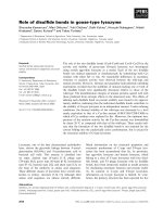
Figure 1 A schematic of the Fluidic NanoPrecipitation System (FNPS). (A) Cartoon of FNPS. Sample inlets are inserted into the dispersing
channel. The inlet channel contains PLGA polymer that precipitates upon contact with the surfactant in the dispersing channel, freezing the
particles in a spherical morphology. (B) Side view of the channel. PLGA droplets are exposed to the hydrodynamic force of the continuous flow.
Xie and Smith Journal of Nanobiotechnology 2010, 8:18
/>Page 2 of 7
of the small size distribution of the nanoparticles gener-
ated using FNPS, filtration is not required prior to use.
The size of PLGA particles generated with the FNPS
can be changed by adjusting the flow rate of the disper-
sing phase. For example, a shift from a flow rate of 35
ml/minute to 50 ml/minute and then to 8 0 ml/minute
decreased particle size from 327 ± 19 nm to 278 ± 35
and then to 193 ± 19 nm (Figure 3A). Similarly, a
decrease in PLGA concentration from 40 mg/ml to 20
mg/ml a nd then to 10 mg/ml resulted in a reduction in
particle diameter from 393 ± 38 nm to 327 ± 19 nm to
231 ± 35 nm (Figure 3B). Since the FNPS is a water/
water miscible solvent system, the composition of the
dispersing phase can also be used to control the size of
the particles. Increasing the concentration of methanol
in the dispersing phase from 20% to 50% and then to
80%, coincided with the reduction in particle size from
512 ± 45 nm to 315 ± 36 nm and then to 148 ± 14 nm
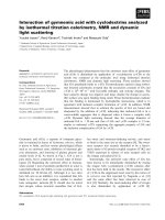
Figure 2 Highly uniform PLGA nanoparticles are fabricated by the Fluidic NanoPrecipitation System (FNPS).ScanningElectron
Microscopy (SEM) images of PLGA nanoparticles fabricated by the (A) FNPS, or the (B) conventional nanoprecipitation method. (C) Diameters of
the particles were measured by using ImageJ. For each sample, the mean diameter was calculated based on the measurements of 200 randomly
chosen particles. White bars indicate the distribution of diameters observed for PLGA nanoparticles fabricated by FNPS (average diameter 148 ±
14 nm). Black bars indicate the distribution of diameters for PLGA nanoparticles fabricated by the traditional nanoprecipitation method (average
diameter 211 ± 70 nm). Samples were imaged without prior filtration.
Xie and Smith Journal of Nanobiotechnology 2010, 8:18
/>Page 3 of 7
(Figure 4). These data suggest that by optimizing all
three of these parameters, the FNPS has the flexibility to
generate uniform particles across a wide range of sizes
from below 100 nm to above 1 μm.
The yield of particles is another important aspect of
any fabrication method. We found that the yield of par-
ticles from the FNPS is typically 80% of the mass of the
PLGA in the inlet solution. Consequently, under the
various co nditions used for this study, the FNPS gener-
ated between two and eight mg of particles/ml/hr. This
compares favorably with the yield of three mgs/ml/hr
fabricated using similar concentrations of PLGA by the
microfluidic system reported by Karnik et al.[34].The
FNPS has many advantages including the ab ility to scal e
up production by simply increasing the number of inlets
entering the dispersing phase. The dispersing stream
could also be recirculated to increase the final concen-
tration of particles in the fluid. In addition, because the
devise has a low risk of clogging, it can be used
continuously.
The mechanism by which the FNPS is able to generate
such small and uniform particles is worthy of discussion.
One factor that influences the final size of the solidified
particles is the size of the monodis pers e droplets from
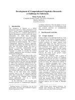
Figure 3 The di amete r of PLGA nanoparticles can be c ontr olled by the flow rates an d PLGA concentrations . (A) SEM images and
diameters of PLGA nanoparticles fabricated at dispersing flow rates of 35 ml/min, 50 ml/min, and 80 ml/min. (B) SEM images and diameters of
PLGA fabricated at PLGA concentrations of 10 mg/ml, 20 mg/ml, and 40 mg/ml. Diameters were measured by using ImageJ. For each sample,
the mean diameter was calculated based on the measurements of 100 randomly chosen particles. Samples are imaged without filtration.
Xie and Smith Journal of Nanobiotechnology 2010, 8:18
/>Page 4 of 7
which they are precipitated. Quevedo et al. [ 30] demon-
strated that the flow in a fluidic system with dimensions
similar to that used here is comparable to a traditional
microfluidic system. They also found that a higher Rey-
nolds number favors the formation of smaller droplets.
So then, parameters like the flow rate in the dispersing
channel, and the liquid compositio n within that channel
will impact Reynolds number and can be used to con-
trolthesizeofdroplets.Theseconclusionsareentirely
consistent with our observation that the flow rate alters
the final particle size.
The actual process of nanoprecipitation will also influ-
ence particle size. This is how our approach differs from
that of Quevedo et al. [30]. They used the T-junction
system to assist in the precipitation of emulsions that
were subsequently made solid by interfacial
polymerization via the action of a cross-linker in the dis-
persing channel. This process creates “hollow” particles
with diameters of several hundred microns. In contrast,
we directly precipitated the PLGA polymer by rapid sol-
vent exchange, also called nanoprecipitation [32]. The
mechanism of particle formation during nanoprecipita-
tion is not entirely understood, meaning that the precise
outcome cannot be predicted. Nevertheless, as has been
previously discussed [32], nanoprecipitation appears to
be governed by the Marangoni effect, wherein move-
ment in an interface is caused by longitudinal variations
of interfacial tension [36]. In such a case, precipitation
is drive n by i) solute transfer out of the phase of higher
viscosi ty, which is influenced by high concentration gra-
dients at the interface; and ii) by interfacial tension,
which, in the case of the FNPS, is determined by
Figure 4 The diameter of PLGA nanoparticles can be controlled by varying the methanol concentrations (v/v) in the dispersing phase.
Diameter of PLGA nanoparticles fabricated using 20%, 50% or 80% v/v methanol in the dispersing phase of the FNPS. The flow rate of the
dispersing channel was maintained at 50 ml/minute. Samples were imaged by SEM without prior filtration. The diameter of the particles was
calculated by using ImageJ. For each sample, the mean diameter was calculated based on the measurements of 100 randomly chosen particles.
Xie and Smith Journal of Nanobiotechnology 2010, 8:18
/>Page 5 of 7
turbulence resulting from flow in the dispersing channel.
Consequently, the size of the final part icle will be influ-
enced not only by features of the dispersing channel
related to Reynolds number, but also by factors that
influence i nterfacial tension. These include the polymer
concentration, the presence and concentration of surfac-
tant [37], and the nature of any payload that is co-preci-
pitated into the particles [37]. The depth of insertion of
the inlet into the dispersing channel might also influ-
ence particle size and geometry due to altered turbu-
lence. However, with this prototype FNPS, it was
impossibletotestthispossibilitybecausewecouldnot
control the depth of insertion with great precision.
Conclusions
In summary, the FNPS described here provides an
approach to produce very sm all and highly uniform
polymeric particles, in the absence of sophisticated
instrumentation or a microfluidic system. The particles
are suitable for multiple uses including drug and ima-
ging agent encapsulation.
Materials and methods
Materials
PLGA Resomer RG502H was purchased from Boehrin-
ger-Ingelheim (Ingelheim, Germany). PL GA sample
solutions were prepared by dissolving PLGA in acetoni-
trile. For example, a 40 mg/ml PLGA solution was pre-
pared by dissolving 40 mg RG502H in 1 ml acetonitri le.
Polyvinyl alcohol (PVA, 87%-89% hydrolyzed) was pur-
chased from Sigma-Aldrich. 1% PVA solution was pre-
pared by dissolving 1 g PVA in 100 ml DI water at
room temperature and filtered to remove any particulate
matter.
Device fabrication and experimental setup
A Fluidic NanoPrecipitation System (FNPS) was fabri-
cated by inserting a stainless steel needle (Hamilton
HA-91039 26s syringe needle) with an inner diameter
0.11 mm, into a Tygon
® tubing (ID 3/32’,OD5/32’ )
that was used to pass the dispersing phase. The needle
was inserted to the interior at 50% of the tubing
diameter.
The PLGA solution fed into the dispersing c hannel
with a 3 ml syringe cont rolled by a single syringe pump
(KDS100, KD Scientific, Massachusetts, USA). A stream
of surfactant (1% PVA solution, 20 ml) passing through
the dispersing channel (Tygon
® tubing with ID 3/32’ ,
and OD 5/32’ ) was controlled by a Fisher Scientific
Variable-Flow Peristaltic Pump.
Nanoparticles were prepared s tarting with 10 and 40
mg/ml of PLGA RG502H polymers in acetonitrile. Sam-
ples (0.2 ml) were injected at a flow rate of 3.2 μl/min.
Nanoparticles were collected into a beaker for analysis.
The nanoparticles were washed by centrifuging for 15
minutes using an Eppendorf 5415R at 13200 rpm at
room temperature and then removing the supernatant.
The nanoparticles were resuspended in DI wate r by bath
sonication (Branson’sModelB200).Thiswasrepeated
three times and the final suspension was sent for analysis.
Scanning Electron Microscope (SEM)
SEM experiments were conducted by depositing the
nanoparticle suspension on freshly cleaved mica and
allowing them to dry. A thin film of Au was sputtered
onto these m ica substrates with sample. Samples were
imaged with scanning electron microscopy (SEM; JEOL
5800LV) without filtration or purification. Particle size
was measured by using ImageJ. For each sample, the
mean diameter was calculated based on the measure-
ments of 100 randomly chosen particles.
Acknowledgements
The work described in this manuscript was supported by a grant from the
U.S. National Institutes of Health (HL080718) awarded to JWS.
Authors’ contributions
JWS and HX conceived and designed the experimental strategy and
interpreted the findings.
HX performed all experiments. All authors read and approved the final
manuscript.
Competing interests
The authors declare that they have no competing interests.
Received: 15 March 2010 Accepted: 13 August 2010
Published: 13 August 2010
References
1. Shive MS, Anderson JM: Biodegradation and biocompatibility of PLA and
PLGA microspheres. Adv Drug Deliv Rev 1997, 28:5-24.
2. Fournier E, Passirani C, Montero-Menei CN, Benoit JP: Biocompatibility of
implantable synthetic polymeric drug carriers: focus on brain
biocompatibility. Biomaterials 2003, 24:3311-3331.
3. Middleton JC, Tipton AJ: Synthetic biodegradable polymers as orthopedic
devices. Biomaterials 2000, 21:2335-2346.
4. Wu L, Ding J: In vitro degradation of three-dimensional porous poly(D,L-
lactide-co-glycolide) scaffolds for tissue engineering. Biomaterials 2004,
25:5821-5830.
5. Giteau A, Venier-Julienne MC, Aubert-Pouessel A, Benoit JP: How to
achieve sustained and complete protein release from PLGA-based
microparticles? Int J Pharm 2008, 350:14-26.
6. Mundargi RC, Babu VR, Rangaswamy V, Patel P, Aminabhavi TM: Nano/
micro technologies for delivering macromolecular therapeutics using
poly(D,L-lactide-co-glycolide) and its derivatives. J Control Release 2008,
125:193-209.
7. Campolongo MJ, Luo D: Drug delivery: Old polymer learns new tracts.
Nat Mater 2009, 8:447-448.
8. Cruz LJ, Tacken PJ, Fokkink R, Joosten B, Stuart MC, Albericio F, Torensma R,
Figdor CG: Targeted PLGA nano-but not microparticles specifically
deliver antigen to human dendritic cells via DC-SIGN in vitro. J Control
Release 2010, 144:118-126.
9. Jaganathan KS, Vyas SP: Strong systemic and mucosal immune responses
to surface-modified PLGA microspheres containing recombinant
hepatitis B antigen administered intranasally. Vaccine 2006, 24:4201-4211.
10. Thomas C, Gupta V, Ahsan F: Influence of surface charge of PLGA
particles of recombinant hepatitis B surface antigen in enhancing
systemic and mucosal immune responses. Int J Pharm 2009, 379:41-50.
Xie and Smith Journal of Nanobiotechnology 2010, 8:18
/>Page 6 of 7
11. Sirsi SR, Schray RC, Wheatley MA, Lutz GJ: Formulation of polylactide-co-
glycolic acid nanospheres for encapsulation and sustained release of
poly(ethylene imine)-poly(ethylene glycol) copolymers complexed to
oligonucleotides. J Nanobiotechnology 2009, 7:1.
12. Zhang YM, Yang F, Yang YQ, Song FL, Xu AL: Recombinant interferon-
alpha2b poly(lactic-co-glycolic acid) microspheres: pharmacokinetics-
pharmacodynamics study in rhesus monkeys following intramuscular
administration. Acta Pharmacol Sin 2008, 29:1370-1375.
13. Jensen DM, Cun D, Maltesen MJ, Frokjaer S, Nielsen HM, Foged C: Spray
drying of siRNA-containing PLGA nanoparticles intended for inhalation. J
Control Release 2010, 142:138-145.
14. Sivadas N, O’Rourke D, Tobin A, Buckley V, Ramtoola Z, Kelly JG, Hickey AJ,
Cryan SA: A comparative study of a range of polymeric microspheres as
potential carriers for the inhalation of proteins. Int J Pharm 2008,
358:159-167.
15. Ungaro F, d’Emmanuele di Villa Bianca R, Giovino C, Miro A, Sorrentino R,
Quaglia F, La Rotonda MI: Insulin-loaded PLGA/cyclodextrin large porous
particles with improved aerosolization properties: in vivo deposition and
hypoglycaemic activity after delivery to rat lungs. J Control Release 2009,
135:25-34.
16. Li X, Xu Y, Chen G, Wei P, Ping Q: PLGA nanoparticles for the oral
delivery of 5-Fluorouracil using high pressure homogenization-
emulsification as the preparation method and in vitro/in vivo studies.
Drug Dev Ind Pharm 2008, 34:107-115.
17. Naha PC, Kanchan V, Manna PK, Panda AK: Improved bioavailability of
orally delivered insulin using Eudragit-L30D coated PLGA microparticles.
J Microencapsul 2008, 25:248-256.
18. Pandey R, Khuller GK: Nanoparticle-based oral drug delivery system for
an injectable antibiotic - streptomycin. Evaluation in a murine
tuberculosis model. Chemotherapy 2007, 53:437-441.
19. Shaikh J, Ankola DD, Beniwal V, Singh D, Kumar MN: Nanoparticle
encapsulation improves oral bioavailability of curcumin by at least 9-
fold when compared to curcumin administered with piperine as
absorption enhancer. Eur J Pharm Sci 2009, 37:223-230.
20. Decuzzi P, Pasqualini R, Arap W, Ferrari M: Intravascular delivery of
particulate systems: does geometry really matter? Pharm Res 2009,
26:235-243.
21. Gratton SE, Ropp PA, Pohlhaus PD, Luft JC, Madden VJ, Napier ME,
DeSimone JM: The effect of particle design on cellular internalization
pathways. Proc Natl Acad Sci USA 2008, 105:11613-11618.
22. Gu F, Zhang L, Teply BA, Mann N, Wang A, Radovic-Moreno AF, Langer R,
Farokhzad OC: Precise engineering of targeted nanoparticles by using
self-assembled biointegrated block copolymers. Proc Natl Acad Sci USA
2008, 105:2586-2591.
23. Siepmann J, Gopferich A: Mathematical modeling of bioerodible,
polymeric drug delivery systems. Adv Drug Deliv Rev 2001, 48:229-247.
24. Tamber H, Johansen P, Merkle HP, Gander B:
Formulation aspects of
biodegradable polymeric microspheres for antigen delivery. Adv Drug
Deliv Rev 2005, 57:357-376.
25. Tracy MA, Ward KL, Firouzabadian L, Wang Y, Dong N, Qian R, Zhang Y:
Factors affecting the degradation rate of poly(lactide-co-glycolide)
microspheres in vivo and in vitro. Biomaterials 1999, 20:1057-1062.
26. Desai MP, Labhasetwar V, Amidon GL, Levy RJ: Gastrointestinal uptake of
biodegradable microparticles: effect of particle size. Pharm Res 1996,
13:1838-1845.
27. Holgadoa MA, Cozar-Bernala MJ, Salasa S, Arias JL, Alvarez-Fuentesa J,
Fernandez-Avevaloa M: Protein-loaded PLGA microparticles angineered
by flow focusing: Physicochemical characterization and protein
detection by reversed-phase HPLC. International Journal of Pharmaceutics
2009, 380:147-154.
28. Almeria B, Deng W, Fahmy T, Gomez A: Controlling the morphology of
electrospray-generated PLGA microparticles for drug delivery. Journal of
Colloid and Interface Science 2010, 343:125-133.
29. Yao J, Lim LK, Xie J, Wang C: Characterization of electrospraying process
for polymeric particle fabrication. J Aerosol Sciences 2008, 39:987-1002.
30. Quevedo E, Steinbacher J, McQuade DT: Interfacial polymerization within
a simplified microfluidic device: capturing capsules. Journal of the
American Chemical Society 2005, 127:10498-10499.
31. Feng S, Huang G: Effects of emulsifiers on the controlled release of
paclitaxel (Taxol) from nanospheres of biodegradable polymers. J Control
Release 2001, 71:53-69.
32. Fessi H, Puixeux F, Devissaguet J P, Ammoury N, Benita S: Nanocapsule
formation by interfacial polymer deposition following solvent
displacement. International Journal of Pharmacy 1989, 55:R1-R4.
33. Gaumet M, Vargas A, Gurny R, Delie F: Nanoparticles for drug delivery: the
need for precision in reporting particle size parameters. Eur J Pharm
Biopharm 2008, 69:1-9.
34. Karnik R, Gu F, Basto P, Cannizzaro C, Dean L, Kyei-Manu W, Langer R,
Farokhzad OC: Microfluidic platform for controlled synthesis of polymeric
nanoparticles. Nano Lett 2008, 8:2906-2912.
35. Gaumet M, Gurny R, Delie F: Fluorescent biodegradable PLGA particles
with narrow size distributions: preparation by means of selective
centrifugation. Int J Pharm 2007, 342:222-230.
36. Sternling CV, Scriven LE: Interfacial turbulence: Hydrodynamic instability
and the marangoni effect. AIChE Journal 1959, 5:514-523.
37. Berg J: Interfacial hydrodynamcis: an overview. Canadian Metallurgy
Quarterly 1982, 21:121-136.
doi:10.1186/1477-3155-8-18
Cite this article as: Xie and Smith: Fabrication of PLGA nanoparticles
with a fluidic nanoprecipitation system. Journal of Nanobiotechnology
2010 8:18.
Submit your next manuscript to BioMed Central
and take full advantage of:
• Convenient online submission
• Thorough peer review
• No space constraints or color figure charges
• Immediate publication on acceptance
• Inclusion in PubMed, CAS, Scopus and Google Scholar
• Research which is freely available for redistribution
Submit your manuscript at
www.biomedcentral.com/submit
Xie and Smith Journal of Nanobiotechnology 2010, 8:18
/>Page 7 of 7