Báo cáo y học: "Treatment of stasis dermatitis using aminaphtone: Spindle cell oncocytoma of the adenohypophysis in a woman: a case report and review of the literature" pdf
Bạn đang xem bản rút gọn của tài liệu. Xem và tải ngay bản đầy đủ của tài liệu tại đây (569.37 KB, 4 trang )
CASE REP O R T Open Access
Spindle cell oncocytoma of the adenohypophysis
in a woman: a case report and review of the
literature
M Mlika
1*
, H Azouz
1
, I Chelly
1
, I Ben Saïd
2
, H Jemel
2
, S Haouet
1
, M Zitouna
1
, N Kchir
1
Abstract
Introduction: Spindle cell oncocytoma of the adenohypophysis is a rare tumour recently reported by Roncaroli
et al. in 2002. This tumour is considered a grade I tumour by the World Health Organization.
Case presentation: We describe what is, to the best of our knowledge, the 14th case of its kind in the literature.
A 45-year-old African woman presented clinical and radiological findings related to a nonfunctioning pituitary
adenoma. The diagnosis was made on the basis of histological and immunohistochemical findings.
Conclusion: The purpose of this work is to report a rare pituitary tumour and to describe its histolo gical and
immunohistochemical features, which were characterized by the expression of thyroid transcription factor 1
antigen by tumour cells. This fact could support the theory of a possible common origin of these tumours in
pituicytomas. In fact, thyroid transcription factor 1 is considered to be a specific marker of pituicytes.
Introduction
Spindle cell oncocytoma (SCO) of the pituitary gland is
a recently described entity which was recognized by the
2007 WHO Classification of Brain Tumours and consid-
ered a WHO grade I tumour [1]. It was initially
described by Roncaroli et al. in 2002 [2], and only 14
cases have been reported in the literature. The histogen-
esis and prognosis of these tumours remain uncertain
and need to be documented more thoroughly in the lit-
era ture. Our aim is to report a new case of SCO and to
describe its histological and immunohistochemical fea-
tures supporting the theory of a possible common origin
with pituicytoma [2].
Case presentation
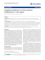
We report the case of a 45-year-old African woman with-
out a particular medical history who presented with
intermittent decrease of visual acuity and headache. Cra-
nial magnet ic resonance imaging (MRI) revealed a solid
adenohypophysis mass of 2 × 1.5 × 1 cm with suprasellar
extension but no invasive growth. This mass showed
contrast enhancing in T1-weighted MRI scans (Figure 1).
Laboratory tests used to explore pituitary disorders
showed normal levels of pituitary hormones, including
follicle-stimulating hormone (FSH) (N > 20 IU/L), lutei-
nizing hormone (LH) (N > 10 IU/L), prolactin (N < 20
μg/L), corticotro pin and thyrotropin. The diagnosis of
nonfunctioning pituitary ma cro adenoma w as suspected,
and the tumour was completely resected via transsphe-
noidal surgery. No adjuvant therapy was administered.
Postoperatively, the patient developed panhypopituitarism
which has been managed by hormone substitution. In
fact, laboratory tests showed marked low levels of FSH
(5 IU/L), LH (2 IU/L), prolactin (0.04 μg/L), corticotropin
(10 nmol/L), thyrotropin (0.01 μU/mL) and somatotropin.
Otherwise, currently there is neither clinical nor radiolo-
gical evidence of a recurrent tumor after a three-month
follow-up period.
Microscopic findings consisted of a solid spindle cell
neoplasm with increased cellularity. Tumour cells were
spindled to epithelioids organized in interlacing fascicles.
The tumour cells had eosinophilic and oncocytic cyto-
plasm (Figure 2A). Nuclear atypia and pleomorphism
were absent. Mitotic count was estimated to 1 per 10
high-power field. There were neither microvascular pro-
liferations nor necrosis.
* Correspondence:
1
Department of Pathology, La Rabta Hospital, Bab Saadoun, Tunis 2037,
Tunisia
Full list of author information is available at the end of the article
Mlika et al. Journal of Medical Case Reports 2011, 5:64
/>JOURNAL OF MEDICAL
CASE REPORTS
© 2011 Mlika et al; licen see BioMed Central Ltd. This is an Open Access article distributed under the terms of the Creative Commons
Attribution License ( which permits unrestricted use, distribution, and reproduction in
any medium, provided the original work is properly cited.
An immunohistochemical study showed that most
tumour cells expressed S-100 protein (Figure 2B).
Vim entin and epit helial membrane antigen (EMA) were
similarly expressed by tumor cells (Figure 2C). There
was no staining either with low-molecular-weight cyto-
keratin or with anterior pituitary hormones, including
somatotropin, corticotropin, thyrotropin, FSH, LH and
prolactin (Figure 2D). Tumour cells expressed the thyr-
oid transcription factor 1 (TTF-1) antigen (Figure 3).
Glial fibrillary acidic protein (GFAP) and CD68 were
not expressed. Therefore, the diagnoses of glial tumour
and granular cell tumour were ruled out. In light of
these histological and immunohistochemical findings,
the diagnosis of SCO was strongly suspected. Ultrastruc-
tural examination showed neoplastic cells filled with
mitochondria and well-formed desmosomes. These find-
ings supported the diagnosis of SCO.
Discussion
SCO is a rare tumour, with only 14 cases reported in
the English-language literature (Table 1). In accord with
the present case, it affects middl e-aged and older adults
of both sexes. It presents as a sellar tumour suspected
to be a functionally inactive macroadenoma.
Imaging findings
The radiological findings are nonspecific and do not
differentiate these tumours from pituitary adenomas.
In our case, they consisted of an enhanced mass of the
sellar region.
Histological and immunohistochemical features
Histological examination is the only means of diagnosis
[2-4]. This tumour is typ ically composed of interlacing
fascicles of spindled to epithelioid cells with oncocytic
cytoplasm. Mild to moderate nuclear atypia and even
focal marked pleomorphism may be seen. The immuno-
profile of these tumours is characterized by simulta-
neous positivity for S-100 protein, vimentin and EMA.
Ultrastructurally, the neoplastic cells contain numerous
mitochondria with lamellar cristae. The neoplastic cells
are linked by intermediate junctions and desmosomes
[1,5].
Pathogenesis
These histological, immunohistochemical and fine struc-
tural features lead most authors to postulate a possible
Figure 1 (A) Coronal magnetic resonance imaging studies
showing a sellar mass with suprasellar extension but no
invasive growth (arrow). (B) T1-weighted image showing the
enhancement of the mass (arrow).
Figure 2 (A) Histology and immunoprofile of spindle cell
oncocytoma. Spindle cell neoplasm with interlacing fascicles of
spindled to epithelioid cells with eosinophilic and oncocytic
cytoplasm (original magnification, ×400; hematoxylin and eosin
stain). (B) Immunohistochemical study showed that most tumor
cells coexpressed S-100 protein (original magnification, ×400;
hematoxylin and eosin stain) and (C) vimentin and epithelial
membrane antigen (original magnification, ×400; hematoxylin and
eosin stain). (D) Tumor cells were negative with pituitary hormones.
Figure 3 Nuclear expression of thyroid transcription factor 1
by tumour cells (original magnification, ×200; hematoxylin and
eosin stain). Inset: A higher-magnification image showing the
nuclear expression (original magnification, ×400; hematoxylin and
eosin stain).
Mlika et al. Journal of Medical Case Reports 2011, 5:64
/>Page 2 of 4
derivation of these tumours from folliculostellate cells.
Very little is known about the functioning of the follicu-
lostellate cells. Some authors have reported that these
cells are implicated in long-distance communication in
the anterior pituitary gland [6-8]. These cells are known
to coexpress the S-100 protein, vimentin and galectin 3.
These findings are shared by the SCO, but in our case
tumour cells also expressed TTF-1. Lee et al. [9]
described the expression of TTF-1 in eight cases of
SCO. They reported that TTF-1 is generally expressed
in fetal neurohypophysis. According to these findings,
this marker could be specific to human pituicytes. The
positivity of TTF-1 in our observation with these eight
reported cases should lead to further research that could
have implications for the classifi cation of these rare
sellar neoplasms and may indicate a similar origin of
SCO and pituicytoma [9].
Differential diagnoses
The diagnosis of these tumours may be challenging. In
fact, they should be distinguished from meningioma in
its oncocytic var iant, granular cell tumo ur, pituicytoma,
oncocytic neoplasm arising from developmental salivary
gland remnants and oncocytic variant of a pituitary ade-
noma [2-4]. Rarely, they should be distinguished from
sellar schwannoma, but this tumour is very rare in that
location [10]. Most meningiomas that are located within
the cranial cavity occur over the cerebral convexities,
but other common sites include para- and suprasellar
regions. The radiological findings may be challenging
when showing an enhancing mass without particular
characteristics. The pathological findings and immuno-
histochemical features show a similar expression
of EMA and vimentin, but S-100 protein is rarely
expressed in meningiomas. Moreover, in opposition to
SCO, tumour cells in meningiomas are filled with inter-
mediate filaments and desmosomal intercellular junc-
tions in ultrastructural examination [2]. Granular cell
tumours and pituicytomas tend to develop in the poster-
ior pituitary gland rather than i n the adenohypophysis.
These tumours are thought to originate from pituicytes.
Granular cell tumors are characterized by a granular
cytoplasm which can be observed in SCO, but, in oppo-
sition to the SCO, granular cell tumour shows a strong
expression of CD68. Besides, the cytoplasm of the gran-
ular cells is filled with phagolysosomes, and there are no
mitochondria. The distinction from pituicytoma relies
on the evidence of oncocytic change in SCO rather than
GFAP staining patterns alone. Oncocytic neoplasms ori-
ginating from salivary gland remnants express epithelial
markers and lack S-100 protein and EMA positivity
[3,4]. The distinction of SCO from an oncocytic variant
ofapituitaryadenomaisbasedontheexpressionof
neurosecretory markers synaptophysin and chromogra-
nin by the adenoma [5]. The differences in immunohis-
tochemical profile between these tumours are illustrated
in Table 2. The treatment of these tumours is based on
surgical resection. Postoperative complications consist
mainly of hypopituitarism as in the case of our patient.
This complication is due to the difficulty of this surgery,
which needs accurate management that is not always
possible in the sellar region.Aconsensualprotocolhas
not been assessed because of the complex issue of these
tumours and the lack of large series. In fact, among the
14 cases reported in the literature, eight patients had a
benign clinical course and six experienced recurrence
despite adjuvant treatment in two cases [6].
Conclusion
SCOs of the pit uitary gland are rare tumours whose
pathogenesis and management remain debated because
of the few numbers of reported cases. These tumours
areconsideredtohaveagoodprognosisdespitethe
early recurrences reported in some cases [8,9]. Addi-
tional clinical follow-up is needed to assess the prognos-
tic features. In our case, the period of follow-up was too
Table 1 Cases reported in the literature
a
Year of
publication
Reference Number of
reported cases
Sex ratio
(M/F)
Mean age
(yr)
Symptoms Evolution
2002 Roncaroli et al. [2] 5 cases 3/2 61.6 Panhypopituitarism, visual defect No recurrence
(35.4 mo)
2005 Dahiya et al. [3] 2 cases 2 F - Panhypopituitarism No recurrence
2005 Kloub et al. [4] 2 cases 1/1 73 - Recurrence after 1 and 11 yr
2006 Vajtai et al. [5] 1 case F 48 Adynamia, decrease of visual
acuity
No recurrence
2009 Borota et al. [6] 1 case F - - Slow regrowth
(30 mo)
2009 Demmsie et al. [7] 1 case M - Visual blurring, weight loss Recurrence after 9 mo
2009 Coiré et al. [8] 1 case F 63 Visual defect Recurrence after 5 mo
a
M, male; F, female.
Mlika et al. Journal of Medical Case Reports 2011, 5:64
/>Page 3 of 4
short, so we can only speculate whether such a tumour
is benign.
Consent
Written, informed consent was obtained from the
patient for publication of this case report and accompa-
nying images. A copy of the written consent is available
for review by the Editor-in-Chief of this journal.
Acknowledgements
We thank Dr Nadia Kourda from Charles Nicolle Hospital for her contribution
in taking the photos.
Author details
1
Department of Pathology, La Rabta Hospital, Bab Saadoun, Tunis 2037,
Tunisia.
2
Department of Neurosurgery, La Rabta Hospital, Bab Saadoun, Tunis
2037, Tunisia.
Authors’ contributions
MM conceived of, coordinated with other coauthors and drafted and revised
the manuscript. HH, IC, IBS, SH, HJ, MZ and NK participated by acquisition
and analysis of literature data and helped to draft the manuscript. All
authors read and approved the final manuscript.
Competing interests
The authors declare that they have no competing interests.
Received: 4 March 2010 Accepted: 14 February 2011
Published: 14 February 2011
References
1. Louis DN, Ohgaki H, Wiestler OD, Cavenee WK: Tumors of the sellar
region. WHO Classification of Tumours of the Central Nervous System. 4
edition. Lyon, France, International Agency for Research on Cancer 2007,
245-246.
2. Roncaroli F, Scheithauer BW, Cenacchi G, Horvath E, Kovacs K, Lloyd RV,
Abell-Aleff P, Santi M, Yates AJ: ’Spindle cell oncocytoma’ of the
adenohypophysis: a tumor of folliculostellate cells? Am J Surg Pathol
2002, 26:1048-1055.
3. Dahiya S, Sarkar C, Hedley-Whyte ET, Sharma MC, Zervas NT, Sridhar E,
Louis DN: Spindle cell oncocytoma of the adenohypophysis: report of
two cases. Acta Neuropathol 2005, 110:97-99.
4. Kloub O, Perry A, Tu PH, Lipper M, Lopes MBS: Spindle cell oncocytoma of
the adenohypophysis: report of two recurrent cases. Am J Surg Pathol
2005, 29:247-253.
5. Vajtai I, Sahli R, Kappeler A: Spindle cell oncocytoma of the
adenohypophysis: report of a case with a 16-year follow-up. Pathol Res
Pract 2006, 202:745-750.
6. Borota OC, Scheithauer BW, Fougner SL, Hald JK, Ramm-Pettersen J,
Bollerslev J: Spindle cell oncocytoma of the adenohypophysis: report of a
case with marked cellular atypia and recurrence despite adjuvant
treatment. Clin Neuropathol 2009, 28:91-95.
7. Demssie YN, Joseph J, Dawson T, Roberts G, de Carpentier J, Howell S:
Recurrent spindle cell oncocytoma of the pituitary, a case report and
review of literature. Pituitary 2009.
8. Coiré CI, Horvath E, Smyth HS, Kovacs K: Rapidly recurring folliculostellate
cell tumor of the adenohypophysis with the morphology of a spindle
cell oncocytoma: case report with electron microscopic studies. Clin
Neuropathol 2009, 28:303-308.
9. Lee EB, Tihan T, Scheithauer BW, Zhang PJ, Gonatas NK: Thyroid
transcription factor 1 expression in sellar tumors: a histogenetic marker?
J Neuropathol Exp Neurol 2009, 68:482-488.
10. Mohammed S, Kovacs K, Munoz D, Cusimano MD: A short illustrated
review of sellar region schwannomas. Acta Neurochir (Wien) 2010,
152:885-891.
doi:10.1186/1752-1947-5-64
Cite this article as: Mlika et al.: Spindle cell oncocytoma of the
adenohypophysis in a woman: a case report and review of the
literature. Journal of Medical Case Reports 2011 5:64.
Submit your next manuscript to BioMed Central
and take full advantage of:
• Convenient online submission
• Thorough peer review
• No space constraints or color figure charges
• Immediate publication on acceptance
• Inclusion in PubMed, CAS, Scopus and Google Scholar
• Research which is freely available for redistribution
Submit your manuscript at
www.biomedcentral.com/submit
Table 2 Immunohistochemical findings in spindle cell oncocytoma and the main differential diagnoses
a
Diagnoses Immunohistochemical markers
Spindle cell oncocytoma S-100 protein, vimentin and EMA are expressed
Oncocytic variant of meningioma EMA is expressed, vimentin is expressed and S-100 protein is negative
Granular cell tumour CD68 is expressed
Pituicytoma GFAP is expressed
Oncocytic neoplasm originating from salivary gland remnants Epithelial markers are expressed, S-100 protein and EMA are negative
Oncocytic variant of a pituitary adenoma Synaptophysin and chromogranin are expressed
a
EMA, epithelial membrane antigen; GFAP, glial fibrillary acidic protein.
Mlika et al. Journal of Medical Case Reports 2011, 5:64
/>Page 4 of 4