báo cáo khoa học: "Development of PEGylated PLGA nanoparticle for controlled and sustained drug delivery in cystic fibrosis" pptx
Bạn đang xem bản rút gọn của tài liệu. Xem và tải ngay bản đầy đủ của tài liệu tại đây (6.19 MB, 18 trang )
RESEA R C H Open Access
Development of PEGylated PLGA nanoparticle for
controlled and sustained drug delivery in cystic
fibrosis
Neeraj Vij
1,2*
, Taehong Min
1
, Rhul Marasigan
1
, Christopher N Belcher
1
, Steven Mazur
1
, Hong Ding
3
, Ken-Tye Yong
3
,
Indrajit Roy
3
Abstract
Background: The mutation in the cystic fibrosis transmembrane conductance regulator (CFTR) gene results in CF.
The most common mutation, ΔF508-CFTR, is a temperature-sensitive, trafficking mutant with reduced chloride
transport and exaggerated immune response. The ΔF508-CFTR is misfolded, ubiquitinated, and prematurely
degraded by proteasome mediated- degradation. We recently demonstrated that selective inhibition of
proteasomal pathway by the FDA approved drug PS-341 (pyrazylcarbonyl-Phe-Leuboronate, a.k.a. Velcade or
bortezomib) ameliorates the inflammatory pathophysiology of CF cells. This proteasomal drug is an extremely
potent, stable, reversible and selective inhibitor of chymotryptic threonine protease-activity. The apprehension in
considering the proteasome as a therapeutic target is that proteasome inhibitors may affect proteostasis and
consecutive processes. The affect on mul tiple processes can be mitigated by nanoparticle mediated PS-341 lung-
delivery resulting in favorable outcome observed in this study.
Results: To overcome this challenge, we developed a nano-based approach that uses drug loaded biodegradable
nanoparticle (PLGA-PEG
PS-341
) to provide controlled and sustained drug delivery. The in vitro release kinetics of drug
from nanoparticle was quantified by proteasomal activity assay from days 1-7 that showed slow drug release from
day 2-7 with maximum inhibition at day 7. For in vivo release kinetics and biodistribution, these drug-loaded
nanoparticles were fluorescently labeled, and administered to C57BL6 mice by intranasal route. Whole-body optical
imaging of the treated live animals demonstrates efficient delivery of particles to murine lungs, 24 hrs post
treatment, followed by biodegradation and release over time, day 1-11. The efficacy of drug release in CF mice
(Cftr
-/-
) lung s was determined by quantifying the changes in proteasomal activity (~2 fold decrease) and ability to
rescue the Pseudomonas aeruginosa LPS (Pa-LPS) induced inflammation, which demonstrates the rescue of CF lung
disease in murine model.
Conclusion: We have developed a novel drug delivery system to provide sustained delivery of CF “correctors” and
“anti-inflammatories ” to the lungs. Moreover, we demonstrate here the therapeutic efficacy of nano-based
proteostasis-modulator to rescue Pa-LPS induced CF lung disease.
Background
The cystic fibrosis transmembrane conductance regula-
tor (CFTR ) encodes a cAMP regulated chloride channel
that is retrieved (25% wild type and 9 9% of ΔF508-
mutated) from the endoplasmic reticulum (ER) during
translation and folding, and targeted to the proteasome
for premature degradation [1]. Alteration of the intracel-
lular fate of mutant CFTR by intervening the protein
processing and/or proteolytic pathway has shown pro-
mise for treating CF but selective inhibition of proteo-
statsis demands the controlled release of optimal
amounts of drug overtime. The latest fast track FDA
approval of first proteasome inhibitor drug, PS-341 for
treatment of refractory multiple myeloma [2-4] has
initiated the examination of protein catabolism for
potential therapeutic intervention in several protein
* Correspondence:
1
Department of Pediatric Respiratory Sciences, Johns Hopkins University
School of Medicine, Baltimore, 21287, USA
Full list of author information is available at the end of the article
Vij et al. Journal of Nanobiotechnology 2010, 8:22
/>© 2010 Vij et al; licensee BioMed Central Ltd. This is an Open Acce ss article distributed under the te rms of the Creative Commons
Attribution License ( which permits unrestricted use, distribution, and reproduction in
any medium, provide d the origin al work is properly cited.
processing disorders. PS-341 (pyrazylcarbonyl-Phe-Leu-
boronate) is an extremely potent, stable, reversible and
selective inhibitor of chymotryptic threonine protease
activity [2]. PS-341 showed encouraging results when
employed in hematological cancers an d solid tumors by
selectively inducing apoptosis in inflammatory cancer
cells while normal cells recover from proteasome i nhibi-
tion [5]. Proteasome inhibitors were recently shown to
have dual therapeutic importance in pharmaco-gene
therapy of CF airway [6]. In this study, proteasome inhi-
bitors- LLnL and doxorubicin enhanced the CFTR gene
delivery and hence CFTR-mediated short-circuit cur-
rents. Moreover, these proteasome inhibitors were also
effective in suppressing functional epithelial sodium
channel (ENaC) activity and currents independent of
CFTR vector administration [6]. We found that PS-341
is highly selective chymotryptic proteasome inhibitor
that rescues ΔF508-CFTR and IBa from proteasomal
degradation [7-9] and hence inhibits NFB-mediated,
IL-8 activation [9]. This ability to ameliorate other pri-
mary aspects of CF disease pathophysiology in addition
to the rescue of misfolded CFTR from proteasomal
degradation is promising for CF therapeutics. A main
concern in considering the proteasome as a therapeutic
target is that proteasome inhibitors may affect the nor-
mal process(es).
Over the past couple of decades, the field of drug
delivery has been revolutionized with the advent of
nanoparticles, wherein these particles act as inert car-
riersfordrugsandgenestotargetcellsortissues[10].
This has resulted in significant improvement in meth-
ods to induce drug acc umulation in target tissues with
subsequent reduction in non-spe cific effects, a major
limitation encountered in conventional therapies for
chronic conditions. However, along with the many
advantages of nanoparticle-mediated drug delivery,
some characteristic drawbacks demand additional stu-
dies to dev elop an ideal formulation for therapeutic.
One such drawback is the persistence of the nano parti-
cle system in the body long after the the rapeutic effect
of the delivered drug has been realized. This has led to
the development of biodegradable nanoparticles, parti-
cularly comprised of the polymer polylactide-coglycolide
(PLGA), where the particle matrix degrades slowly in
vivo and the by-products like lactic and glycolic acid are
easily metabolized and excreted [11]. Therefore, PLGA
nanoparticles, due to their ability to entrap both water-
soluble and water-insoluble molecules, are in process of
extensive evaluation for the delivery of drugs, genetic
materials and proteins to cultured cells and experimen-
tal animals. These nanoparticulate systems are rapidly
endocytosed by cells followed by release of t heir thera-
peutic payload by both passive diffusion and slow
matrix degradation [12,13].
The nano-drug delivery system used here provides con-
trolled and sustained PS-341 delivery for selective inhibi-
tion of proteasome mediated homeostatic process
(proteostasis). This study was designed to standardize the
toxicity and efficacy of nano-drug delivery system in both
in vitro and in vivo (WT mice) systems, and evaluate the
efficacy of PLGA-PEG mediated PS-341 lung delivery in
controlling inflammatory CF lung disease. The long term
goal of this study was to test the efficacy of the novel
nano-system to control CF lung disease for future pre-
clinical development of 2
nd
generation targeted delivery
system that can selectively d eliver drugs to lung epithe-
lium. Recent studies have identified several novel “correc-
tors” and molecular targets for functional rescue of
misfolded ΔF508-CFTR protein or chronic inflammator y
statebutthechallengeistoprovidesustainedandcon-
trolled drug delivery to CF subjects. We are developing
methods to encapsulate selected known CF correctors,
potentiators and antim icrobials, in PLGA-PEG based
nanoparticles to develop this nanosystem as a therapeutic
delivery vehicle for variety of CF drugs. We anticipate
that therapeutic development of this novel nano-based
biodegradable therapeutic vehicle will have enormous
applications in treatment of chronic pathophysiology of
obstructive lung diseases like CF and COPD as these sys-
tems are designed to bypass the mucus barrier and slowly
release the drug to the lung tissue or cell that warrants
further preclinical evaluation and standardization.
Results
Characterization of PLGA-PEG
PS-341
nanoparticles
The multiple batches of PS-341 or fluorescent marker
dye, nile-red, loaded PLGA nanoparticles were synthe-
sized using non-polar core of oil-in-water microemulsion
technique with PEGylated phospholipid DSPE-mPEG
2000
as the emulsifier. In this formulation, the hydrophobic
phospholipid part of the emulsifier remain embedded in
the PLGA matrix by hydrophobic interactions, whereas
the hydrophilic PEG part point outwards on the nanopar-
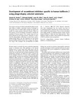
ticle surface, forming a polymeric brush (Fig 1A). This
brush effect is implicated in the in vivo stability of such
nanoparticles against opsonic capture by (a) shielding the
high negative charge o f the polymer and (b) forming a
steric barrier against approaching opsonins and prevent-
ing agglomeration of nanoparticles [10]. Therefore, by
using a molecule like DSPE-mPEG
2000
as emulsifier, we
achieve both stability and PEGylation of PLGA nanopar-
ticles. The dynamic laser scattering (DLS) results show
that the average radius of PLGA-PEG
PS341
nanoparticles
used in this study is 121.5 ± 15 nm (PDI = 0.106; Fig 1B).
The diameter of nanoparticles, varied by less than 15%,
suggesting that their colloidal stability is not affected
under physiological pH. Transmission electron micro-
scopy (TEM) verifies that the size of the PLGA-PEG
PS341
Vij et al. Journal of Nanobiotechnology 2010, 8:22
/>Page 2 of 18
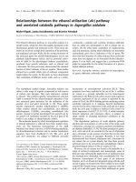
Figure 1 Synthesis and c haracte rizatio n of PLG A-PEG
PS341
nanopartic les .ThePS-341orfluorescentmarkerdye,nilered,loadedPLGA
nanoparticles were synthesized using non-polar core of oil-in-water microemulsion technique with PEGylated phospholipid DSPE-mPEG
2000
as
the emulsifier. Dynamic laser scattering (DLS) was employed to measure the size, distribution and colloidal stability of the PLGA-PEG
PS341
nanoparticles while transmission electron microscopy (TEM) was used to characterize the size and shape of the nanoparticles. (A) Schematic
shows that PS-341 and/or nile red dye is encapsulated in PLGA nanoparticles. The hydrophobic phospholipid part of the emulsifier remains
embedded in the PLGA matrix by hydrophobic interactions, whereas the hydrophilic PEG part point outwards on the nanoparticle surface,
forming a polymeric brush. (B) The DLS results show that radius of PLGA-PEG
PS341
nanoparticles is 121.5 ± 15 nm (PDI = 0.106). The radius of
nanoparticles varied by less than 15%, suggesting that their colloidal stability is not affected under physiological pH. (C) TEM shows that PLGA-
PEG
PS341
nanoparticles are mono-dispersed, spherical and are ~200 nm in size. DLS and TEM based size and surface characterization of
nanoparticles confirms size distribution and colloidal stability of mono-dispersed particles.
Vij et al. Journal of Nanobiotechnology 2010, 8:22
/>Page 3 of 18
nanoparticles is ~200 nm. Moreover, data also verifies
that PLGA-PEG
PS341
nanoparticles are mono-dispersed
and spherical in shape (Fig 1C). The results were repro-
ducible in multiple batches.
PLGA-PEG based nano drug-delivery exhibits sustained
release and activity
We determined the in vitro efficacy of the nanoparticle
system by evaluating the release kinetics of short-lived
dye (30 mins), nile red, from PLGA-PEG nanoparticles
by quantifying the absorption of released dye at 525 nm.
Short-lived nile red dye was selected to determine the
efficacy of sustained release from nanoparticles. We
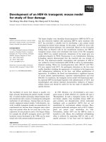
observed a sinusoidal-like, sustained release of the dye
from day 1 to 15, with a maximum release at day 10
(Fig 2A). Next, we quantified the release kinetics of the
drug- PS-341 from PLGA- PEG in vitro,onceeveryday
for 7 days, using Proteasomal Activity Assay. During
this experiment, we recorded proteasome inhibitory
activity (Relative Luminescence Units, RLU) of room
temperature incubated PLGA-PEG
PS341
-andDSPE-
PEG
PS341
- (control, non-PLGA) nanoparticles for day 1
to 7 and observed sustained release of PS341 from
PLGA-PEG (Fig 2B). We also observed that PLGA-
PEG
PS341
provides more effective drug activity compared
to DSPE-PEG
PS341
. Next, we compared the efficacy of
PLGA-PEG
PS341
drug delivery in CFBE41o- cells to PS-
341 treatment by Proteasome-Glo Chymotrypsin Cell
Based Assay (Promega). We observed a significantly bet-
ter decrease (~1.2 fold, p < 0.05) in proteasome activity
when using the PLGA-PEG mediated PS341 delivery as
compared to PS341 tre atment (non-nanoparticle) at
similar concentrations (Fig 2C). Thus, the PLGA-PEG
nanoparticle en hances the drug delivery and therapeutic
effectiveness. We verified these results with microscopy
of PLGA-PEG
PS341/NileRed
treated cells (described below).
As a funct ional parameter for the in vivo treatment effi-
cacy of PLGA-PEG
PS341
we quantified proteasomal
activity in murine lung tissues. We observed significant
reduction (~2 fold, p < 0.01) in proteasomal activity of
Cftr
-/-
-andCftr
+/+
- mice lungs by day-3 of intranasal
PLGA-PEG
PS341
(10 μg) treatment (Fig 3). Next, nile red
labeled PLGA-PEG nanoparticles were insufflated in
Cftr
+/+
(n = 4) mice airways at indicated doses to stan-
dardize the biodistribution and release kinetics. Live ani-
mals were imaged by Xenogen IVIS 200 optical imaging
device (Ex 465 nm and Em 525 nm) from day 1 to 11
under constant supply of isoflurane using an automated
anesthesia machine in accordance with our JHU ACUC
approved protocol. We observed significant amount of
PLGA-PEG
PS341-NileRed
particles in murine lungs by 24
hrs and observed its sustained release from days 1 to 11
given the short half-life of the nile red (Fig 4). Bladder
shows the significant amounts of excreted nanoparticles
demonstrating the efficient clearance of biodegradable
nanoparticles overtime.
PLGA-PEG nanoparticles mediated intracellular delivery
and efficacy
The indicated concentrations of PLGA-PEG
PS341-NileRed
was added to CFBE41o- c ells and incubated for 24 hrs
followed by fluorescence microscopy to detect the nano-
particle mediated nile red delivery to CF cells. We
observed the cytosolic release of nile red in perinuclear
space (Fig 5) that verifies the effica cy of our therapeutic
vehicle for bronchial epithelial cell delivery. For reporter
assay, CFBE41o- cells were treated for 24 hours with
indicated doses of PLGA-PEG
PS-341
aft er 6 hrs of NFB
or IL-8 and renila luciferase reporter plasmid transfec-
tions. The TNF-a (10 ng/ml) was used to induce proin-
flammatory signaling overnight. NFBandIL-8
luciferase activity was quantified using the Dual Lucifer-
ase® Reporter Assay System (Promega). We observed
that treatment with the 10 μl of PLGA-PEG
PS341
(10 ng/
μl) significantly decreased TNF-a induced NFB(Fig
6A) and IL-8 (Fig 6B) promoter activities (*p < 0.05).
The data verifies the efficacy of PLGA-PEG mediated
drug delivery and NFB inhibitory activity.
PLGA-PEG
PS341
controls NFB mediated proinflammatory
response in CF lungs
To test the efficacy of PS-341 in controlling p roinflam-
matory response, the age and sex matched Cftr
-/-
mice
(n = 3, each group) were injected (i.p.) with 15 mg/kg
body weight Pseudomonas aeruginosa (Pa )-LPS, 24 hr s
after first PS-341 treatment (0.6 mg/kg/day). Control,
untreated group, was injected with 100 μl saline. Second
PS-341 treatment was also given together with LPS or
saline treatment and after 24 hrs, serum was collected
(day-3) for ELISA. The serum cytokine levels were
quantified by sandwich ELISAs. We obse rved that treat-
ment with the PS-341 significantly decreased Pa-LPS
induced IL1-b and IL-6 levels (Fig 7), demonstrating the
ability of PS-341 to refrain both basal and Pa-LPS
induced inflammatory response (*p < 0.05). Since sys-
temic administration of PS-341 significantly inhibits the
basal cytokine response, it may have immunosuppressive
adverse effects. We concluded that a irway delivery of
PS-341 will be more effective in treating CF lung disease
as compared to the intraperitoneal treatment due to
increased bioavailability and reduced side effects. A
main concern in conside ring the pr oteasome as a thera-
peutic target is that proteasome inhibitors may affect
normal protein-processing machinery (pr oteostasis). The
nano-drug delivery system used here provides a feasible
alternative for controlled and sustained PS-341 delivery
to lungs for selective inhibition of proteostasis to miti-
gate the consequences. The Cftr
-/-
mice (n = 3, each
Vij et al. Journal of Nanobiotechnology 2010, 8:22
/>Page 4 of 18
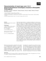
Figure 2 Release kinetics of PLGA-PEG nanoparticles shows sustained release and drug activity overtime. A) Release kinetics of nile red
from PLGA-PEG nanoparticles (n = 3) was quantified by recording absorption of released dye at 525 nm. We observed a sinusoidal-like,
sustained release of the dye from day 1 to 15, with a maximum release at day 10. Triplicate samples are shown by different symbols. B) We
quantified the release kinetics of PS-341 from PLGA-PEG and DSPE-PEG, once daily for 7-days, using the proteasomal activity assay. We recorded
proteasome inhibitory activity (Relative Luminescence Units, RLU) of room temperature incubated PLGA-PEG
PS341
and DSPE-PEG
PS341
nanoparticles for day 1 to 7, and observed more effective and sustained drug activity of PS341 from PLGA-PEG compared to DSPE-PEG. C) We
compared the efficacy of PLGA-PEG
PS341
drug delivery in CFBE41o- cells as compared to PS-341 by Proteasome-Glo Chymotrypsin Cell Based
Assay (Promega). We observed a significantly enhanced decrease in proteasome activity when using the PLGA-PEG mediated PS341 delivery as
compared to the PS341 treatment at similar concentrations. The PLGA-PEG nanoparticle system provides sustained release and drug activity, and
enhances therapeutic effectiveness.
Vij et al. Journal of Nanobiotechnology 2010, 8:22
/>Page 5 of 18
group) were treated with Pa-LPS and/or PLGA-
PEG
PS341
(10 μg). Control, untreated group, was treated
with 10 μl saline and all mice were euthanized on day-3
as described above. The bronchoalveolar lavage fluid
(BALF) cytokine and myeloperoxidase (MPO) levels
were quantified by sandwich ELISAs to determine the
efficacy of drug in controlling neutrophil mediated
inflammatory respo nse. We observed that treatm ent
with the PLGA-PEG
PS341
sig nificantly decreases Pa-LPS
induced IL1-b (Fig 8A), IL-6 (Fig 8B) and MPO (Fig 8C)
levels confirming that PLGA-PEG mediated PS-341
delivery controls Pa-LPS induced inflammatory response
and neutrophil levels, *p < 0.05.Thedataverifiesthe
efficacy of PLGA-PEG mediated PS-341 drug delivery in
controlling Pa-LPS induced lung disease in CF mice.
We verified that PLGA-PEG
PS341
treatment controls Pa-
LPS induced NFB protein lev els (Fig 9), indicating
towards its ability to control CF lung disease.
PLGA-PEG
PS341
inhibits P. aeruginosa LPS induced CF lung
disease
The age and sex matched Cftr
-/-
mice (n = 3, each
group) were treated with Pa-LPS and/or PLGA-
PEG
PS341
(10 μg) by insufflations and lung tissues were
processed for immunostaining as described above. The
PLGA-PEG
PS341
treated mice exhibited significant
increase (day 3) in Nrf2 (major antioxidant response
transcription factor) expression and nuclear localization
leading to decrease in LPS induced oxidative stress as
seen by NOS2 immunostaining (Fig 10). The PLGA-
PEG
PS341
treated mice exhibited significant decrease
(day 3) in LPS induced NFB expression and nuclear
localization, and decline in number of inflammatory,
macrophages (Mac-3
+
) and neutrophil (NIMP-R14
+
),
cells (Fig 11). H&E staining verified the rescu e from Pa-
LPS induced inflammation by PLGA-PEG
PS341
(Fig 12).
The PLGA-PEG mediated PS341 lung delivery controls
Pa-LPS induced inflammation and oxidative stress and
has a potential to provide sustained drug delivery to
control chronic CF lung disease.
Discussion
Nanotechnology is having an increasing impact in the
health care industry, offering unprecedented capability of
not only carrying multiple diagnostic or therapeutic pay-
loads in the same “package,” but also facilitating the
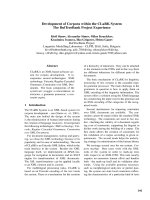
Figure 3 Proteasomal activity in murine lung after proteasomal inhibition. The proteasomes were immunoprecipitated from Cftr
-/-
- and Cftr
+/+
- mice lungs (n = 3), treated with PLGA-PEG
PS341
(10 μg, intranasal), and 200 μM Suc-LLVY-AMC was used as a substrate to quantify the
proteasomal activity in a 96-well plate, in triplicate. Fluorescence intensities were measured at 360 nm excitation and 440 nm emission by
SpectraMax Pro fluorescence plate reader. Recombinant purified proteasome was used as a positive control while no IP served as a negative
control. The data shows that PLGA-PEG mediated PS341 delivery significantly inhibits the proteasomal activity (~2 fold, p < 0.01). The data verifies
the efficacy of PLGA-PEG mediated PS-341 delivery to murine lungs.
Vij et al. Journal of Nanobiotechnology 2010, 8:22
/>Page 6 of 18
Figure 4 Sustained delivery of nile red by PLGA-PEG nanoparticles. The nile red loaded PLGA-PEG nanoparticles were insufflated in Cftr
+/+
(n = 4) mice airway. Live animals were imaged by Xenogen IVIS 200 optical imaging device (Ex 465 nm and Em 525 nm) from day 1 to 11. All
animals were kept under constant supply of isoflurane using an automated anesthesia machine attached to imaging device and handled in
accordance with our JHU ACUC approved animal protocol. We observed significant amount of PLGA-PEG
PS341-NileRed
particles in murine lungs and
bladder (excreted nanoparticles) by 24 hrs and observed its sustained release from days 1 to 11 given the short half-life of the nile red.
Vij et al. Journal of Nanobiotechnology 2010, 8:22
/>Page 7 of 18
targeted delivery into specific sites and across complex
biological barriers. The development of novel nano-sys-
tems for pulmonary gene or drug delivery may provide
a convenient, noninvasive method for the administration
of gene or drugs to the lungs. Such a system can also
facilitate s ustained site directed delivery to specific dis-
ease cell type or tissue bypassing the obstructive patho-
physiological barriers. Mucous hypersecre tion is a
hallmark of chronic obstructive pulmonary disease
(COPD) and cystic fibrosis (CF) [14]. We have pre-
viously shown that proteasomal inhibition by extremely
potent, s table, reversible, and selective inhibitor of chy-
motryptic threonine protease activity, PS341 (Velcade/
Bortezomib) rescues the CF pathophysiology of bron-
chial epithelial cells [9,15].
We and others have recently reported that selective
inhibition of proteasome activity helps in rescue of mis-
folded or partially folded protein by induction of folding
machinery [8,9,16-19] and it is not possible to traffic or
rescue the misfolded protein by inhibiting its ubiquitina-
tion due to presence of redundant ubiquitination path-
ways and lack of enhanced chaperone activity. The
molecular mechanisms by which proteasome inhibitors
or proteostatic re gulators can help in rescue of trans-
membrane proteins have been recently described
[9,16-19]. Moreover, our recent data suggests that selec-
tive proteasome inhibition also helps in controlling
chronic inflammation that will be required for treating
the patients with chronic lung disease, as rescuing mis-
folded CFTR may not be sufficient for favorable
Figure 5 PLGA-PEG mediated cystosolic delivery. The indicated concentrations of PLGA-PEG
PS341-NileR ed
was added to CFBE41o- cells and
incubated for 24 hours. Cells were fixed with 10% neutral buffer formalin and stained with Hoechst dye for nuclear staining. Fluorescence
microscopy was used to capture images of Hoechst staining (DAPI filter) and nile red (Texas Red filter) that shows perinuclear cytosolic
localization of released dye. We show the cytosolic release of nile red in perinuclear space using the PLGA-PEG nanoparticles containing 1000 or
2000 ng dye. The nile red dye added directly to the media at similar concentrations as a negative control did not show any cytosolic delivery
after 24 hrs. The data verifies the efficacy of our novel therapeutic vehicles for bronchial epithelial cell delivery.
Vij et al. Journal of Nanobiotechnology 2010, 8:22
/>Page 8 of 18
Figure 6 Treatment with PLGA-PEG
PS341
attenuates NFB and IL-8 promoter activities. CFBE41o- cells were treated for 24 hours with 100
ng PLGA-PEG
PS-341
and transfected with NFB, IL-8 and/or renila luciferase reporter plasmids. After six hrs of transfection, TNF-a (10 ng/ml) was
used to induce proinflammatory signaling overnight. NFB- and IL-8- firefly and renila luciferase activities were quantified using the Dual
Luciferase® Reporter Assay System. We observed that treatment with the 10 μl of PLGA-PEG
PS341
(10 ng/μl) significantly decreases TNF-a induced
A) NFB and B) IL-8 luciferase activities (*p < 0.05). Data is shown as RLU (Relative Luminescence Intensity) of firefly luciferase promoter activity
normalized to renilla luciferase internal control. The data verifies the efficacy of PLGA-PEG mediated drug delivery and activity.
Vij et al. Journal of Nanobiotechnology 2010, 8:22
/>Page 9 of 18
therapeutic outcome. We confirmed that proteasome
inhibition restrain the IB a degradation [7,8] and hence
NFB-mediated, IL-8 activation [9]. PS-341 can enter
mammalian cells and inhibit NFB activation and
NFB-dependent gene expression. PS-341 is known to
inhibit TNF-a-induced gene expression of the cell-sur-
face adhesion molecules E-selectin, ICAM-1, and
VCAM-1 on primary human umbilical vein endothelial
cells [20,21]. In a rat model of streptococcal cell wall-
induced polyart hritis [22], PS-341 attenuates the
neutrophil-predominant acute phase and markedly inhi-
bits the progression of the T cell-dependent chronic
phase of the inflammatory response [20]. Clearly, thi s
warrants further evaluation and selective delivery of this
class of compounds for treatment of CF lung disease.
We evaluated the efficacy of PLGA based nano-sys-
tems for selective drug delive ry. A major draw back of
PLGA nanoparticles is that when formulated with the
commonly us ed emulsifier polyvinyl alcohol (PVA), they
arehydrophobicandhaveahighnegativechargeon
Figure 7 Systemic treatment with PS-341 attenuates Pa-LPS induced pro -inf lammatory response and neutrophil levels. Cftr
-/-
mice
(n = 3, each group) were treated with Pa-LPS and/or PS-341 by intraperitoneal injection. Control, untreated group, was injected with 100 μl
saline. The serum cytokine levels were quantified by sandwich ELISAs. We observed that treatment with the PLGA-PEG
PS341
decrease Pa-LPS
induced A) IL1-b and B) IL-6 levels indicating that PS-341 can control Pa-LPS induced inflammatory response (*p < 0.05) if delivered efficiently to
the airway. The data indicates that PS-341 can control Pa-LPS induced inflammatory response.
Vij et al. Journal of Nanobiotechnology 2010, 8:22
/>Page 10 of 18
Figure 8 Treatment with PLGA-PEG
PS341
attenuates Pa-LPS induced proinflammatory response and neutrophil levels.TheCftr
-/-
mice
(n = 3, each group) were treated with Pa-LPS and/or PLGA-PEG
PS341
. Control, untreated group, was treated with 10 μl saline. The
bronchoalveolar lavage fluid (BALF) cytokine and myeloperoxidase (MPO), levels were quantified by sandwich ELISAs. The treatment with the
PLGA-PEG
PS341
significantly decreased Pa-LPS induced A) IL1-b, B) IL-6 and C) MPO levels confirming that PLGA-PEG mediated PS-341 delivery
controls Pa-LPS induced inflammatory response and neutrophil chemotaxis (*p < 0.05). The data verifies the efficacy of PLGA-PEG mediated PS-341
drug delivery in controlling Pa-LPS induced lung disease.
Vij et al. Journal of Nanobiotechnology 2010, 8:22
/>Page 11 of 18
their surface. As a result, such a system, when adminis-
tered in experimental animals, is rapidly opsonized by
the de fense system of the body (Reticuloendothelial Sys-
tem, RES or Mononuclear Phagocyte System, MPS; sys-
temic circulation or airway) [10,11]. The most common
waytoovercomethischallengeiscoatingofthedrug
delivery system with the outer layer of polyethylenegly-
col (PEG) that endow these nanoparticles with ‘stealth’ ,
or RES/MPS evading properties [10]. PEGylation also
increases the circulation time of the nanoparticles,
thereby enhancing their propensity of accumulation in
target organs or cells by passive diffusion, taking aid of
the enhanced permeability and retention (EPR) effect
[23]. P EG chai ns, covalently attached with PLGA nano-
particles using ring-opening polymerization method,
results in increased residence in blood (intravenous) or
airway (intranasal) and enhanced accumulation in target
tissues or cells [24]. Nanoparticle mediated drug delivery
presents with the added advantag e of targeting the drug
to specific organs or cells in the body, for example by
conjugating it with a monoclonal antibody that will tar-
get the system speci fically to the CF bronchi al epithelial
cells which over express the complementary antigen
(our ongoing studies). However, until date, the use of
drug loaded PLGA nanoparticles synthesized using the
popular emulsifier PVA has resulted in poor in vivo
drug delivery efficie ncy. It has also been found that such
a formulation ca n never be completely purified of the
emulsifier PVA, which is sus pected of non-specific toxi-
city [25].
In order to develop an improved, clinically viable for-
mulation of PLGA nanoparticles over existing PVA
based o nes, we adopted a strategy used in the synthesis
of PEGylated liposomes and PEGylated immunolipo-
somes, and employed commercially available PEGylated
phospholipids (like Distereolylphos- phatidylethanola-
mine-mPEG2000, or DSPE-mPEG2000) as emulsifiers
[26]. Such molecules have surfactant-like properties, and
spontaneously self-aggregate in aqueous solutions form-
ing micelles [27]. We anticipate based on our studies
that they can function as exc ellent emulsifiers for a
hydrophobic polymeric system like PLGA. The DSPE-
mPEG
2000
emulsifier provides stabilization of PLGA
nanoparticles. We have designed here a novel PLGA-
PEG based biodegradable therapeutic vehicle to provide
sustained release of drug to the airway. The major chal-
lenge in delivery and therapeutic efficacy of nano-deliv-
ery systems in chronic obstructive airway conditions is
severe inflammation and mucous hypersecretion [14,28].
Mucous hypersecretion is a hallmark of several chronic
obstructive airway diseases, including COPD and CF.
Distinct etiologies and inflammatory responses drive
mucous hypersecretion in these diseases. In CF and
COPD, the in flammatory response is neutrophilic and
may be induced by infection or co mponents in cigarette
smoke. Controlling inflammation is at the root of treat-
ment using corticost eroids, antibiotics or other available
drugs in these chronic obstructive inflammatory condi-
tions. Yet despite therapy, challenge is the sustained
delivery of drugs to target cells or tissues. In spite of
wide application of nano-based drug delivery systems in
chronic obstructive airway diseases and variety of other
pulmonary conditions like allergy, asthma, lung cancer
etc, very few are tested till date [14,29-31]. To test the
efficacy of our novel therapeutic drug delivery vehicle
we have tested the sustained release and delivery of
FDA approved proteasome inhibitor drug, PS341 in
murine lungs by its ability to control Pseudomonas aeru-
ginosa LPS induced CF lung disease in murine model. In
this study, we determined that our PLGA-PEG drug
delivery system can not only provide sustained drug
release (day-3) to murine lungs but also control NFB
mediated neutrophil levels and inflammation. Our con-
trol studies using same amount of drug by insufflation,
did not control neutrophil levels indicative of poor bioa-
vailability. Our data s uggest that nanoparticle mediated
Figure 9 Treatment with PLGA-PEG
PS341
attenuates NFB mediated inflammatory response.TheCf tr
-/-
mice (n = 3, each group) were
treated with Pa-LPS and/or PLGA-PEG
PS341
. Control, untreated group, was treated with 10 μl saline. The lung tissue was isolated on day-3 and
total protein extract was used for immunoblotting. The treatment with the PLGA-PEG
PS341
significantly decreases Pa-LPS induced NFB levels
confirming that PLGA-PEG mediated PS-341 delivery controls NFB mediated inflammatory response in murine model. b-actin shows the equal
loading. The data verifies the efficacy of PLGA-PEG mediated PS-341 drug delivery in controlling NFB mediated lung disease.
Vij et al. Journal of Nanobiotechnology 2010, 8:22
/>Page 12 of 18
Figure 10 The PLGA-PEG mediated PS-341 delivery to murine lungs controls Pa-LPS induced oxidative stress.TheCftr
-/-
mice (n = 3,
each group) were treated with Pa-LPS and/or PLGA-PEG
PS341
. The PLGA-PEG
PS341
treated mice exhibited significant increase in Nrf2 (major
antioxidant response transcription factor) expression and nuclear localization leading to decrease in LPS induced oxidative stress as seen by
NOS2 immunostaining. Changes in nuclear (Nrf2) and total protein (NOS2) expression levels are shown in bottom panels (densitometry units).
The PLGA-PEG mediated PS-341 lung delivery controls Pa-LPS induced oxidative stress. Scale: white bar = 50 μm, red bar = 10 μm.
Vij et al. Journal of Nanobiotechnology 2010, 8:22
/>Page 13 of 18
Figure 11 The PLGA-PEG mediated PS-341 delivery to murine lungs controls Pa-LPS induced CF lung inflammation.TheCftr
-/-
mice
(n = 3, each group) were treated with Pa-LPS and/or PLGA-PEG
PS341
. The PLGA-PEG
PS341
treated mice exhibited significant decrease in LPS
induced NFB expression and nuclear localization, and decline in number of inflammatory, macrophages (Mac-3
+
) and neutrophil (NIMP-R14
+
)
cells. Changes in nuclear (NFB) and total protein (Mac-3
+
and NIMP-R14) expression levels are shown in bottom panels (densitometry units).
The PLGA-PEG mediated PS-341 lung delivery controls Pa-LPS induced inflammation. Scale: white bar = 50 μm, red bar = 10 μm, black = 100 μm.
Vij et al. Journal of Nanobiotechnology 2010, 8:22
/>Page 14 of 18
intranasal drug delivery helps in improving the efficacy
of drug by assisting in its lung delivery and
biodistribution.
The PLGA-P EG
PS341
pro vide s con trolled and targeted
drug delivery with selective inhibition of proteasome
mediated homeostatic processes (proteostasis) in lung
epithelia. We observed that inhibition of the proteasome
with PS341 not only rescue ΔF508-CFTR but also IB
from proteasomal degradation [7-9]; hence inhibiting
the NFB mediated- IL-8 secretion in CF [9]. We have
standardized the PLGA-PEG based PS341 delivery to CF
(Cft r
-/-
, F ABP-CFT R gut corre cted) murine l ungs based
on its ability to control Pa-LPS induced lung disease
(Fig 8, 9, 10, 11 and 12) and inhibition of proteasomal
activity (Fig 3). We found that PLGA-PEG mediated
intranasal PS341 de livery, at indicated dose, results in
~2-fold inhibition of proteasomal activity in murine
lungs. In addition, we have verified that intra nasal deliv-
ery of fluorescently labeled PLGA-PEG
NileRed
particles to
murine lungs provide sustain ed release from da y 1-11
(Fig 4). We observed that significant amount of particle
is delivered to murine lungs by 24 hrs of inoculation.
We also evaluated the release chemistry and kinetics of
PLGA-PEG
PS341
(Fig 2A, 2B and 2C) followed by verifi-
cation of functional efficacy (Figs 6, 8, 9, 10, 11 and 12).
Conclusions
We demonstrate here the nanoparticle mediated lung
delivery for treatment of CF. We anticipate that this
studywillhaveahighimpactonthedevelopmentof
novel targeted drug-delivery therapeutics for CF and
other airway diseases like COPD and asthma. The nano-
drug delivery system here provides controlled and sus-
tained PS-341 delivery for selective inhibition of pro-
teostasis. Recent studies have identified several novel
“correctors” and molecular targets for functional rescue
of misfolded CFTR protein or chr onic inflammatory
state in CF but delivery of these drugs to CF epithelia is
a challenge. Thus, furthe r pre-clinical development of
this novel nano -based biodegradable therapeutic vehicle
and verification of its human (CF & COPD) mucus-
penetration ability will have enormous applications in
treatment of chronic pathophysiology of obstructive
lung diseases.
Materials and methods
Cell Culture and Reagents
The CFBE41o- ( cystic fibrosis bronchial epithelia l cell
lines, from Dr. Dieter Gruenert [32,33]) cells were main-
tained in MEM Earl’ s salt L-Glutamine (200 mM L-
Glutamine) medium containing 100 units/ml penicillin,
Figure 12 The PLGA-PEG mediated PS-341 delivery to murine lungs controls Pa-LPS induced CF lung disease.TheCftr
-/-
mice (n = 3,
each group) were treated with Pa-LPS and/or PLGA-PEG
PS341
. H&E staining verifies the rescue from Pa-LPS induced CF lung disease by PLGA-
PEG
PS341
in triplicate samples. The PLGA-PEG mediated PS-341 lung delivery controls Pa-LPS induced CF lung disease. Scale: black = 100 μm.
Vij et al. Journal of Nanobiotechnology 2010, 8:22
/>Page 15 of 18
100 μg/ml streptomycin, 0.25 μg/ml amphotericin B and
10% fetal bovine serum. MEM and other components
were purchased from Invitrogen, Carlsbad, CA. TNF-a
(R&D Systems Inc., Minneapolis, MN), nile red (Invitro-
gen), PS-341 (Millenium Pharmaceuticals, Cambridge,
MA), PLGA (Avanti Polar Lipids, Alabaster, AL), DSPE-
PEG
2000
(Avanti) and Pseudomonas aeruginosa LPS
(Sigma, St. Louis, MO) were added to cells or injected
in mice as indicated. All other common laboratory che-
micals were from Sigma or Fisher Scientific.
PLGA-PEG synthesis
We dissolved calculated amounts of PLGA and PS-341
and/or nile red in acetone and injected it in DSPE-
mPEG
2000
emulsifier dissolved in water or PBS followed
by immediate rigorous emulsification by a high power
sonicator. This result i n the synthesis of PEGylated
nanoparticles (PNPs) of PLGA dispersed in the aqueous
solution, w ith the water-insoluble drug (PS-341) or dye
(nile red) entrapped in the hydrophobic PLGA matrix.
We removed acetone by rotary vacuum evaporation and
purified drug-loaded nanoparticles by ultracentrifugation
followed by rigorous washing (3x) with water or PBS
and resuspension in PBS.
Transmission Electron Microscopy (TEM)
Transmission electron microscopy (TEM) was used to
determine the size, shape and dispersion of PLGA-
PEG
PS341
nanoparti cles using a JEOL JEM-10 0cx micro-
scope at an accelerating voltage of 100 kV. The speci-
mens were prepared by drop-coating the sample
dispersion onto a carbon -coated 300 mesh copper grid,
which was placed on filter paper to absorb excess
solvent.
Dynamic laser scattering (DLS)
Dynamic laser scattering (DLS) was employed to mea-
sure the size distribution and colloidal stability of the
PLGA-PEG
PS341
nanoparticles dispersion in water using
a Brookhaven Instrument 90Plus Particle Size Analyzer
at a wavelength of 633 nm and scattering angle o f 90°.
DLS was also used to examine the colloidal stability of
nanoparticles dispersed in PBS (pH 7.4) over three days.
Release Kinetics and Proteasome Activity Assay
Release kinetics of nile red from PLGA-PEG nanoparti-
cles was quantified by recording absorption of released
dye in resuspension buffer (PBS, 100 μl) at 525 nm
using the VERSAMAX plate reader and SoftMax Pro
software from molecular devices. Nanoparticle samples
were aliquoted and incubated at room temperature in
triplicate for indicated time points and analyze d for nile
red release. We quanti fied the re lease kinetics of PS-341
from PLGA-PEG in resuspension buffer (PBS, 100 μl),
once daily for a period of 7 days, using Proteasomal
Activity Assay from Drug Discovery (BioMol). We
recorded proteasome inhibitory activity of room tem-
perature incubated PLGA-PEG
PS341
nanoparticles from
day 1 to 7 following the manufacturer’ sprotocol.We
similarly quantified the efficacy of drug delivery to
CFBE41o- cells by quantifying proteasomal activities of
cell lysates after 24 hrs of P LGA-PEG
PS341
, PLGA-PEG
(control) or PS341 treatment as indicated. We also
quantified proteasomal activities in murine lungs by
immunoprecipitating (IP) proteasome from lung extracts
(1000 μg) using the proteasome isolation kit (Calbio-
chem) following the manufacturer’s instruc tions. The
200 μM Suc-LLVY-AMC (Calbiochem) was used as a
substrate to estimate chymotrypsin-like proteasomal
activity in a 96-well plate. Fluorescence intensities were
measure d at 360 nm excitation and 440 nm emissio n by
VERSAMax fluorescence plate reader (Molecular
Devices) using the SoftMax Pro software. Recombinant
purified proteasome (BIOMOL) was used as a positive
control while no IP served as a negative control.
Animal Experiments
All animal experiments were carried out in accordance
with the Johns Hopkins University (JHU) Animal Care
and Use Committee (ACUC) approved protocol. To
induce inflammatory lung disease in vivo,theage(~16
weeks) and sex matched, B6- 129S6- Cftr
-/-
(Cftrtm
1Kthc
-
TgN
(FABPCFTR)
) [34,35] inbred mice (n = 3) were treated,
intratracheally (i.t., 10 μg in 100 μl PBS) or intraperito-
neally (i.p., 15 mg/kg/bw in 100 μl PBS) with Pseudomo-
nas aeruginosa (Pa)-LPS, 24 hrs post- PLGA-PEG
PS341
nanoparticle (intranasal, 5 μl/nost ril of 1 μg/μl) or
PS341 (i.p., 0.6 mg/kg/bw in 100 μlPBSfor2days)
administration. Based on a previous report [36,37] and
pilot experiments on the release kinetics and in vivo effi-
cacy of the drug, day-3 time point was selected for eval-
uating the functional efficacy of the drug. Moreover, we
have previously standardized that L PS induced lung
inflammation, at the selected dose, is a t its peak in
Cftr
-/-
mice at 24 hrs [38]. Serum and total lung protein
extracts were isolated at day- 3 after euthanasia in the
presence of anesthesia following our JHU ACUC
approved protocol. The quantification of protein levels
by Western blotting of total lung protein extracts (as
described below), and cytokine levels by ELISA of bro-
choalveolar lavage fluid (BALF)/serum (as described
below) was used to identify the changes in pro-inflam-
matory signaling. For live animal imaging exper iments,
Cftr
+/+
mice insufflated with PLGA-PEG
NileRed
nanopar-
ticles were imaged from day 1-11 using Xenogen IVIS
200 optical imaging device (Ex 465 nm and Em 525 nm)
that was directly connected to automatic anesthesia
machine providing constant supply of isoflurane.
Vij et al. Journal of Nanobiotechnology 2010, 8:22
/>Page 16 of 18
Immunoblotting
Lung tissues were lysed by sonication (three 5 sec
pulses) on ice in cold room using the T-PER (Pierce
Biotech. Inc., Rockfo rd, IL) protein lysis buffer contain-
ing protease-inhibitor cocktail (Pierce). The protein
extracts were suspended in Laemmli’ s sample buffer
(Invitrogen) containing b-mercaptoethanol (Invitro gen),
resolved by 4-10% SDS-PAGE 12-wel l gel (lane- 1, mar-
ker; 2-4, control; 5-7, LPS; 8-9, PLGA-PEG
PS341
loaded
in duplicate to accommodate all samples in single 12-
well gel; 10-12, LPS + PLGA-PEG
PS341
) a nd transferred
to a 0.45 μm pore size nitrocellulose membrane (Invitro-
gen). The b-actin (Sigma) and NFB(SantaCruzBio-
tech Inc., Santa Cruz, CA) p rimary antibod ies, and anti-
rabbit-HRP secondary antibody (Amersham, Piscat away,
NJ) were used for immunoblotting.
Immunostaining
Six-week-old mice (n = 3 per genotype) were euthana-
tize d as described above and lungs were collected. Lung
was fixed in 1 ml 10% neutral buffered formalin over-
night (Fisher Scientific, Pittsburgh, PA), embedded in
paraffin, sectioned, and prepared fo r immunostaining.
Macrophages and neutro phils were immunostained with
the rabbit polyclonal Mac-3 or NIMP-R14 (2 μg/ml) pri-
mary antibody (Abca m, Inc., Cambridge, UK), respec-
tively, followed by a second ary goat anti-rat Alexa Fluor
488, 5 μg/ml (Molecular Probes, Eugene, OR) antibody.
Nrf2, NOS2 and NFB levels were similarly quantified
using polyclonal antibodies from Santa Cruz Biotech
Inc. Negative controls consisted of identical treatments
with the omission of the primary antibody. Hoechst dye,
1 μg/ml (Molecular Probes, Invitrogen) was used for
nuclear staining. The slides were then mounted (Vecta-
shield; Vector Laboratories Inc., Burlingame, CA), and
images were captured as described below. Nuclei were
detected by Hoechst (Invitr ogen) while H&E was used
to evaluate lung morphology and inflammatory state.
Images were captured by Axiov ert 200 Carl Zeiss Fluor-
escence microscope using the Zeiss Axiocam HRC cam-
era and Axiovision software with appropriate filter
settings for FITC and DAPI. All fluorescent images were
captured at room temperature w ith oil (63X, fluores-
cence) and air (20X and 40X) as the imaging medium.
Themagnificationsforthefluorescencemicroscope
were LD Plan- Achroplan (20X/0.40 Korr Phz), Neo
Fluar (40X/0.6X Phz Korr) and Achromat (63X/1.4 oil),
respectively with 1.6X optivar.
IL-1b, IL-6 and MPO Immunoassay
At the indicated time points, BALFs or serum were col-
lected from each mouse as reported earlier [38-40] and
storedat-80Cuntiluse.BALForserumIL-1b levels
were measured using solid-phase ELISA (R&D
Biosystems, Minneapolis, MN). Standards, and high and
low cytokine controls were included. The plates were
read at 450 nm on 96-well microplate reader (Mole cular
Devices, Sunnyvale, CA) using SOFT-MAX-Pro software
(Molecular Devices). The mean blank reading was sub-
tracted from each sample and control reading. The
amount of sub strate turnover was determined calorime-
trically by measuring the absorbance, which is propor-
tional to IL-1b concentration. A standard curve was
plotted and an IL-1b concentra tion in each sample was
determined by interpo lation from standard curve. The
data represents the mean ± SD of triplic ate samples.
The IL-6 cyt okine and myeloperoxidase (MPO) levels
were similarly quantified using an ELISA system (R&D
Biosystems and Hycult Biotech, Canton, MA) as
described before [15].
NFB or IL-8 Reporter Assay
CFBE41o- cells were transfected with NFB- or IL-8-
firefly luciferase promoter (pGL-2) and renila luciferase
(pRLTK) control. Cells were induced with 10 ng/ml of
TNF-a and/or 100 ng/ml PLGA-PEG
PS341
nanoparticles
and luciferase activities were measured after overnight
treatment. Dual-Luciferase® Reporter (DLRTM) Assay
System (Promega) was used to measure NFB- or IL-8-
reporter (firefly luciferase) and renila luciferase activities
from CFBE41o- cell extracts. Data was normalized with
internal renila luciferase control for each sample and
the changes in reporter activities were calculated.
Statistical Analysis
Representative data is shown as the mean ± SD of at
least three experiments. The one-way ANOVA with a
Dunnett planned comparison was run for each sample
versus control. A * p < 0.05 was considered to have sta-
tistical significance. The murine and human microscop y
data was analyzed by densitometry (Matlab R2009b,
Mathworks Co.) and spearman’s correlation coefficient
was used to calculate the significance among the indi-
cated groups.
Acknowledgements
The authors were supported by R025-CR07 and VIJ07IO grants from the
Cystic Fibrosis Foundation, FAMRI, NASA grant NNJ06HI17G, and NIH grants
CTSA UL RR 025005 and RHL096931 (NV). The funders had no role in
decision to publish or preparation of the manuscript.
Author details
1
Department of Pediatric Respiratory Sciences, Johns Hopkins University
School of Medicine, Baltimore, 21287, USA.
2
Institute of NanoBioTechnology,
Johns Hopkins University, Baltimore, 21218, USA.
3
Department of Chemistry,
State University of New York, Buffalo, 14260, USA.
Authors’ contributions
Conceived and designed the experiments: NV. Performed the experiments:
NV, TM, RM, SM, HD, KTY and IR. Analyzed the data: NV & TM. Contributed
reagents, materials and analysis tools: NV & IR. Wrote the paper: NV. Helped
Vij et al. Journal of Nanobiotechnology 2010, 8:22
/>Page 17 of 18
with the editing of the paper: KTY & IR. All authors read and approved the
final manuscript.
Competing interests
The authors declare that they have no competing interests.
Received: 20 June 2010 Accepted: 24 September 2010
Published: 24 September 2010
References
1. Ward CL, Omura S, Kopito RR: Degradation of CFTR by the ubiquitin-
proteasome pathway. Cell 1995, 83:121-127.
2. Mitchell BS: The proteasome–an emerging therapeutic target in cancer.
N Engl J Med 2003, 348:2597-2598.
3. Bross PF, Kane R, Farrell AT, Abraham S, Benson K, Brower ME, Bradley S,
Gobburu JV, Goheer A, Lee SL, et al: Approval summary for bortezomib
for injection in the treatment of multiple myeloma. Clin Cancer Res 2004,
10:3954-3964.
4. Kane RC, Bross PF, Farrell AT, Pazdur R: Velcade: U.S. FDA approval for the
treatment of multiple myeloma progressing on prior therapy. Oncologist
2003, 8:508-513.
5. Adams J: Proteasome inhibition in cancer: development of PS-341. Semin
Oncol 2001, 28:613-619.
6. Zhang LN, Karp P, Gerard CJ, Pastor E, Laux D, Munson K, Yan Z, Liu X,
Godwin S, Thomas CP, et al: Dual therapeutic utility of proteasome
modulating agents for pharmaco-gene therapy of the cystic fibrosis
airway. Mol Ther 2004, 10:990-1002.
7. Dai RM, Chen E, Longo DL, Gorbea CM, Li CC: Involvement of valosin-
containing protein, an ATPase Co-purified with IkappaBalpha and 26 S
proteasome, in ubiquitin-proteasome-mediated degradation of
IkappaBalpha. J Biol Chem 1998, 273:3562-3573.
8. Hideshima T, Chauhan D, Richardson P, Mitsiades C, Mitsiades N, Hayashi T,
Munshi N, Dang L, Castro A, Palombella V, et al: NF-kappa B as a
therapeutic target in multiple myeloma. J Biol Chem 2002,
277:16639-16647.
9. Vij N, Fang S, Zeitlin PL: Selective Inhibition of Endoplasmic Reticulum-
associated Degradation Rescues {Delta}F508-Cystic Fibrosis
Transmembrane Regulator and Suppresses Interleukin-8 Levels:
THERAPEUTIC IMPLICATIONS. J Biol Chem 2006, 281:17369-17378.
10. Davis SS: Biomedical applications of nanotechnology–implications for
drug targeting and gene therapy. Trends Biotechnol 1997, 15:217-224.
11. Panyam J, Labhasetwar V: Biodegradable nanoparticles for drug and gene
delivery to cells and tissue. Adv Drug Deliv Rev 2003, 55:329-347.
12. Qaddoumi MG, Ueda H, Yang J, Davda J, Labhasetwar V, Lee VH: The
characteristics and mechanisms of uptake of PLGA nanoparticles in
rabbit conjunctival epithelial cell layers. Pharm Res 2004, 21:641-648.
13. Cartiera MS, Johnson KM, Rajendran V, Caplan MJ, Saltzman WM: The
uptake and intracellular fate of PLGA nanoparticles in epithelial cells.
Biomaterials
2009, 30:2790-2798.
14. Roy I, Vij N: Nanodelivery in airway diseases: challenges and therapeutic
applications. Nanomedicine 2010, 6:237-244.
15. Vij N, Amoako MO, Mazur S, Zeitlin PL: CHOP transcription factor mediates
IL-8 signaling in cystic fibrosis bronchial epithelial cells. Am J Respir Cell
Mol Biol 2008, 38:176-184.
16. Vij N: AAA ATPase p97/VCP: cellular functions, disease and therapeutic
potential. J Cell Mol Med 2008, 12:2511-2518.
17. Vij N, Mazur S, Zeitlin PL: VCP is involved in ERAD and aggresome
formation of ΔF508-CFTR. Pediatric Pulmonology 2006, 41:209.
18. Balch WE, Morimoto RI, Dillin A, Kelly JW: Adapting proteostasis for
disease intervention. Science 2008, 319:916-919.
19. Mu TW, Ong DS, Wang YJ, Balch WE, Yates JR, Segatori L, Kelly JW:
Chemical and biological approaches synergize to ameliorate protein-
folding diseases. Cell 2008, 134:769-781.
20. Palombella VJ, Conner EM, Fuseler JW, Destree A, Davis JM, Laroux FS,
Wolf RE, Huang J, Brand S, Elliott PJ, et al: Role of the proteasome and NF-
kappaB in streptococcal cell wall-induced polyarthritis. Proc Natl Acad Sci
USA 1998, 95:15671-15676.
21. Read MA, Neish AS, Luscinskas FW, Palombella VJ, Maniatis T, Collins T: The
proteasome pathway is required for cytokine-induced endothelial-
leukocyte adhesion molecule expression. Immunity 1995, 2:493-506.
22. Cromartie WJ, Craddock JG, Schwab JH, Anderle SK, Yang CH: Arthritis in
rats after systemic injection of streptococcal cells or cell walls. J Exp Med
1977, 146:1585-1602.
23. Fang J, Sawa T, Maeda H: Factors and mechanism of “EPR” effect and the
enhanced antitumor effects of macromolecular drugs including
SMANCS. Adv Exp Med Biol 2003, 519:29-49.
24. Gref R, Minamitake Y, Peracchia MT, Trubetskoy V, Torchilin V, Langer R:
Biodegradable long-circulating polymeric nanospheres. Science 1994,
263:1600-1603.
25. Sahoo SK, Panyam J, Prabha S, Labhasetwar V: Residual polyvinyl alcohol
associated with poly (D, L-lactide-co-glycolide) nanoparticles affects their
physical properties and cellular uptake. J Control Release 2002, 82:105-114.
26. Huwyler J, Wu D, Pardridge WM: Brain drug delivery of small molecules
using immunoliposomes.
Proc Natl Acad Sci USA 1996, 93:14164-14169.
27. Lukyanov AN, Torchilin VP: Micelles from lipid derivatives of water-soluble
polymers as delivery systems for poorly soluble drugs. Adv Drug Deliv Rev
2004, 56:1273-1289.
28. Wine JJ: The genesis of cystic fibrosis lung disease. J Clin Invest 1999,
103:309-312.
29. Rytting E, Nguyen J, Wang X, Kissel T: Biodegradable polymeric
nanocarriers for pulmonary drug delivery. Expert Opin Drug Deliv 2008,
5:629-639.
30. Yang W, Peters JI, Williams RO: Inhaled nanoparticles–a current review. Int
J Pharm 2008, 356:239-247.
31. Zhang W, Yang H, Kong X, Mohapatra S, San Juan-Vergara H, Hellermann G,
Behera S, Singam R, Lockey RF, Mohapatra SS: Inhibition of respiratory
syncytial virus infection with intranasal siRNA nanoparticles targeting
the viral NS1 gene. Nat Med 2005, 11:56-62.
32. Cozens AL, Yezzi MJ, Kunzelmann K, Ohrui T, Chin L, Eng K, Finkbeiner WE,
Widdicombe JH, Gruenert DC: CFTR expression and chloride secretion in
polarized immortal human bronchial epithelial cells. Am J Respir Cell Mol
Biol 1994, 10:38-47.
33. Bruscia E, Sangiuolo F, Sinibaldi P, Goncz KK, Novelli G, Gruenert DC:
Isolation of CF cell lines corrected at DeltaF508-CFTR locus by SFHR-
mediated targeting. Gene Ther 2002, 9:683-685.
34. Van Heeckeren AM, Scaria A, Schluchter MD, Ferkol TW, Wadsworth S,
Davis PB: Delivery of CFTR by adenoviral vector to cystic fibrosis mouse
lung in a model of chronic Pseudomonas aeruginosa lung infection. Am
J Physiol Lung Cell Mol Physiol 2004, 286:L717-726.
35. van Heeckeren AM, Schluchter MD, Xue W, Davis PB: Response to acute
lung infection with mucoid Pseudomonas aeruginosa in cystic fibrosis
mice. Am J Respir Crit Care Med 2006, 173:288-296.
36. Elliott PJ, Ross JS: The proteasome: a new target for novel drug therapies.
Am J Clin Pathol 2001, 116:637-646.
37. Lee SW, Kim JH, Park YB, Lee SK: Bortezomib attenuates murine collagen-
induced arthritis. Ann Rheum Dis 2009, 68:1761-1767.
38. Vij N, Mazur S, Zeitlin PL: CFTR is a negative regulator of NFkappaB
mediated innate immune response. PLoS ONE 2009, 4:e4664.
39. Wu F, Vij N, Roberts L, Lopez S, Joyce S, Chakravarti S: A novel role of the
lumican core protein in bacterial lipopolysaccharide-induced innate
immune response. J Biol Chem 2007, 7;282(36):26409-17.
40. Singh G, Katyal SL:
An immunologic study of the secretory products of
rat Clara cells. J Histochem Cytochem 1984, 32:49-54.
doi:10.1186/1477-3155-8-22
Cite this article as: Vij et al.: Development of PEGylated PLGA
nanoparticle for controlled and sustained drug delivery in cystic
fibrosis. Journal of Nanobiotechnology 2010 8:22.
Vij et al. Journal of Nanobiotechnology 2010, 8:22
/>Page 18 of 18