báo cáo khoa học: " Absorption and translocation to the aerial part of magnetic carbon-coated nanoparticles through the root of different crop plants" potx
Bạn đang xem bản rút gọn của tài liệu. Xem và tải ngay bản đầy đủ của tài liệu tại đây (1.41 MB, 8 trang )
SHOR T COMM U N I C A TION Open Access
Absorption and translocation to the aerial part of
magnetic carbon-coated nanoparticles through
the root of different crop plants
Zuny Cifuentes
1
, Laura Custardoy
2,3,4
, Jesús M de la Fuente
4
, Clara Marquina
2,3
, M Ricardo Ibarra
3,4
,
Diego Rubiales
5
, Alejandro Pérez-de-Luque
1*
Abstract
The development of nanodevices for agriculture and plant research will allow several new applications, ranging
from treatments with agrochemicals to delivery of nucleic acids for genetic transformation. But a long way for
research is still in front of us until such nanodevices could be widely used. Their behaviour inside the plants is not
yet well known and the putative toxic effects for both, the plants directly exposed and/or the animals and
humans, if the nanodevices reach the food chain, remain uncertain. In this work we show that magnetic carbon-
coated nanoparticles forming a biocompatible magnetic fluid (bioferrofluid) can easily penetrate through the root
in four different crop plants (pea, sunflower, tomato and wheat). They reach the vascular cylinder, move using the
transpiration stream in the xylem vessels and spread through the aerial part of the plants in less than 24 hours.
Accumulation of nanoparticles was detected in wheat leaf trichomes, suggesting a way for excretion/detoxification.
This kind of studies is of great interest in order to unveil the movement and accumulation of nanoparticles in plant
tissues for assessing further applications in the field or laboratory.
Background
Several areas, such as medicine, materials science and
electronics, have begun to benefit and apply nanotech-
nology for their research since some decades ago. How-
ever, only during the recent years, researchers from
other disciplines start to see the potential applications of
nanoscience, as it is the case of agriculture [1]. Nano-
sensors, smart delivery systems and nanomaterials (as
for example, nanoparticles) appear as the most promis-
ing devices for application in agriculture and food
industry. For example, using smart delivery systems in
agriculture and plant research will open up new possibi-
lities for multiple applications, from agrochemical treat-
ments to genetic transformation [2,3]. However, it is not
easy to adapt a technology developed for animals and
humans to the plant kingdom. Effective means of nano-
particles application should be identified, and the
behaviour, and their movem ent and accumulation
within the plants should be understood.
During the last years, some works have been published
about absorption and uptake of nanoparticles by plants,
but mainly dealing with and focused on their putative
adverse effects [4-8]. Nevertheless, in order to use nano-
particles as potential smart delivery systems, more sys-
tematic studies are needed to unveil the transport routes,
the organs and tissues where nanoparticles tend to accu-
mulate, and if there are differences regarding plant spe-
cies and the kind of nanoparticles used. Such studies are
important not only from the point of view of the applica-
tion of nanoparticles in plants, but also for understanding
putative toxic effects on plants and the possibilities of
such nanodevices to accumulate in fruits and grains for
further entry into the food chain.
In a previous research, we analyzed the penetration and
transportation of magnetic carbon-coated nanoparticles
through the leaves and aerial part of the plant in cucum-
ber (Cucurbita pepo) [9,10]. The magnetic nature of our
nanoparticles would allow further multiple applications
once the nanopartic les are inside the plants. For example,
* Correspondence:
1
IFAPA, Centro Alameda del Obispo, Área de Mejora y Biotecnología, Avda.
Menédez Pidal s/n, PO Box 3092, Córdoba, 14004 Spain
Full list of author information is available at the end of the article
Cifuentes et al. Journal of Nanobiotechnology 2010, 8:26
/>© 2010 Cifuentes et al; licensee BioMed Central Ltd. This is an Open Access article distributed under the terms of the Creative
Commons Attribution License (http://creativ ecommons.org/licenses/by/2 .0), which permits unrestricted use, distribution, and
reprodu ction in any m edium, provided the original work is properly cited.
the nanoparticles could be moved or immobilized in cer-
tain areas or tissues [9] applying a magnetic field, for
delivering substa nces (drugs,DNA,etc.).Inaddition,
they could work as contrast agents for magnetic reso-
nance imaging (MRI) and be used for in vivo monito ring
the movement and distribution of the nanoparticl es (and
their eventual load) inside the plant, enhancing such kind
of studies [11]. Furthermo re, hyperthermi a [12] might be
used for treatment of, for example, localized parts of
trees affected by diseases or insect attacks. However,
prior to th e development of such applications a deep
understanding on nanoparticle penetration and move-
ment within the plant is needed.
In the present work, we have studied the absorption
and translocation of magnetic carbon-coated nanoparti-
cles through t he root in four crop plants belonging to
different families: sunflower (Helianthus annuus)from
the family Compositae; tomato (Lycopersicum sculen-
tum)fromtheSolanaceae;pea(Pisum sativum), from
the Fabaceae; and wheat (Triticum aestivum), from the
Triticeae.
Methods
The same kind of carbon-coated iron nanoparticles used
in previous studies [9,10] were produced in an arc-dis-
charge furnace [13] based on the previously designed by
Krätschmer-Huffman in 1990 [14]. Our arc-discharge
furnace c onsist of a cylindrical chamber, in which there
are two graphite electrodes: a stationary anode contain-
ing 10 μm diameter iron powders, and a moveable gra-
phite c athode. An arc is produced between the graphite
electrodes in a helium atmosphere. The graphite elec-
trode is sublimed and builds up a powder deposit (soot)
on the inner surface of the chamber. In this material we
found: carbon nanostructures (as for example carbon
nanotubes, amorphous carbon etc) and iron and iron
oxides nanoparticles encapsulated in graphitic layers
(leading to a par ticle- diameter size distribution centred
at approximately 10 nm), together with a small amount
of non-coated or partially coated metallic particles.
These particles (which are not biocompatible) were
eliminated by chemical etching, washing the soot with
HCl 3M at 8 0°C. This procedure favours the formation
of carboxylic groups on the graphitic shell, which, due
to their hydrophobic nature, will contribute to the stabi-
lity of the final particle suspension. In order to eliminate
the amorphous carbon and therefore increase the con-
centration of magnetic nanoparticles, a magnetic purifi-
cation of the powder is carried out. For this purpose,
stable suspensions of the particles are prepared in a sur-
factant solution: 2.5 g of SDS in 500 ml of distilled
water. A field gradient produced by 3KOe permanent
magnet was used for magnetic separation of this suspen-
sion. The resultant powder was several times washed in
water, before proceeding to nanoparticles suspension i n
manitol solution (1%) and further application.
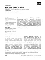
HR-TEM images of the powder samples produced by
arc discharge show spherical magnetic nanoparticles
encapsulated in several layers of graphitic carbon, and sur-
rounded by amorphous carbon (Figure 1a). HR-TEM also
makes it possible to view the atomic planes of the nano-
particle metallic core (Figure 1b). The diameter of the par-
ticles has also been obtained. By analysing several images,
the diameter probability distr ibution function can be
obtained and plotted as a size distribution histogram. The
powder sample produced by arc discharge contains parti-
cles of diameters ranging from 5 nm up to 50 nm, with
the centre of the distribution at 10 nm. HR-TEM shows
that after chemical etching the coating of the magnetic
particles is complete. Hydrodynamic size was measured by
Dynamic Light Scattering technique (Beckman Coulter N5
particle size analyser). The measurements showed that the
carbon-coated magnetic particles in solution form aggre-
gates ranging from 5 nm to several hundred nanometers,
being the average hydrodynamic diameter 200 nm (See
Additional file 1: Hydrodynamic size).
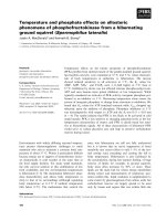
The plants were grown in vi tro using a Petri dish sys-
tem (rhizotron) allowi ng visualizing the roots [15].
When the plantlets developed the second pair of leaves
in each species, nanoparticles [16,17] w ere applied to
the roots as a suspension in manitol solutio n (1%) (Fig-
ure 2) immersing only some roots of each plantlet in
the bioferrofluid. This allowed later to check if the
nanoparticles could move to other roots. Samples of tis-
sues from different parts of the plants (Figure 2) were
taken after 24 and 48 hours and fixed for further micro-
scopic analysis. Sections of samples were obtained either
by using a vibratome or by hand cut, avoiding embed-
ding and washing of nanoparticles from the tissues. Tak-
ing advantage of the black colour that present the
bioferrofluid, a conventional light micro scopy technique
was used to follow its distribution, without observation
of single nanoparticles or small aggregates, which
requires electronic microscopy.
Results and discussion
Firstly we assessed that bioferrofluid was able to pene-
trate into the treated roots and to reach the vascular
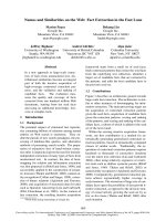
cylinder in a short period of time. Study of the samples
taken at the point of application showed that after only
24 hours of exposure to the bioferrofluid, nanoparticles
were able to leak into the vascular tissues of the tested
crops (Figure 3). This i ndicates t hat applicati on by
immers ing the roots into nanoparticle solutions is faster
and more reliable in order to get big amounts o f nano-
particles inside the plant, than applying the bioferrofluid
through the le aves and aerial parts by pulverization or
injection [9,10].
Cifuentes et al. Journal of Nanobiotechnology 2010, 8:26
/>Page 2 of 8
At this point, there are no studies about the real
mechanism by which nanoparticles can penetrate into
the plant cells. However, there is a recent work dealing
with internalization of gold nanopa rticles using tobacco
protoplasts [18]. In such paper, the authors describe
how gold nanoparticles penetrated into the protoplasts
by endocytosis and were linked to different pathways
upon their charge, incl uding a clath rin- dependent path-
way. So endocytosis appears as a reasonable way for
internalization of nanoparticles. In fact, in a previous
work [10] we found that internalized nanoparticles accu-
mulate in clusters inside the cells, and despite cell mem-
branes were not observed because the fixation method
didn’ t preserve them, probably the nanoparticles are
inside vesicles or cell organelles. In addition, the nano-
particles were suspended in mannitol, a solution more
suitable for plants than gelafundin, and there are reports
about enhancement of endocytosis by mannitol [19].
A recent paper [20] deals with penetration of gold nano-
particles through lipid membranes bypassing endocyto-
sis. However, this entry way, although possible in the
case of our carbon coated nanoparticles, is likely not
common, because in such case a strong cytotoxicity
(and probably phytotoxicity) should be observed.
Nanoparticles were detected easily in the xylem vessels
of the four crops studied, but some differences were
observed among species. Pea roots accumulated higher
contents of bioferrofluid (Figure 3a) than sunflower or
wheat, for example. This difference still remained after
48 hours of exposure to bioferrofluid (Figure 3d-f), sug-
gesting that pea roots could be more permeable to nano-
particle penetration or that there is a lower transportation
Figure 1 TEM images at 300 kV using the cs image corrector (CEOS). a) Nanoparticles encapsulat ed in several layers of graphitic carbon,
and surrounded by amorphous carbon. b) Detail showing the atomic planes of the nanoparticle metallic core.
Figure 2 Schematic representation of the Petri dish rizhotron with the four crops: a) pea; b) sunflower; c) tomato; d) wheat.Squares
indicates sampling points of plant tissues.
Cifuentes et al. Journal of Nanobiotechnology 2010, 8:26
/>Page 3 of 8
rate towards other plant parts, involving higher accumula-
tion of nanoparticles at the application point.
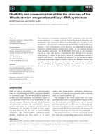
After a successful uptake of the nanoparticles by the
plant roots, we monitored the translocation of such
nanoparticle s into the aerial part. Figure 4a-h shows
sections of the plant crow n belonging to th e four crop
species after 24 and 48 hours of expo sure to the biof er-
rofluid. The black deposit corresponding to the nano-
particles was clearly visible in the xylem vessels after 24
hours (Figure 4a-d). It implies that the nanoparticles
had quickly mo ved towards the aerial part of the plants
following the transpiration stream. Differential response
among crop species was also noticed for nanoparticle
translocation. Pea and wheat showed a high c oncentra-
tion of nanoparticles in the vascular tissues of the
crown, whereas the presence of the bioferrofluid was
less intense in tomato and sunflower. After 48 hours the
nanoparticles were detected in cortical tissue from the
crown of pea and wheat (Figure 4e, 4h) and even some
cells in the cortex of tomato (Figure 4g), whereas no
bioferrofluid was detected outside the vascular tissues of
sunflower. This fact supports the idea that high amounts
of nanoparticles penetrate quickly in the pea root and
move into the aerial part, not being accumulated in the
roots by a high transportation rate as suggested above.
In the case of sunfl ower, it seems that the nanoparticles
uptake through the roots is much slower than in the
other species, and for that reason there is a lower accu-
mulation after 24 hours of treatment. In addition, the
bioferrofluid seems to be more restricted to the vascular
tissues than in the other species.
Subsequent sections of upper parts of the plants con-
firmed that nanopartic les had spread and reached most
of the aerial part after 24 hours of exposure to the bio-
ferrofluid. Following the same pattern, accumulation of
nanoparticles was detected in xylem vessels correspond-
ing to the first (Figure 4i-k) and second (Figure 4o-q)
internodes of the crops. Again, a higher presence of
Figure 3 Longitudinal sections of roots of pea (a, d), sunflower (b, e) and wheat (c, f). Arrows indicate accumulation of bioferrofluid in the
cells. *, xylem containing ferrofluid; #, parenchimatic cell containing ferrofluid; p, parenchimatic cells; x, xylem vessels. Scale bars: a) and f),50μm;
b) and e), 100 μm; c) and d), 25 μm.
Cifuentes et al. Journal of Nanobiotechnology 2010, 8:26
/>Page 4 of 8
Figure 4 Sections from different samples of the aerial parts of pea (a,e,i,l,o,r), sunflower (b,f,j,m,p,s), tomato (c,g,k,n) and wheat (d,h,q,
t). a) Detail of the crown of pea after 24 h of exposure to bioferrofluid. b) Idem in sunflower. c) Idem in tomato. d) Crown of wheat after 24 h of
exposure, showing an intense accumulation of bioferrofluid in tissues. e) Detail of the crown of pea after 48 h of exposure to bioferrofluid.
f) Idem in sunflower. g) Idem in tomato. h) Detail of a longitudinal section in wheat after 48 h of exposure to bioferrofluid. i) Detail of a cross
section of the first internode of pea after 24 h of exposure to bioferrofluid. j) Idem in sunflower. k) Idem in tomato. l) Detail of a cross section of
the first internode of pea after 48 h of exposure to bioferrofluid. m) Idem in sunflower. n) Idem in tomato. o) Detail of a cross section of the
second internode of pea after 24 h of exposure to bioferrofluid. p) Idem in sunflower. q) Detail of a longitudinal section of the second internode
in wheat after 24 h of exposure to bioferrofluid. r) Detail of a cross section of the second internode of pea after 48 h of exposure to
bioferrofluid. s) Idem in sunflower. t) Detail of a longitudinal section of the second internode in wheat after 48 h of exposure to bioferrofluid.
Scale bars represent 100 μm, except in g), q) and t) whereas it represents 50 μm. Arrows indicate accumulation of nanoparticles in vascular
tissues in a-c), f), i-t), and in cortical cells in e), g), h). Arrowheads indicate accumulation of nanoparticles in cortical cells in a), l), r), and in
trichomes in q). Asterisks (*) indicate localization of vascular bundles.
Cifuentes et al. Journal of Nanobiotechnology 2010, 8:26
/>Page 5 of 8
bioferrofluid was detected in pea and wheat compared
with tomato and sunflower. However, such difference
tends to disappear after 48 hours o f exposure, s howing
an intense accumulation of nanoparticles in all the
crops (Figure 4l-n, r-t). The bioferrofluid moved also
towards the leaves and was detected in leaf petioles
(Figure 5a).
It is remarkable that nanoparticles strongly accumu-
lated in leaf trichomes of wheat plants (Figure 5b). The
presence of nanoparticles in this kind of stru ctures (tri-
chomes) has been previously reported [10], but never in
such a high amount, nor in the other crop species.
Because trichomes can play a secretory function [21], it
is possible that this accumulation of nanoparticles inside
them indicates a putative detoxifying pathway in wheat.
The reasons for the differences in accumulation of
nanoparticles in trichomes are unclear, but we think
that should be due to differences in the physiology of
the plants: wheat belongs to the monocot group of
plants, whereas the other three crops are dicots. It is
known that different plant species show different beha-
viour regarding accumulation and excretion of heavy
metals [22], so it is not surprising that such differences
can also be found regarding metal nanoparticles.
Finally, the presence of nanoparticles in roots
not exposed directly to the bioferrofluid was checked
(Figure 5c-e). The characteristic black deposit was
detected within the central cylinder of roots located
diametrically opposite to the treated roots. These data
suggest that nanoparticles had moved not only
upwards through the xylem vessels following the tran-
spiration stream, but also downwards, probably
through the phloem and using the source-sink pressure
gradient [23]. In fact, previous works have shown the
Figure 5 Sections from a pea petiole (a), wheat leaf (b), and pea (c), sunflower (d) and tomato (e) roots. a) Detail of a cross section of a
petiole from the first internode of pea after 24 h of exposure to bioferrofluid. Arrows indicate accumulation of nanoparticles in vascular tissues.
b) Detail of a longitudinal view of wheat leaf showing accumulation of bioferrofluid in trichomes. c) Detail of a longitudinal section of a root of
pea not immersed into the bioferrofluid and after 48 h of exposure to bioferrofluid of opposite roots. Arrows indicate accumulation of
nanoparticles in vascular tissues. d) Idem in sunflower. e) Idem in tomato. *, xylem containing ferrofluid; #, parenchimatic cell containing
ferrofluid; p, parenchimatic cells; x, xylem vessels. Scale bars represent 100 μm, except in d) whereas it represents 50 μm.
Cifuentes et al. Journal of Nanobiotechnology 2010, 8:26
/>Page 6 of 8
translocation of nanoparticles applied on the aerial
part of the plants into the roots [9], and there are evi-
dences that radial transport from cell to cell occurs
[10], which may involve the trafficking pathway to
plasmodesmata. Once the nanoparticles are inside the
cells, they can be transported via endosomes toward
other areas and discharged outside the cells by exocy-
tosis. In that case, Rab proteins should be involved in
the process and direct the cargo to specific areas near
plasmodesmata locations [24]. This mechanism allows
transportation through the cell and would secure a
pass through the endodermal cells, avoiding the Cas-
parian strip. However, movement via apoplast of the
nanoparticles is compatible with the previous mechan-
ism, but the nanoparticles should enter the symplast
way once they reach the endodermis and the Casparian
strip.
Because these microscopic techniques allow observation
only with low resolution, the bioferrofluid was usually
visualized inside xylem vessels where big accumulations of
nanoparticles took place. However, 48 hours after roots
exposure to bioferrofluid, nanoparticles were also detected
in vascular and cortical parenchimatic cells of the plants
(Figure 4e, g, h, l, r). As stated above, this is also in accor-
dance with previous reports about radial transport of car-
bon-coated magnetic nanoparticles between neighbouring
cells [10], and indicates that radial transport allows the
movement of nanoparticles outside the vascular tissues.
Detailed studies using electronic microscopy are underway
in order to unveil the nature of this transportation.
In summary, in this work we have presented results
showing how carbon-coated magnetic nanop articles can
be absorbed by the root system of four different crop
plants and spread using the vascular system to reach the
wholeplant.Therearedifferencesinthespeedof
absorption and distribution of the nanoparticles depend-
ing on the species, being faster in pea and wheat than in
tomato and sunflower. In addition, it seems that sun-
flower shows a lower capability for radial movement of
bioferrofluid outside the vascular tissues than the other
crops. Within the first 24 hour of exposure to the sus-
pension, the nanoparticles can reach the upper part of
the plants, and in the case of wheat they accumulate
inside leaf trichomes. After 48 hours of exposure, the
bioferrofluid is located outside the vascular tissues (pea,
tomato and wheat) and has moved downwards t o non
treated roots. This fast movement of the nanoparticles
inside the plants can have an important impact for the
development of nanoparticles as smart delivery systems
inside the plant and further studies about their distribu-
tion and accumulation. It seems clear that root applica-
tion is faster and more reliable than leaf treatments
[9,10]. This might have implications in toxicity studies,
because the way the nanoparticles are applied to the
plants can strongly affect the final result. Further studies
are needed to assess the effects of plant organs like
flowers or fruits which te nd to act as strong sink of
plant resources (water and nutrients). There is a recent
report [8] showing that fullerene nanoparticles can pass
into the next generation of rice plants, which necessarily
implies accumulation within the rice grains. Would that
happen with bigger nanoparticles or nanomaterials
synthesized with other components (i.e. starch, chitin,
other metals )? In addition, despite the fact that plants
could toler ate the presence of nanoparticles insid e their
tissues, an important question to be addressed is what
happens with such nanoparticles if they move into th e
food chain. Could nanoparticles accumulated in a fruit/
grain survive and pass through the digestive system of
animals into the bloodstream?
Additional material
Additional file 1: Hydrodynamic size. The data show the
hydrodynamic size of the nanoparticles measured by Dynamic Light
Scattering technique.
Acknowledgements
This research was supported by the projects granted by the Spanish Ministry
of Science and Innovation (MICINN) AGL2008-01467 and EUI2008-00114, and
by ARAID fundation.
Author details
1
IFAPA, Centro Alameda del Obispo, Área de Mejora y Biotecnología, Avda.
Menédez Pidal s/n, PO Box 3092, Córdoba, 14004 Spain.
2
Instituto de Ciencia
de Materiales de Aragón (ICMA), CSIC-Universidad de Zaragoza, Pedro
Cerbuna 12, Zaragoza, 50009 Spain.
3
Departamento de Física de la Materia
Condensada, Universidad de Zaragoza, Pedro Cerbuna 12, Zaragoza, 50009
Spain.
4
Instituto de Nanociencia de Aragón, Universidad de Zaragoza,
Campus Rio Ebro, Edificio i+d+i, Mariano Esquillor s/n. Zaragoza, 50018
Spain.
5
CSIC, Instituto de Agricultura Sostenible, Alameda del Obispo s/n, PO
Box 4084, Córdoba, 14080 Spain.
Authors’ contributions
ZC carried out the nanoparticle treatments to the plants and the microscopy
study, the processing of plant samples, and wrote the first manuscript draft.
LC carried out the synthesis of nanoparticles and the bioferrofluid
suspension. CM and MRI participated in the design of the nanoparticle
synthesis and preparation of the suspension, in the design of the study and
to the writing of parts of the manuscript. JMF contributed to the
experimental design of nanoparticle synthesis and to the writing of parts of
the manuscript. DR participated in the design of the study and helped in
experiments of nanoparticle treatments to the plants. APL conceived the
study, participated in the design and coordination of the work and helped
to draft the manuscript. All authors read and approved the final manuscript.
Competing interests
The authors declare that they have no competing interest s.
Received: 27 July 2010 Accepted: 8 November 2010
Published: 8 November 2010
References
1. Robinson DKR, Morrison M: Nanotechnology Developments for the
Agrifood sector- Report of the ObservatoryNANO. Institute of
Nanotechnology, UK; 2009 [ />Cifuentes et al. Journal of Nanobiotechnology 2010, 8:26
/>Page 7 of 8
2. Pérez-de-Luque A, Rubiales D: Nanotechnology for parasitic plant control.
Pest Manag Sci 2009, 65:540-545.
3. Torney F, Trewyn BG, Lin VSY, Wang K: Mesoporous silica nanoparticles
deliver DNA and chemicals into plants. Nature Nanotechnol 2007,
2:295-300.
4. Lin D, Xing B: Phytotoxicity of nanoparticles: Inhibition of seed
germination and root growth. Environ Pollut 2007, 150:243-250.
5. Lin D, Xing B: Root uptake and phytotoxicity of ZnO nanoparticles.
Environ Sci Technol 2008, 42:5580-5585.
6. Zhu H, Han J, Xiao JQ, Jin Y: Uptake, translocation, and accumulation of
manufactured iron oxide nanoparticles by pumpkin plants. J Environ
Monitor 2008, 10:713-717.
7. Khodakovskaya M, Dervishi E, Mahmood M, Xu Y, Li Z, Watanabe F, Biris AS:
Carbon nanotubes are able to penetrate plant seed coat and
dramatically affect seed germination and plant growth. ACS Nano 2009,
3:3221-3227.
8. Lin S, Reppert J, Hu Q, Hudson JS, Reid ML, Ratnikova TA, Rao AM, Luo H,
Ke PC: Uptake, translocation, and transmission of carbon nanomaterials
in rice plants. Small 2009, 5:1128-1132.
9. González-Melendi P, Fernández-Pacheco R, Coronado MJ, Corredor E,
Testillano PS, Risueño MC, Marquina C, Ibarra MR, Rubiales D, Pérez-de-
Luque A: Nanoparticles as smart treatment-delivery systems in plants:
assessment of different techniques of microscopy for their visualization
in plant tissues. Ann Bot 2008, 101:187-195.
10. Corredor E, Testillano PS, Coronado MJ, González-Melendi P, Fernández-
Pacheco R, Marquina C, Ibarra MR, de la Fuente JM, Rubiales D, Pérez-de-
Luque A, Risueño MC: Nanoparticle penetration and transport in living
pumpkin plants: in situ subcellular identification. BMC Plant Biol 2009,
9:45.
11. Scheenen TWJ, Vergeldt FJ, Heemskerk AM, Van As H: Intact plant
magnetic resonance imaging to study dynamics in long-distance sap
flow and flow-conducting surface area. Plant Physiol 2007, 144:1157-1165.
12. Gallego O, Puntes V: What can nanotechnology do to fight cancer? Clin
Transl Oncol 2006, 8:788-795.
13. De Teresa JM, Marquina C, Serrate D, Fernández-Pacheco R, Morellon L,
Algarabel PA, Ibarra MR: From magnetoelectronic to biomedical
applications based on the nanoscale properties of advanced magnetic
materials. Int J Nanotech 2005, 2:3-22.
14. Kratschmer W, Lamb LD, Fostiropoulos K, Huffman DR: Solid C
60
: a new
form of carbon. Nature 1990, 347:354-358.
15. Pérez-de-Luque A, Jorrín JJ, Cubero JI, Rubiales D: Orobanche crenata
resistance and avoidance in pea (Pisum spp.) operate at different
developmental stages of the parasite. Weed Res 2005, 45:379-387.
16. De Teresa JM, Marquina C, Serrate D, Fernández-Pacheco R, Morellon L,
Algarabel PA, Ibarra MR: From magnetoelectronic to biomedical
applications based on the nanoscale properties of advanced magnetic
materials. Int J Nanotech 2005, 2:3-22.
17. Kratschmer W, Lamb LD, Fostiropoulos K, Huffman DR: Solid C60: a new
form of carbon. Nature 1990, 347:354-358.
18. Onelly E, Prescianotto-Baschong C, Caccianiga M, Moscatelli A: Clathrin-
dependent and independent endocytic pathways in tobacco protoplasts
revealed by labelling with charged nanogold. J Exp Bot 2008,
59:3051-3068.
19. Etxeberria E, Gonzalez P, Pozueta-Romero J: Mannitol-enhanced, fluid-
phase endocytosis in storage parenchyma cells of celery (Apium
graveolens; Apiaceae) petioles. Am J Bot 2007, 94:1041-1045.
20. Lin J, Zhang H, Chen Z, Zheng Y: Penetration of lipid membranes by gold
nanoparticles: insights into cellular uptake, cytotoxicity, and their
relationship. ACS Nano 2010, 4:5421-5429.
21. Wagner GJ: Secreting glandular trichomes: more than just hairs. Plant
Physiol 1991, 96:675-679.
22. Sarret G, Harada E, Choi YE, Isaure MP, Geoffroy N, Fakra S, Marcus MA,
Birschwilks M, Clemens S, Manceau A: Trichomes of tobacco excrete zinc
as zinc-substituted calcium carbonate and other zinc-containing
compounds. Plant Physiol 2006, 141:1021-1034.
23. Oparka KJ, Santa Cruz S: The great escape: phloem transport and
unloading of macromolecules. Annu Rev Plant Physiol Plant Mol Biol 2000,
51:323-347.
24. Oparka KJ: Getting the message across: how do plant cells exchange
macromolecular complexes? TRENDS Plant Sci 2004, 9:33-41.
doi:10.1186/1477-3155-8-26
Cite this article as: Cifuentes et al.: Absorption and translocation to the
aerial part of magnetic carbon-coated nanoparticles through the root of
different crop plants. Journal of Nanobiotechnology 2010 8:26.
Submit your next manuscript to BioMed Central
and take full advantage of:
• Convenient online submission
• Thorough peer review
• No space constraints or color figure charges
• Immediate publication on acceptance
• Inclusion in PubMed, CAS, Scopus and Google Scholar
• Research which is freely available for redistribution
Submit your manuscript at
www.biomedcentral.com/submit
Cifuentes et al. Journal of Nanobiotechnology 2010, 8:26
/>Page 8 of 8