báo cáo khoa học: " Simultaneous detection of Human Immunodeficiency Virus 1 and Hepatitis B virus infections using a dual-label time-resolved fluorometric assay" potx
Bạn đang xem bản rút gọn của tài liệu. Xem và tải ngay bản đầy đủ của tài liệu tại đây (331.54 KB, 6 trang )
SHORT COMMU N I C A TION Open Access
Simultaneous detection of Human
Immunodeficiency Virus 1 and Hepatitis B virus
infections using a dual-label time-resolved
fluorometric assay
Tiina Myyryläinen
1†
, Sheikh M Talha
2†
, Sathyamangalam Swaminathan
2
, Raija Vainionpää
3
, Tero Soukka
1
,
Navin Khanna
2
, Kim Pettersson
1*
Abstract
A highly specific and novel dual-label time-resolved immunofluorometric assay was developed exploiting the
unique emission wavelengths of the intrinsically fluorescent terbium (Tb
3+
) and europium (Eu
3+
) tracers for the
simultaneous detection of human immunodeficiency virus 1 (HIV-1) and hepatitis B virus (HBV) infections, respec-
tively. HIV-1 infection was detected using a double antigen sandwich format wherein anti-HIV-1 antibodies were
captured using an in vivo biotinylated version of a chimeric HIV-1 antigen and revealed using the same antigen
labeled with Tb
3+
chelate. Hepatitis B surface antigen (HBsAg), which served as the mark er of HBV infection, was
detected in a double antibody sandwich using two monoclonal antibodies (mAbs), one chemically biotinylated to
capture, and the other labeled with Eu
3+
nanoparticles, to reveal. The performance of the assay was evaluated
using a collection (n = 60) of in-house and commercially available human sera panels. This evaluation showed the
dual-label assay to possess high degrees of specificity and sensitivity, comparable to those of commercially avail-
able, single analyte-specific kits for the detection of HBsAg antigen and anti-HIV antibodies. This work demonstrates
the feasibility of developing a potentially time- and resource-saving multiplex assay for screening serum samples
for multiple infections in a blood bank setting.
Findings
The World Health Organization recommends screening
for infect ions by human immunodefic iency virus (HIV),
hepatitis B virus (HBV), hepatitis C virus (HCV) and
Treponema pallidum (syphilis) for the provision of a
safe blood supply [1]. Currently these infections are
detected using independent tests. In a step towards a
multiplex assay for blood bank screening, we have
explored the feasibility of developing an integrated dual-
label assay designed to identify infections by HIV and
HBV.
We have exploited the inherent fluorescence of
lanthanide chelates to develop a screening assay for the
simultaneous detection of HIV and HBV infections
based on time resolved fluorometry (TRF) of terbium
(Tb
3+
)andeuropium(Eu
3+
) labels. TRF technology
using lanthanide chelates with high fluorescence inten-
sity coupled to very low background signals, made possi-
ble by the temporal separation of long-lived emission
signals, has the potential for achieving very high levels
of sensitivity [2-5]. Consequently, lanthanide chelate-
based TRF assays are available commercially for the
detection of hormones, tumor markers, celiac disease
markers and for neonatal screening. A recombinant
HIV-1 env (r-HIV-1env) antigen and two HBsAg speci-
fic monoclonal antibodies (mAbs), 21B and 5 S, were
created first (unpublished data). The principle of the
dual-label TRF assay is depicted pictorially in Figure 1A.
Serum analytes were captured efficiently using specific
biotinylated binders immobilizedathighdensityon
streptavidin (SA)-coated plates. We used an in vivo
biotinylated version of the r-HIV-1 env protein (r-Bio-
* Correspondence:
† Contributed equally
1
Department of Biotechnology, University of Turku, Turku, Finland
Full list of author information is available at the end of the article
Myyryläinen et al. Journal of Nanobiotechnology 2010, 8:27
/>© 2010 Myyryläinen et al; licensee BioM ed Central Ltd. This is an Open Access article distributed under the terms of the Creative
Commons Attribution License (http://cre ativeco mmons.org/licenses/by/2.0), which permits unrestricted us e, distribu tion, and
reproduction in any medium, provided the original work is properly cited.
HIV-1 env) and chemically biotinylated mAb 21B (Bio-
mAb 21B), immobilized on SA-coated microtiter wells,
to capture anti-HIV-1 antibodies and HBsAg, respec-
tively. Captured anti-HIV-1 antibodies were detected
with Tb
3+
chelate-labeled r-HIV-1env antigen. For the
detection of captured HBsAg, we utilized the F(ab)
2
fragment of 5 S mAb. The Fc portion of the antibody
molecule can frequently give rise to falsely positive or
negative results through interaction with other reagents
of the test or normal constituents of patient samples. Its
elimination enzymatically or through recombinant
expression of antib ody fragment has been shown to s ig-
nificantly decrease this source of error [6,7]. Therefore,
we cleaved 5 S mAb with bromelain to produce 5 S F
(ab)
2
fragment, and covalently coupled it to carboxyl-
activated Fluoro-Max™polystyrene nanoparticles, doped
with Eu
3+
chelate and used it as the tracer to detect
HBsAg. In contrast to Tb
3+
,Eu
3+
is available commer-
cially in a nanoparticle format, which has been shown to
improve the detection sensitivity greatly [8-10]. The TRF
assay described here differs from those reported earlier.
It utilizes labels that provide optimal fluorescence with-
out the need for a separate di ssociation-based fluores-
cence enhancement of the D ELFIA assays [2,4,5] or a
non-dissociative signal development step of the LANFIA
procedure [3] and permits measurement of the fluores-
cence directly from the dry surface of the microtiter
wells.
We first evaluated the potential for cross-talk between
the two fluorescent labels. The emission spectra of Eu
3+
chelate-doped nanoparticles and Tb
3+
chelate, recorded
using a Cary Eclipse spectrofluorometer (Varian, USA)
are shown in Figure 1B. T he data show that while Eu
3+
fluorescence at the emission maximum of Tb
3+
is negli-
gible (< 0.02% at 545 nm), Tb
3+
fluorescence at 615 nm,
the emission maximum of the Eu
3+
, was almost 3%. In
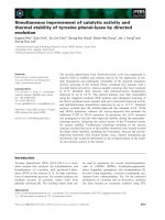
order to determine the magnitude of this cross-talk in
the actual assay setting, a dilution series of Tb
3+
labeled
r-Bio-HIV-1env was immobilized on to SA-coated
microtiter wells, followed by washing and measurement
of fluorescence, using a Victor 1420 multilabel counter
(Perkin Elmer Life and Analytical Sciences, Singapore),
at 545 nm and 615 nm. The results of this experiment,
showninFigure2A,indicatethemagnitudeofTb
3+
cross-talk one may expect while measuring Eu
3+
fluores-
cence at 615 nm in a dual-label assay. Depending on the
instrument used, Tb
3+
cross-talk was determined to
range from 1.2-2.5%. Thus, all Eu
3+
fluorescence data
were corrected using the measuring instrument-specific
Tb
3+
cross-talk. Eu
3+
cross-talk in Tb
3+
fluorescence
1
2
3
4
5
6
7
8
0.01
0.1
1
10
100
400 450 500 550 600 650 700 75
0
Normalized fluorescence
Wavelen
g
th
(
nm
)
AB
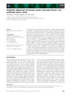
Figure 1 Design of the dual-label time-resolved immunofluorometric assay. (A) A schematic illustration of the assay for simultaneous
detection of HIV and HBV infections. The Arabic numerals indicate individual assay components: (1) microtitre well surface; (2) streptavidin; (3) r-
Bio-HIV-1 Env; (4) Bio mAb 21B; (5) anti-HIV-1 antibodies in infected serum; (6) HBsAg in infected serum; (7) r-HIV-1env labeled with Tb
3+
chelate
(which is measured at 545 nm); (8) 5 S F(ab)
2
coated Eu
3+
nanoparticles (which is measured at 615 nm). (B) The emission spectra of Tb
3+
chelate
(green line) and Eu
3+
nanoparticles (orange line).
Myyryläinen et al. Journal of Nanobiotechnology 2010, 8:27
/>Page 2 of 6
measur ement was determined as shown in Figure 2B. In
this experiment, chemically biotinylated rHBsAg was
immobilized on SA-coated microtiter wells, and incu-
bated with a dilution series of Eu
3+
doped 5 S F(ab)
2
nanoparticles. As before, fluorescence was mea sured at
both wavelengths (545 nm and 615 nm). The results
showed that Eu
3+
is unlikely to manifest significant
cross-talk during the measurement of Tb
3+
fluorescence
at 545 nm.
Prior to deploying the dual-label assay for the simulta-
neous detection of both HIV-1 and HBV infections, we
evaluated its sensitivity to detect each of the two ana-
lytes (anti-HIV-1 antibody and HBsAg) in the absence
(single-label) and presence (dual-label) of the binders of
the other analyte. Using rHBsAg (subtype adw), ranging
from 0.02-200 ng/mL, the dual-label assay was per-
formed in the absence and presence of the anti-HIV-1
antibody binders, r-Bio-HIV-1 env and Tb
3+
chelate
labeled r-HIV-1 env. Unlike in the case of rHBsAg, it is
not possible to use ‘known’ concentrations of anti-HIV-
1 antibodies, given their polyclonal nature and inherent
differences in affinity and specificity for HIV-1 antigens.
Thus, to explore the sensitivity of detection of anti-HIV-
1 antibodies, the dual-label assay was p erformed using
serial dilutions (as a surrogate for a range of known
concentrations) of an anti-HIV-1 antibody-containing
serum sample in the absen ce and presen ce of the
HBsAg binders, Bio-mAb 21B and 5 S F(ab)
2
coated
Eu
3+
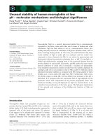
nanoparticles. The data shown in Figure 3 reveal
that there was very good correlation between t he single
and dual-label formats of the assay with respect to each
of the two analytes tested. There was essentially no dis-
cernible difference in the lowest limits of detection
(LLOD) of either analyte when the two assay formats
were compared. For HBsAg, the LLOD was 0.011 and
0.013 ng/mL, respectively, in the absence and presence
of anti-HIV-1 antibody binders. The corresponding
LLOD for anti-HIV-1 antibody detection cannot be
designated for the r eason mentioned above. Neverthe-
less, it is evident from Figure 3B th at antibodies present
in as low as 0.01 to 0.001 μloftheHIV-1positive
serum (used in this experiment) are detected in this
assay, which reaches saturation at 1 μlofthisserum.
Overall, the data justify the conclusion that combining
the anti-HIV-1 antibody- and HBsAg-binders in a dual-
label assay will not compromise the sensitivity of detec-
tion of either analyte. This is further borne out by the
analysis of sera that contain HBsAg as well as anti-HIV-
1 antibodies (see below).
Next, we tested the feasibility o f the dual-label assay
for detecting HIV and HBV infections in human serum
samples. First, we used an in-house panel of 100 ‘ nor-
mal’ serum samples that were confirmed to be negative
for both HIV and HBV infections (HIV
-
/HBV
-
), using
Vidas HIV Duo Quick and HBsAg Ultra kits (bioMér-
ieux SA, Marcy I’Etoile, France). The mean Tb
3+
and
Eu
3+
fluorescence readouts of these normal serum sam-
ples plus 5× standard deviation (SD) of the correspond-
ing means were used as the cut-offs for anti-HIV
antibodies and HBsAg determinations, respectively.
Next, we t ested a set of 37 serum samples (Department
of Virology, University of Turku). These represented
AB
10
-1
10
0
10
1
10
2
10
3
10
0
10
2
10
4
10
6
10
8
Fluorescence
(
counts
)
Tb-labeled r-Bio-HIV-1env (ng/well)
0
20
40
60
80
100
Coefficient of variation (%)
10
6
10
7
10
8
10
9
10
0
10
2
10
4
10
6
10
8
Fluorescence (counts)
5S F(ab)
2
- Eu-nanoparticles (pcs/well)
0
20
40
60
80
100
Coefficient of variation (%)
Figure 2 Cross-talk between the two lanthanide labels used in the assay. (A) Tb
3+
cross-talk. (B) Eu
3+
cross-talk. The filled symbols represent
the fluorescence and the empty symbols represent the co-efficient of variation, with circles and squares representing data points pertaining to
Tb
3+
and Eu
3+
, respectively.
Myyryläinen et al. Journal of Nanobiotechnology 2010, 8:27
/>Page 3 of 6
infected samples of which 25 were HBV
+
and 12 HIV
+
,
using the Vidas commercial assays mentioned above.
For a given analyte, signal/cut-off (S/Co) ratios <1 and
≥1 were considere d as negative and positive , respec-
tively. The results are summarized in Table 1. An analy-
sis of these serum samples using the dual-label assay
showed that while all 12 HIV
+
serum samples were
identified to contain anti-HIV-1 a ntibodies, HBsAg
antigen could be detected in 23 of the 25 HBV
+
serum
samples. Of the two remaining HBV
+
serum samples,
onewasaborderlinesample(seeAdditionalfile1:
Figure S1). Thes e two serum samples tested negative for
HBsAg using the single label assay also (data not
shown), suggesting that the dual-label assay format per
se did not compromise sensitivity of HBsAg detection.
To examine the performance of the dual-label assay in
the background of other infections, w e tested it against
a BBI viral co-infection panel PCA 201 (from B oston
Biomedica Inc., now SeraCare Life Sciences Inc., Milford
MA). This panel was characterized for HIV-1, HBV,
HCV and HTLV infections using standard commercially
available reference tests (see Additional file 1: Table S1).
Twenty-three of the 25 panel members were available
for this study. One member of this panel was seronega-
tive for both HIV-1 and HBV infections (sample# 24).
The dual-label assay identified this c orrectly as HIV
-
/
HBV
-
. Of the remaining 22 serum samples, 16 and 19
samples were designated as HIV
+
and HBV
+
, respec-
tively, with 13 samples seropositive for both HIV-1 and
HBV (Table 1). Out of these 13 HIV
+
/HBV
+
serum
samples, 6 were positive for HCV, and two for HTLV as
well. The remaining three HIV
+
serum samples were
negative for HBV but positive for HCV and HTLV. The
dual-label assay could identify 16 out of 16 HIV
+
serum
samples (100%). It is noteworthy that one borderline
serum sample (sample# 20, S/Co ratio = 1.1) was also
picked up unambiguously by the dual-label test (S/Co
ratio = 14.8). This essentially is indicative of enhanced
A
B
10
-3
10
-2
10
-1
10
0
10
1
10
3
10
4
10
5
0
20
40
60
80
100
Tb fluorescence (counts)
HIV-1 positive serum
(
μl/well
)
Coefficient of variation
(
%
)
10
-2
10
-1
10
0
10
1
10
2
10
3
10
4
10
0
10
1
10
2
10
3
10
4
10
5
10
6
10
7
Eu fluorescence (counts)
HBsAg (ng/ml)
0
20
40
60
80
100
Coefficient of variation (%)
Figure 3 Comparison o f the sensitivity of analyte detection in single versus dual-label assay formats. (A) HBsAg detection. Eu
3+
fluorescence data for the single label and dual-label assays are shown by the empty star and filled square symbols, respectively. Corresponding
coefficients of variation for the single and dual-label assays are represented by the filled star and empty square symbols, respectively. (B) Anti-
HIV-1 antibody detection. Tb
3+
fluorescence data for the single label and dual-label assays are shown by filled circles and squares, respectively.
Corresponding coefficients of variation for the single and dual-label assays are represented by the empty circles and squares, respectively.
Table 1 Evaluation of the dual-label TRF assay for
simultaneous detection of HIV-1 and HBV infections
Grp n Infection profile
(Ref assay)
a
Dual-label assay
(HIV-1
+
/HBV
+
)
b
In-house sera panel
1 25 HIV-1
-
/HBV
+
0/23
c
2 12 HIV-1
+
/HBV
-
12/0
BBI co-infection panel
3 13 HIV-1
+
/HBV
+
13/10
d
4 6 HIV-1
-
/HBV
+
0/6
5 3 HIV-1
+
/HBV
-
3/0
6 1 HIV-1
-
/HBV
-
0/0
a
The Reference assays for the in-house sera panel were Vidas HIV Duo Quick
and HBsAg Ultra assays, for anti-HIV-1 antibody and HBsAg detection,
respectively; the Reference assays for the BBI co-infection panel are
mentioned in the Supplementary Information (see Additional file 1). The “+”
and “-” superscripts indicate positive and negative tests, respectiv ely.
b
This column indicates the results obtained using the dual-label assay
described in the text. The numbers shown indicate the serum samples that
scored positive for both analytes in the dual-label assay.
c
missed two HBsAg
+
serum samples
d
missed three HBsAg
+
serum samples
Myyryläinen et al. Journal of Nanobiotechnology 2010, 8:27
/>Page 4 of 6
sensitivity of the dual-label test and is in agreement with
the conclusions based on Figure 3B. Our data show that
the dual-label assay is capable of identifying HIV
+
serum samples regardless of the presence or absence o f
HBV, HCV and HTLV co-infectio ns, with high sensitiv-
ity and specificity. However, it is to be noted that we
have used r-HIV-1env as the antigen to detect anti-
HIV-1 antibodies to obtain a technical proof-of-concept.
Of the 19 HBV
+
serum samples, 13 samples were also
HIV
+
, as m entioned already, and the rest ( n = 6) were
HIV
-
. Many of these serum samples were co-infected
with HCV, HTL V or both. The dual-label assay ident i-
fied 16 of the 19 HBV
+
serum samples, regardless of
HIV,HCVorHTLVinfectionstatus.Ofthe3HBV
+
serum samples that were missed by the dual-label assay,
onewasaborderlinesample(sample#9,S/Coratio=
1). As with the in-house serum samples, these 3 mem-
bers of panel PCA 201 also turned out to be false-nega-
tive in the single label HBV assay. This rules out the
possibility that Tb
3+
cross-talk may have masked Eu
3+
signals and interfered with HBsAg detection. The data
show that the concordance of the dual-label assay with
regard to HBsAg detection using the reference assay
(Abbott EIA) is 84%. This presumably stems from low
sensitivity of the mAbs used for detection of HBsAg,
despite the use of a tracer F(ab)
2
-Eu
3+
nanoparticle for
the detection of this analyte in the dual-label assay.
In conclusion, we have developed a lanthanide fluores-
cent reporter-based dual-label assay for the simulta-
neous detection of HIV-1 and HBV infections in
donated blood samples. The high sensitivity of this
approach derives from the temporal resolution of the
long lifetime high intensity fluorescence of Eu
3+
and
Tb
3+
lanthanide tracers measured by TRF. Qdots have
emerged recently as highly efficient fluorescent probes.
However, these have short-lived fluorescence. Therefore,
TRF cannot be employed to measure their signals and
their detection is limited by autofluorecence. Further,
the Eu
3+
and Tb
3+
tracers used in this study are inher-
ently fluorescent, obviating the need for additional signal
development steps as in the DELFIA and LANFIA
methods [2-5], and can be measured directly from the
dry surface of the microtiter wells. The simultaneous
detection of two analytes combined with a relatively
simpler assay format eliminating the extra signal devel-
opment step, will contribut e to both cost and time
saving.
To our knowledge, this work, which represents the
first report of a dual-label HIV/HBV assay, demonstrates
in principle, the feasibility of developing a multiplex
assay for screening samples for multiple infections in a
blood bank setting. However, a limitation is the poten-
tial for interference among the reporters, as illustrated
by the Tb
3+
cross-talk in Eu
3+
measurements in this
study.Itmaybeachallengingtasktoeliminatethe
interference arising out of cross-talk among multiple
fluorescent reporters. One way of circumventing the
cross-talk problem in multiplexing would be to design
an array-in well strategy in which multiple analyte-cap-
turing reagents are spatially isolated in the same well
andusedinconjunctionwithasinglereporter,for
example Eu
3+
nanoparticles. In this set up, analytes can
be identified based on the specific locations from which
signals are detected.
Additional material
Additional file 1: Myyrylainen et al (Addl files). The file is organized
into three sections. Section 1 describes essential Methods. Section 2
provides S/Co data on the evaluation of in-house sera panel using the
dual-label TRF assay (Figure S1). Section 3 provides S/Co data on the
evaluation of the BBI viral co-infection panel PCA 201 using the dual-
label TRF assay (Table S1)
Acknowledgements
This work was supported by grants from the Department of Biotechnology,
Government of India and Academy of Finland (grant #115524) under a joint
Indo-Finnish collaborative research programme. SMT was the recipient of a
research fellowship from the University Grants Commission, Government of
India.
Author details
1
Department of Biotechnology, University of Turku, Turku, Finland.
2
Recombinant Gene Products Group, International Centre for Genetic
Engineering & Biotechnology, Aruna Asaf Ali Marg, New Delhi-110067, India.
3
Department of Virology, University of Turku, Turku, Finland.
Authors’ contributions
TM and SMT performed experiments. RV collected and characterized the
human sera samples. SS and NK designed the HIV antigen and generated
the monoclonal antibodies. TS, NK and KP conceived and designed the
experiments. SS, TS, NK and KP interpreted the data and prepared the
manuscript. All authors read and approved the manuscript.
Competing interests
The authors declare that they have no competing interests.
Received: 11 August 2010 Accepted: 26 November 2010
Published: 26 November 2010
References
1. World Health Organization: Screening donated blood for transmission
transmissible infections: recommendations. 2009 [ />bloodsafety/ScreeningTTI.pdf], (accessed 6th August 2010).
2. Siitari H: Dual-label time-resolved fluoroimmunoassay for the
simultaneous detection of adenovirus and rotavirus in faeces. J Virol
Methods 1990, 28:179-188.
3. Meriö L, Pettersson K, Lövgren T: Monoclonal antibody-based dual-label
time-resolved fluorometric assays in a simplified one-step format. Clin
Chem 1996, 42:1513-1517.
4. Eriksson S, Vehniäinen M, Jansén T, Meretoja V, Saviranta P, Pettersson K,
Lövgren T: Dual-label time-resolved immunofluorometric assay of free
and total prostate-specific antigen based on recombinant Fab
fragments. Clin Chem 2000, 46:658-666.
5. Ankelo M, Westerlund A, Blomberg K, Knip M, Ilonen J, Hinkkanen AE:
Time-resolved immunofluorometric dual-label assay for simultaneous
detection of autoantibodies to GAD65 and IA-2 in children with type I
diabetes. Clin Chem 2007, 53:472-479.
Myyryläinen et al. Journal of Nanobiotechnology 2010, 8:27
/>Page 5 of 6
6. Vaidya HC, Beatty BG: Eliminating interference from heterophilic
antibodies in a two-site immunoassay for creatine kinase MB by using F
(ab’)
2
conjugate and polyclonal IgG. Clin Chem 1992, 38:1737-1742.
7. Väisänen V, Peltola , Lilja H, Nurmi M, Pettersson K: Intact free prostate-
specific antigen and free and total human glandular kallikrein 2.
Elimination of assay interference by enzymatic digestion of antibodies
to F(ab’)
2
fragments. Anal Chem 2006, 78:7809-7815.
8. Härmä H, Soukka T, Lövgren T: Europium nanoparticles and time-resolved
fluorescence for ultrasensitive detection of prostate-specific antigen. Clin
Chem 2001, 47:561-568.
9. Huhtinen P, Soukka T, Lövgren T, Härmä H: Immunoassay of total
prostate-specific antigen using europium(III) nanoparticle labels and
streptavidin-biotin technology. J Immunol Methods 2004, 294:111-122.
10. Valanne A, Huopalahti S, Vainionpää R, Lövgren T, Härmä H: Rapid and
sensitive HBsAg immunoassay based on fluorescent nanoparticle labels
and time-resolved detection. J Virol Methods 2005, 129:83-90.
doi:10.1186/1477-3155-8-27
Cite this article as: Myyryläinen et al.: Simultaneous detection of Human
Immunodeficiency Virus 1 and Hepatitis B virus infections using a dual-
label time-resolved fluorometric assay. Journal of Nanobiotechnology 2010
8:27.
Submit your next manuscript to BioMed Central
and take full advantage of:
• Convenient online submission
• Thorough peer review
• No space constraints or color figure charges
• Immediate publication on acceptance
• Inclusion in PubMed, CAS, Scopus and Google Scholar
• Research which is freely available for redistribution
Submit your manuscript at
www.biomedcentral.com/submit
Myyryläinen et al. Journal of Nanobiotechnology 2010, 8:27
/>Page 6 of 6