Báo cáo y học: " Multifocal invasive ductal breast cancer with osteoclast-like giant cells: a case report" doc
Bạn đang xem bản rút gọn của tài liệu. Xem và tải ngay bản đầy đủ của tài liệu tại đây (2.97 MB, 5 trang )
CAS E REP O R T Open Access
Multifocal invasive ductal breast cancer with
osteoclast-like giant cells: a case report
Georg Richter
1*
, Christoph Uleer
2
, Thomas Noesselt
3
Abstract
Introduction: To the best of our knowledge, this is the first case report of a multifocal (trifocal) invasive carcinoma
of the breast containing osteoclast-like giant cells.
Case presentation: A 64-year-old Caucasian woman presented for routine mammography screening with three
radiodense lesions in the lower inner quadrant of the right breast, a primary breast cancer. Microscopic
examination showed three foci of invasive ductal carcinoma with multinucleate d osteoclast-like giant cells.
Osteoclast-like giant cells in breast cancer are a rare phenomenon. They are described in less than two percent of
all breast cancers and occur in association with invasive ductal cancer and invasive lobular can cer. In addition,
osteoclast-like giant cells have been described in several sarcomas and metaplastic carcinomas of the breast.
Conclusion: To the best of ou r knowledge, this is the first report of a multifocal infiltrating ductal carcinoma of the
breast containing osteoclast-like giant cells. This could be an indication for a possible early event in carcinogenesis
associated with a biological event or secretion that indicates the differentiation and/or migration of stromal cells or
macrophages.
Introduction
Carcinoma of the breast containing osteoclas t-like giant
cells is uncommon and described in less than 2% of
breast cancer patients [1-3]. In addition, osteoclast-like
giant cells are described in a ductal carcinoma in situ
and metaplastic carcinomas of the breast [4,5], although
the stromal origin of the giant cells is unknown. Immu-
nohistochemical and ultrastructural studies suggest that
the osteoclast-like giant cells are of stromal histiocytic
origin or might be differentiated from macrophages
[6-9]. The characteristic multinucleated giant cells are
found at the periphery of the tumor cells and within the
glandular luminal spaces in primary in situ, invasive
breast cancers and in metastases. We report the first
case of a multifocal invasive ductal breast cancer with
osteoclast-like giant cells.
Case presentation
A 64-year-old Caucasian woman presented for routine
mammography screening within the National Mammo-
graphy Screening Program. She had no known family
history of breast cancer and denied recent signs or
symptoms of breast disease on her intake questionnaire.
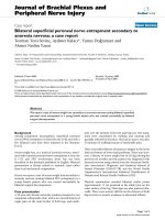
The digital mammogram showed three radiopaque
lesions in the lower inner quadrant of the right breast,
which were readily detectable in both the me diolateral
oblique and craniocaudal projection views (Figure 1).
The density of the breast tissue was estimated as type 2
according to the classification system of the American
College of Radiology (low-density, fibroglandular tissue).
Round microcalcifications were found to be diffusely
distributed in both breasts.
Each of the three lesions in the right lower inner
quadrant had slightly irregular margins and measured
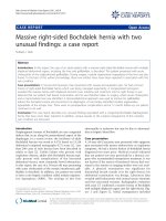
0.7 cm × 0.9 cm. Since these lesions w ere absent in
the previous screening mammogram performed two
years earlier (Figure 2), th ey were conside red suspi-
cious for multifocal breast cancer (Breast Imaging
Reporting and Data System (BI-RADS) category 4B).
Therefore, the woman was called back into the screen-
ing center for further evaluation. A craniocaudal spot
compression view focused on the three lesions was
obtained. On this view, the radiodense lesions with
irregular margins were easily distinguished from the
surrounding fat tissue ( Figure 3).
* Correspondence:
1
Institute of Pathology, 31785, Hameln, Germany
Full list of author information is available at the end of the article
Richter et al. Journal of Medical Case Reports 2011, 5:85
/>JOURNAL OF MEDICAL
CASE REPORTS
© 2011 Richter et al; licensee BioMed Cent ral L td. This is an Open Access article distributed under the terms of the Creative Commons
Attribution License ( which pe rmits unrestricted use, distribution, and reproduction in
any medium, provided the original work is properly cited.
A breast ultrasound was performed, and in the right
inner lower quadrant the lesions were visible as complex
masses with irregular margins and inhomogeneous
internal echoes (BI-RADS analogue 4). The left breast as
well as the ipsilateral and contralateral axillary lymph
nodes were normal.
Since there was a good correlation between the suspi-
cious mammographic lesions and the ultrasound image,
an ultrasound-guided c ore needle biopsy was perf ormed
for each of the three tumors. Five specimens were
thereby obtained confirming the diagnosis of multifocal
invasive cancer. Because of the multifocal character of
the breast can cer, a bilateral breast magnetic resonance
imaging (MRI) scan was o btained to exclude further
lesions. Eleven days after the woman’ sfirstcontactwith
the screening center, the interdisciplinary tumor board
recommended breast-conserving surgery and sentinel
node biopsy following preoperative needle localization of
the tumor.
As the foci were lying close together in one quadrant,
a breast-preserving operation could be performed. Addi-
tionally, a sentinel node marking a nd a sentinel node
biopsy were induced by clinically and sonographically
negative axillary results.
For the operation, the three foci were portrayed preo-
peratively using sonography with a needle marking.
First, the sentinel node b iopsy was carried out. After
marking with Nanocol l technetium-99 m (Gipharma Sri,
Saluggia Vercelli, Italy) a sentinel node was portrayed in
the right axilla by lymphscintigraphy. Intraoperative 1.5
ml Acid Blue solution (Guerbet, Sulzbach, Taunus, Ger-
many) was additionally injected peritumorally, and the
axilla was examined using a gamma probe. A focus of
heightened activity showed up in the right lower axilla.
A radioactively mark ed lymph node was found during
the preparation of the axilla. There were no other foci
of heightened activity . The frozen section examination
of the sentinel node was negative.
Figure 1 Digital mammography (mediolateral projection).
Figure 2 Screening mammogram performed two years earlier
than 2009.
Figure 3 Craniocaudal spot compression in digital
mammogram view focused on the three lesions.
Richter et al. Journal of Medical Case Reports 2011, 5:85
/>Page 2 of 5
Afterward a breast-preserving excision including a skin
spindle was performed. The excision contained all the
invasive foci and presented clear margins. For reconstruc-
tion, intramammary wound closure with an advancement
plastic of breast tissue was in stalled into the defect. Post-
operative proper wound healing was observed.
An intraoperative investigation of a breast specimen
weighing 31 g a nd measuring 8 cm × 5 cm × 3 cm was
undertaken to examine the resection margins. Also, one
sentinel node was examined to exclude metastases. In the
macroscopic examination, three neighboring foci show-
ing a brown incision surface and measuring 1.2 cm, 0.8
cm and 0.6 cm were found (Figure 4). The specimens
were routinely fixated in 4% buffered formalin, embedded
in paraffin and sectioned into 3 μmto4μm thick sec-
tions. Then the specimens were routinely stained with
hematoxylin and eosin. Also, they were immunohisto-
chemically stained with the primary antibodies Cytokera-
tin 5/6 (Cell Marque) (Roche Ventana Medical Systems,
Illkirch, France), Cytokeratin 7 (Roche Ventana), Vimen-
tin (Roche Ventana), CD68 (Roche Ventana), Estrogen
Receptor (Roche Ventana), Progesterone Receptor
(Roche Ventana), human epidermal growth factor recep-
tor 2 (HER2) (Roche Ventana) and Ki-67 antigen (Roche
Ventana) using the ultraView™ Universal Alkaline Phos-
phatase Red Detection Kit (Roche Ventana) on the Roche
Ventana benchmark with on-slide positive controls. All
Ventana kits are ready to use.
Microscopic examination showed three foci of an inva-
sive ductal carcinoma with a moderate amount tubule
formations, moderate nuclear pleomorphism with visible
nucleoli and 8 mitoses/10 high-power fields following the
grading of Elston and Ellis [12] (Figures 5 and 6). No
squamous cells or other metaplasia were exhibited in any
of the foci. On the basis of immunohistochemistry, we
detected a positive reaction for cytokeratin 7 and a nega-
tive reaction for cytokeratin 5/6 and vimentin in the
epithelial tumor cells (Figure 7). Using the Allred score,
Figure 4 Macrophotography of the greatest focus.
Figure 5 Microphotograph y (hematoxylin and eosin staining;
original magnification, × 200).
Figure 6 Microphotograph y (hematoxylin and eosin staining;
original magnification, × 400).
Figure 7 Immunohistochemical positive reaction of the stromal
cells for vimentin (original magnification, × 400).
Richter et al. Journal of Medical Case Reports 2011, 5:85
/>Page 3 of 5
the estrogen and progesterone receptors were similarly
positive in all three foci (Proportion Score 5 + Intensity
Score 3 = Total Score 8) (Figures 8 and 9), and in accor-
dan ce with the Dako score, we detected a HER2 score of
0 (negative) (Figure 10). The Nottingham grade for inva-
sive cancer was 0.2 × 1.2 + (G) 2 + 0 = 3.4; Nottingham
Prognostic Index score 3.4 (intermediate). The giant cells
contained numerous uniform nuclei and eosinophilic
cytoplasm and had an appearance identical to an osteo-
clast. Immunohistochemically, the giant cells showed a
positive reaction for vimentin and CD68 ( Figure 11) and
a negative reaction for the cytokeratin s and the hormone
receptors.
Owing to the tumor entity, there is a heightened risk
of a systemic recurrence. Anthracycline-based che-
motherapy with four cycles of epirubicin 90 mg/m
2
and
cyclophosphamide 600 mg/m
2
wasaddedastherewas
an overexpression of plasminogen activator inhibitor-1
(PAI-1, 5 4 ng/mg; urokinase plasminogen activator, 1.1
ng/mg) as a prediction of the effectiveness for adjuvant
chemotherapy. Our patien t was treated with continua-
tion of adjuvant therapy with aromatase inhibitor and
radiation of the breast with 50.4 dye plus local boost
radiotherapy of the tumor bed.
Conclusion
To the best of our knowledge, we present the first case
report of a multifocal invasive ductal breast cancer with
osteoclast-like giant cells. Osteoclast-like giant cells are
rare in breast cancer, and the prognostic significance of
their presence is uncertain [10,11]. Immunohistochem-
ical and ultrastructural studies suggest that the osteo-
clast-like giant cells are of stromal hist iocytic origin or
possibly are terminally differentiated from macrophages.
We detected three neighboring foci of an invasive ductal
Figure 8 Immunohistochemical positive reaction with antibody
against the estrogen receptor in the tumor cells (original
magnification, × 400).
Figure 9 Immunohistochemical positive reaction with antibody
against the progesterone receptor in the tumor cells (original
magnification, × 400).
Figure 11 Immunohistochemical positive reaction with
antibody against CD68 in the giant cells (original
magnification, × 400).
Figure 10 Immunohistochemical negative reaction with
antibody against human epidermal growth factor receptor 2
(HER2)/neu in the tumor cells (original magnification, × 400).
Richter et al. Journal of Medical Case Reports 2011, 5:85
/>Page 4 of 5
breast cancer with giant cells containing numerous uni-
form nuclei and eosinophilic cytoplasm adjacent to the
epithelial tumor cells, an appearance identical to osteo-
clasts. This could be an indication for a possible e arly
event in carcinogene sis associated with a biological
event or secretion that indicates the differentiation and/
or migration of stromal cells or macrophages.
Consent
Written informed consent was obtained from the patient
for publication of this case report and accompanying
images. A copy of the written consent is available for
review by the Editor-in-Chief of this journal.
Acknowledgements
The authors thank the patient described in this study. Also, the authors
would like to thank C. E. Noble-Pyott for her relentless and excellent work
on this case report.
Author details
1
Institute of Pathology, 31785, Hameln, Germany.
2
Mammography Screening
Unit Lower Saxony South, D-31134 Hildesheim-Hameln-Göttingen, Germany.
3
Department of Gynecology, District Hospital Hameln, D-31785 Hameln,
Germany.
Authors’ contributions
UC analyzed and interpreted the mammography and ultrasound. TN
performed the operation and administered chemotherapy. GR performed
the histological examination and was a major contributor in writing the
manuscript. All authors read and approved the final manuscript.
Competing interests
The authors declare that they have no competing interests.
Received: 17 May 2010 Accepted: 27 February 2011
Published: 27 February 2011
References
1. Rosen PP: Mammary carcinoma with osteoclast-like giant cells. Rosen’s
Breast Pathology Philadelphia: Lippincott Williams & Wilkins; 2001, 517-526.
2. Holland R, van Haelst UJ: Mammary carcinoma with osteoclast-like giant
cells: additional observations on six cases. Cancer 1984, 53:1963-1973.
3. Cai N, Koizumi J, Vazquez M: Mammary carcinoma with osteoclast-like
giant cells: a study of four cases and a review of literature. Diagn
Cytopathol 2005, 33:246-251.
4. Krishnan C, Longacre T: Ductal carcinoma in situ of the breast with
osteoclast-like giant cells. Hum Pathol 2006, 37:369-372.
5. Wargotz ES, Norris HJ: Metaplastic carcinomas of the breast: V.
Metaplastic carcinoma with osteoclastic giant cells. Hum Pathol 1990,
21:1142-1150.
6. Pettinato G, Petrella G, Manco A, di Pisco B, Salvatore G, Angrisani P:
Carcinoma of the breast with osteoclast-like giant cells: fine needle
aspiration cytology, histology and electron microscopy of 5 cases. Appl
Pathol 1984, 2:168-178.
7. Sano M, Kikuchi K, Zhao C, Kobayashi M, Nakanishi Y, Nemoto N:
Osteoclastogenesis in human breast carcinoma. Virchows Arch 2004,
444:470-472.
8. Athanasou NA, Wells CA, Quinn J, Ferguson DP, Heryet A, McGee JO: The
origin and nature of stromal osteoclast-like multinucleated giant cells in
breast carcinoma: implications for tumour osteolysis and macrophage
biology. Br J Cancer 1989, 59:491-498.
9. Shishido-Hara Y, Kurata A, Fujiwara M, Itoh H, Imoto S, Kamma H: Two
cases of breast carcinoma with osteoclastic giant cells: are the
osteoclastic giant cells pro-tumoural differentiation of macrophages?
Diagn Pathol 2010, 5:55.
10. Agnantis NT, Rosen PP: Mammary carcinoma with osteoclast-like giant
cells: a study of eight cases with follow up. Am J Clin Pathol 1979,
72:383-389.
11. Saimura M, Fukutomi T, Tsuda H, Tanaka SA, Nanasawa T: Breast carcinoma
with osteoclast-like giant cells: a case report and review of the Japanese
literature. Breast Cancer 1999, 6:121-126.
12. Elston CW, Ellis IO: Pathological prognostic, factors in breast cancer. 1.
The value of histological grade in breast cancer: experience from a large
study with long term follow up. Histopathology 1991, 19:403-410.
doi:10.1186/1752-1947-5-85
Cite this article as: Richter et al.: Multifocal invasive ductal breast cancer
with osteoclast-like giant cells: a case report. Journal of Medical Case
Reports 2011 5:85.
Submit your next manuscript to BioMed Central
and take full advantage of:
• Convenient online submission
• Thorough peer review
• No space constraints or color figure charges
• Immediate publication on acceptance
• Inclusion in PubMed, CAS, Scopus and Google Scholar
• Research which is freely available for redistribution
Submit your manuscript at
www.biomedcentral.com/submit
Richter et al. Journal of Medical Case Reports 2011, 5:85
/>Page 5 of 5