báo cáo khoa học: "In vivo observation of gold nanoparticles in the central nervous system of Blaberus discoidalis" potx
Bạn đang xem bản rút gọn của tài liệu. Xem và tải ngay bản đầy đủ của tài liệu tại đây (3.65 MB, 9 trang )
RESEARC H Open Access
In vivo observation of gold nanoparticles in the
central nervous system of Blaberus discoidalis
Aracely Rocha
1
, Yan Zhou
1,2
, Subrata Kundu
1,2
, Jorge M González
3
, S BradleighVinson
3
, Hong Liang
1,2*
Abstract
Background: Nanoparticles (NPs) are widely studied for biomedical applications. Understanding interactions
between NPs and biomolecules or cells has yet to be achieved. Here we present a novel in vivo method to study
interactions between NPs and the nervous system of the discoid or false dead-head roach, Blaberus discoidalis. The
aims of this study were to present a new and effective method to observe NPs in vivo that opens the door to new
methods of study to observe the interactions between NPs and biological systems and to present an inexpensive
and easy-to-handle biological system.
Results: Negatively charged gold nanoparticles (nAuNPs) of 50 nm in diameter were injected into the central
nervous system (CNS) of the insect. By using such a cost effective method, we were able to characterize nAuNPs
and to analyze their interactions with a biological system. It showed that the charged particles affected the insect’s
locomotion. The nAuNPs affected the insect’s behavior but had no major impacts on the life expectancy of the
cockroach after two months of observation. This was apparently due to the encapsulation of nAuNPs inside the
insect’s brain. Based on cockroach’s daily activity, we believed that the encapsulation occurred in the first 17 days.
Conclusions: The method proposed here is an inexpensive and reliable way of observing the response of
biological systems to nanoparticles in-vivo. It opens new windows to further understand how nanoparticles affect
neural communication by monitoring insect activity and locomotion.
Background
Due to their small size, nanoparticles (NPs) have been
used to probe biological systems [1-3]. Common biologi-
cal systems, mainly mice, currently used to study, ana-
lyze, and test in vivo treatments for neuron damage and
repair are expensive and many times difficult to main-
tain. It is necessary to find a suitable biological system
that is inexpensive, easy to maintain, and handle. As
early as in 1990, Huber et a l. reported cockroaches as
good candidates for neurobiology studies [4]. This idea
was later applied by Scharrer for endocrine studies [5].
There are reports proving the simila rities between verte-
brate and invertebrate brains [6]. In particular, non-
vertebrate systems such as cockr oaches were ideal
models for neurotoxicology studies [7]. The comparison
between invertebrate (like cockroaches) a nd vertebrate
(like mice) has been made in terms of their behavior,
anatomy, biology, and physiology. Invertebrate subjects
are not only cost effective and readily available, but also
they do not feel pain [8]. This opens new avenues for
experimental protocols and controls curren tly imple-
mented in vertebrate animals and humans.
Cockroaches have been used as model systems for
neurological research. Early neurobiology cockroach
resear ch has been focused on octopamine and serotonin
response in the nervous system (NS). Previous studies
were to observe how chemicals were distributed in the
brain and how they affected the ner vous system [9,10].
In more recent wo rk by Brown et al., roaches have been
used to study the effects of age on memory and brain
integrity [11].
The use of nanoparticles in biological systems is a
subject that has been under scrutiny for some time. The
use of nanoparticles for imaging and drug delivery has
been extensively studied in mice. Hainfeld and collea-
gues have used gold nanoparticles to enhance radiother-
apy in mice and as a contrast agent for X-ray imaging
[12,13]. Functionalized gold nanoparticles have also
* Correspondence:
1
Department of Mechanical Engineering, Texas A&M University, College
Station, Texas, USA
Full list of author information is available at the end of the article
Rocha et al . Journal of Nanobiotechnology 2011, 9:5
/>© 2011 Rocha et al; licensee BioMed Central Ltd. This is an Ope n Access article distributed under the terms of the Creative Commons
Attribution License ( which permits unrestricted use, distribution, and reproduction in
any medium, provided the original work is properly cited .
been used to investigate targeted drug delivery [14-16].
However, these in vivo methods have not been applied
for simpler and inexpensive biological systems like
insects.
Inthepresentwork,weuseBlaberus discoidalis,a
neotropical cockroach, as the model system. We study
the effects and interactions of negatively charged gold
nanoparticles (nAuNPs) with the cockroaches CNS
in vivo. The authors refer to the nervous system as the
brain and the nerve cord as described in the American
Cockroach by Bell [17]. Negatively charged nanoparti-
cles were selected to enhance nanoparticle interaction
with the nervous system during signal transfer i.e. dur-
ing a nerve impulse.
Methods
A new method to introd uce nanoparticles into the ner-
vous system (NS) of Blaberus discoidalis roaches was
used.Thismethodallowedustostudyeffectsofnano-
particles on the roach’s CNS in vivo. Two groups of roa-
ches were selected for t his study. Each group had 9
individuals. The selected groups were separated for
24 hour s prior to the treatment. Group 1 served as con-
trol; no nanoparticles were injected into this group.
Group 2 was treated with negatively charged spherical
gold nanoparticles (nAuNPs) of 50 nm in diameter.
Male Blaberus discoidalis (weight = 2.1 ± 0.3 g) grown
in-house were used in this study. These roaches were
maintained in hard plastic containers (9 × 18”)insidean
environment controlled room with a temperature of 28 ±
2°C and a 12/12 h day/night cycle. They were fed with
Dry dog chow. Food and water were supplied ad libitum.
The cockroaches were kept in isolation to minimize
stressors like noise, wind, and vibration that could alter
their behavior. A two-minute video was taken daily at
8:00 am, only 10 minutes into the light cycle, to record
their activity. Although the insect is most active during
the dark cycle , light was needed to record their activity.
The first hour was selected for recording since slightly
over one third or 38.1% of the cockroaches show activity
during the first hour of the light cycle [17,18].
The nanoparticles were ~50 nm in diameter. They
were synthesized using the well known Turkevich
method [19]. The synthesized Au particles were stabi-
lized and separated from each other by the negatively
charged tri-sodium-citrate molecule. Their size was con-
trolled by the reaction time and the amount of gold
atoms present in the solution. This method delivers 95%
of spherical particles and no further treatment was done
to eliminate the remaining 5% of non-spherical nanopar-
ticles. The average particle size is 46.7 nm ± 5.47 nm as
verified by JEOL-JEM 2010 TEM and analyzed with
Image J. The particle size distribution image and analy-
sis is summarized in Figure 1. The particles were
suspended in DI wat er with a concentration of 1 × 10
11
nanoparticles/mL. They were then coated with tri-
sodium citrate molecules to create a negatively charged
surface. The charge was to avoid agglomeration, ensure
suspension in the solution, and to promote their interac-
tions with the CNS.
According to Patil and colleagues [20] and Tim and
colleagues [21], the zeta potential values for gold nano-
particles prepared by this method are stable and
strongly dependent on nanoparti cle size. The zeta
potential for a 47.1 nm gold nanoparticle prepared by
this method is -32.65 mV [21]. The negatively charged
gold nanopar ticles are also fluo rescent. The 50 nm par-
ticles used absorb a light wave of 510 nm and emit at
560 nm [22-24]. T his allows for fluorescent and spectral
imaging to identify the presence of nAuNPs in the tissue
without adding fluorescent tags.
Nanoparticle introduction to the CNS
The nAuNPs were introduced in the CNS through an
injection between the brain and the sub esophageal gang-
lion (SEG) through the neck in the direction shown in
Figure 2. A 1 cc syringe with 30 gauge needle was used to
inject the nAuNPs suspended in DI water. The cockroach
Figure 1 Negatively charged gold nanoparticles ( nAuNP) size
distribution & analysis.
Rocha et al . Journal of Nanobiotechnology 2011, 9:5
/>Page 2 of 9
was immobilized by exposing it to a CO
2
atmosphere until
no signs of motion were observed (approximately 30 s).
The needle was inserted 1.5 to 2 mm into the neck in the
location and direction shown in Figure 2, allowing to
reach the brain of the insect. A stepper motor with speed
and time control was used to inject 7 μLofnAuNPs/DI
water solution, giving 7 × 10
11
nanoparticles injected into
each co ckroach.
The roaches were placed in the plastic container
immediately after treatment and were closely monitored
for the first 4 hours to ensure activity had been
resumed. The insects were monitored daily to verify
activity. The roaches that did not show signs of activity
were considered dead and were removed and placed in
a-80°Cfreezertopreventtissuedamageandallow
further analysis. After two months, 7 cockroaches from
the control and 6 cockroaches from the treated group
were alive, giving 78% and 67% survival rates respec-
tively. The activity recording was stopped at two months
and two cockroaches from the nAuNPs treated group
and two from the control group we re sacrificed and
their brains dissected for analysis. The remaining cock-
roaches from each group were sacrificed by freezing at
-80°C.
Imaging and testing
Four instruments were used to analyze the presence of
nAuNPs in the cockroach’s brain and to study the interac-
tions between nAuNPs and the brain tissue: hyperspectral
imaging, XPS, confocal microscopy, and TEM imaging.
The Hyperspectral imaging from CytoViva was used to
identify the organs affected by the nAuNPs. The XPS was
used to verify the presence of nAuNPs embedded in the
brain tissue. The confocal microscope and TEM were
used to gain insights into the interaction of nAuNPs and
the insect’sCNS.
Sample preparation
Sample preparation varied with each test system. The two
nAuNPs treated cockroaches prepared for Hyperspectral
imaging were dissected to remove the organs in the
thorax and head. The organs removed included the brain,
ant ennae, fat bodi es, esophagus, malphigian tubules, and
haemolymph. The organs were fixed with Zamboni’s fixa-
tive (Newcomer Supply) for 10 minutes and rinsed with
DPBS 3 times for 5 minutes. The samples were allowed
to air dry over a 25 mm glass cover slip.
The samples prepared for XPS, Confocal microscopy,
and TEM imaging were obtained from frozen sections.
The cockroach’s head was removed and the brain
extracted. The brain was rinsed with DPBS and fixed with
FrozFix (Newcommer Supply) for two hours to allow thor-
ough diffusion of the fixative in the brain tissue. The brain
was then mounted in Optical coherence tomography
(OCT, Fischer Scientific) and allowed to harden at -17°C.
The samples were sliced to 12 μm thickness with a cryo-
cutter. The slices were collected on 1in
2
quartz micro-
scope slides for XPS analysis. The samples prepared for
confocal microscopy were mounted on positively charged
microscope slides under DPBS media and covered with a
glass cover slip. The samples for TEM imaging were
placed on copper grids and allowed to dry for imaging.
Results
Cockroach activity
The cockroach activity was recorded by measuring the
total distance walked by each group daily. Two-minute
video recordings were performed at the beginning of the
light cycle at 8:00 am for six weeks. This time is chosen
because it is when the insects are most active under light.
The motion of each cockroach was traced with Image
Tool and the distance walked was calculated by comparing
with a fixed reference of known size in the container. The
results of cockroach activity are summarized in Figure 3.
The days not shown in the summary are due to video
recording device failure or due to corrupt video files.
There are several possible factors affecting insects’
activity. Reproductive cycle, age, temperature, humidity,
wind, noise, vibration, and changes in weather are just a
few examples [17]. The variation due to the reproduc-
tive cycle and age was eliminated by using only young
males in this study. The effects of temperature, humid-
ity, and wind were dim inished by mainta ining them i n a
controlled environment. However, the fluctuations in
Figure 2 Nanoparticle injection site and direction is indicated
with the red arrow.
Rocha et al . Journal of Nanobiotechnology 2011, 9:5
/>Page 3 of 9
noise, vibration and changes in weather affect the activ-
ity of both g roups. The effects of these variables are
diminished by presenting the activity ratio of the treated
to the untreated group. Although the treated/untreated
ratio still shows variations (days 4, 11, and 13 in particu-
lar), Figure 3 indicates an increased activity for the
nAuNPs treated group for 17 days following treatment.
After 17 days, their activity falls below that of the
control group. After two months, 7 cockroaches from
the control and 6 cockroaches from the treated group
were alive, giving 78% and 67% survival rate respectively.
The observation period was terminated at 2 months
since there were no visible differences in the cock-
roaches’ behavior after day 17.
What is the reason behind this? To understand the
effects of nAuNPs on the insects’ behavior, we con-
ducted a series of characterization experiments for NPs
with surrounding tissues. Spectroscopic and morpholo-
gic analyses were conducted using hyperspectral ima-
ging, XPS, Confocal microscopy, and TEM. Using these
tools we identified the location and interactions of the
nAuNPs with the cockroach’s CNS.
Spectroscopic analysis
The hyperspectral imaging system from CitoViva was
used to identify the location of the nAuNPs particles in
the tested roach. This imaging system identified the pre-
sence of gold in th e tissues by comparison. A sample of
nAuNPs/DI water solu tion was scanned to identify the
emitted fluorescence of the nanoparticles. The hyper-
spectral imaging, as shown in Figure 4a, provided a
range of emitted signal due to the variations in size dur-
ing nanoparticle fabrication and possible agglomeration
Figure 3 Normalized (nAuNPs treated/untreated) activity.
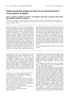
Figure 4 Hyperspectral imaging of NP solution and treated nervous system. (a) Negative gold nanoparticle hyperspectral imaging.
(b) Spectral scan of brain and nerve cord. (c) Scan areas for nAuNPs/DI water solution spectra. (d) Scan area of treated nerve cord.
Rocha et al . Journal of Nanobiotechnology 2011, 9:5
/>Page 4 of 9
once in cont act with the CNS. A signal library was gen-
erated from this scan, Figure 4a. The nAuNPs treated
tissue was then scanned an d the spectral imaging was
compared to that of the library. From t he scanned tis-
sues, only the spectra recorded from the brain and
nerve cord matched to that of the library generated
from the nAuNPs/DI water solution. Results are shown
in the Figure 4b. The optical images of the scanned
regions are shown in t he Figures 4c and 4d and corre-
spond to Figures 4a and 4b respectively.
A Kratos Axis Ultra Imaging X-ray photoelectron
spectrometer (XPS) with a spherical mirror analyzer was
used in this study. It was operated with a Mg-Ka
(1253.6 eV) X-ray radiation at a power of 350 W and a
base pressure of 10
-10
Torr. The XPS system was used
to verify the presence of the nanoparticles inside the
brain by scanning the cryocut and fixed cockroach bra in
slices mounted on quartz slides. A control and a
nAuNPs treated brains were scanned for comparison.
Figure 5a shows t he results for the contro l sample and
Figure 5b for the nAuNPs treated brain. The binding
energy for gold is at 85 eV.
The high signal-to-noise ratio of the XPS scans was
caused by too few particles on the scanned surface. The
samples used for these scans were 12 μm thick slices that
were cryocut from the cockroach brain. The XPS could
only scan to a few nanometers (<10 nm) deep from the
surface. This limited t he number of nAuNPs present in
the scanned region since only a few nanoparticles were
exposed within 10 nm from the surface. Interestingly, the
difference between the control and the nAuNPs treated
samples were seen around 85 eV. The curve fitting
obtained for Figure 5b was obtained by averaging of 21
consecutive intensity readings (10 above and 10 below) for
each binding energy value recorded. This allows for a
moving average and smoothing of the fitted curve. The
XPS results indicated that the gold nanoparticles were dis-
persed inside the insect’s brain.
Morphological analysis
Microscopic imaging
An Olympus FV1000 Confocal Microscope equipped
with a 510 nm argon laser was used to detect where the
nAuNPs were located within the brain. The samples
were fixed and cryocut to 12 μm thickness and mounted
with DPBS (Dulbecco’ s Phosphate Buffered Sali ne).
The gold nanoparticles used in this study fluoresced at
560 nm with an excitation wavelength of 510 nm. In the
transmission images, Figure 6a and 6b, exhibited visible
differences in the tissues of the nAuNPs treated and
untreated brains respectively. The darke r regions were
an indication of nanoparticle dispersion within the
tissue.
The electro n transmission microscopic image showed
a clear difference between the treated and untreated
cockroach brains. The nAuNPs treated brain had an
abnormal tissue (dark) due to the embedded nanoparti-
cles. This further proved the existence nAuNPs inside
the cockroach’s brain. Figure 6c and 6d show the fluor-
escence of the treated and un treated brains respectively.
The main challenge of the fluorescent images was the
self fluorescence of the cockroach brain tissues. The self
fluorescence was absorbed and emitted at a wavelength
close to that of the gold nanoparticles. However, it was
clear that the nAuNPs treated brain had stronger fluor-
escence intensity than the control. The horizontal yellow
line on the top images of Figures 6c and 6d showed the
location of the intensi ty profile below. These locations
were selected because they exhibit the highest intensity.
The fluorescence of the treated brain was significantly
higher than that of the untreated brain. The intensity
difference was further enhanced by the fact that the
laser power was set at 30% for the treated brain and
50% for the untreated brain, i.e. the fluorescent signal
recorded for the untreated brain was partially due to the
higher laser power and the self fluorescence of the
tissue.
Nanoscopic imaging
Upon closer inspection of the treated brain tissue, there
was evidence of nanoparticle encapsulation. Figure 7a
Figure 5 Gold has a bonding energy of 85 eV. (a) XPS results for
control cockroach brain. (b) XPS results for nAuNPs treated
cockroach brain.
Rocha et al . Journal of Nanobiotechnology 2011, 9:5
/>Page 5 of 9
showed well-defined 2-5 μm (2000 to 5000 nm) dia-
meter spheres. Upon inspection of the fluorescent image
of this view, Figure 7b, hundreds of small nanoparticles
were found dispersed or agglomerated (indicated with
green arrows) inside these spheres. Figure 7c, an overlay
of the transmission (6a) and fluorescent (6b) images
further proved th e agglomeration of nanoparticles inside
the spherical structures. A JEOL-JEM 2010 TEM was
used to characterize the morphology of NPs in the cock-
roaches’ brain. Figure 8 showed nAuNPs (in dar k) sur-
rounded by light colored spheres, i.e., the nanoparticles
were encapsulated. The spheres in Figure 8 ranged from
Figure 6 TEM of treated and untreated brains . Transmission light image of (a) nAuNPs treated dissected cockroach brain and ( b) control.
Darker tissue is a sign of nanoparticles. A clear difference can be observed in the treated tissue (a) while the untreated (b) shows no difference
in the tissue. Fluorescent image of (c) nAuNPs trated and (d) untreated samples. The lower window shows the fluorescent intensity at the
location of the yellow line on the upper windows.
Rocha et al . Journal of Nanobiotechnology 2011, 9:5
/>Page 6 of 9
200 to 500 nm in diameter. This value disagreed with by
one order of magnitude to that observed in Figure 7. In
Figure 8, we observed a single nanoparticle embedded in
asphereof200-500nmindiameterwhileFigure7
shows an agglomeration of these smaller spheres into
larger ones of approximately 2-5 μm in diameter. This
indicates a multi-level self-arrangement of embedded
nanoparticles. Based on studies by Cedervall et al. [25]
and Lundqvist and colleagues [26], it is known that the
nanoparticles will interact with the proteins present in
the b iological system, i.e. the material surrounding the
nanoparticles are proteins present in the nervous system
of the cockroach.
Discussion
The results of characterization have repeatedly proven
that the nAuNPs were encapsulated. How did this pro-
cess occur? There are two possible reasons [1], a defense
mechanism of the immune system of the cockroach
against a foreign object, or [2] as a protein corona that
surrounds the nanoparticles due to its negative surface
charge. In terms of defense mechanisms, when a foreign
object e nters the biological system, the response of the
immune system is to block further damage by encapsu-
lating the object. This response has been readily found
and studied in insects [27,28]. The immune system sur-
rounds the foreign object by phagocytes to then be
digested and/or destroyed. Some parasites avoid encap-
sulation due to an ionic surface. When these parasites
were rinsed to remove th e ions from the surface, encap -
sulation happened [29]. Once encapsulated, the foreign
objects were expected to either reduce in size or change
morphologically. In the present rese arch, the nanoparti-
cles are small enough (50 nm) to be encapsulated by
phagocytosis. Through this process the immune system
will excrete the nanoparticle from the cell. It is evi-
denced in Figure 6b that the nAuNPs nanoparticles
remain inside the cells after 2 months of injection. In
the present research, we only observed nanoparticle
encapsulati on with no visi ble changes in particle size or
morphology, as shown in Figure 8. It is seen that parti-
cles are well defined spheres of approximately 50 nm
diameter. It has been reported that a protein corona is
the encapsulation of charged particles by the polar
amino acids in proteins [25,27,30]. When the charged
nanoparticles come in contact with live tissue , the pro-
teins or amino acids of opposite charge will be attracted
to the surface of the particle. This immediate attraction
might affect the normal behavior of other proteins
whose function or processes depended on the protein
now in contact with the nanoparticle. This chain reac-
tion may continu e until equilibrium i s reached. Accord-
ing to our results of roaches’ behavior, the nAuNPs
treated roaches had a sudden increase in their activity
Figure 7 (a) Transmission, (b) fluorescent, and (c) overly image of nAuNPs treated brain. Particle encapsulation is evident. The arrows in
(b) indicate particle agglomerations.
Figure 8 TEM image of nAuNPs treated brain confirms
nanoparticle encapsulation by the brain tissue. The arrows
indicate the nanoparticle inside the protein capsule.
Rocha et al . Journal of Nanobiotechnology 2011, 9:5
/>Page 7 of 9
during 17 days after treatment, followed by a decrease in
their activity for the remaining of the observation per-
iod. This might be due to the affected signal transfer in
the nervous system. Similar change in behavior based
on ion transfer was reported by Hoyle [31] and Luo
et al [32]. This correlation of activity and the effect of
the nAuNPs on the CNS of the insect are due to how
the brain of the cockroach controls its muscle response
and locomotion [6]. There is a significant decrease in
activities after 23 days which can be attributed to
changes in noise and vibration in the building. Although
proteins do not break into ions, introducing charged
particles into the nervous system causes an imbalance in
the signal transmission that links to the insect’ s
locomotion.
Conclusions
We injected nAuNPs into Blaberus Discoidalis in order
to study the interactions between particles and the
roach’s nervous system. In vivo studies showed that the
nAuNPs were adapted by the roach and transferred
inside the nerve cord within 17 days. After that the
nAuNPs were encapsulated by the proteins present in
the nervous system.
The method proposed here is an inexpensive and re li-
able way of observing how biological systems respond to
nanoparticles in-vivo. It opens new avenues to further
understand how nanoparticles affect neural communica-
tion and to treat and repair damaged nerves.
The methodology used here was proven effective to
introduce nanoparticles into the nervous system and to
conduct in situ characterization. There were 67% of
treat ed roaches and 78% of untreated roaches alive after
two months of treatment which indicates no major
impact on the life expectancy of the cockroach for the
two-month duration of this study. A longer observation
period would be necessary in the future to assess the
impact of nAuNPs on the average cockroach life.
Abbreviations
CNS means the entral nervous system. The nAuNPs is for short as negatively
charged gold nanoparticles. The SEG is the sub esophageal ganglion.
Acknowledgements
This research was partially funded by NSF 0515930. Authors wish to thank
Jerry H. Houl for his assistance in cryocutting, to CitoViva for performing the
hyperspectral imaging, to Ke Wang for the XPS analysis, and to Carlos
Sanchez for his assistance in cockroach activity recording. The use of the FV
1000 and TEM at the Microscopy and Imaging Center facility at Texas A&M
University was acknowledged. The Olympus FV1000 confocal microscope
acquisition was supported by the Office of the Vice President for Research at
Texas A&M University. Assistance provided by the Materials Characterization
Facility at Texas A&M University was greatly appreciated.
Author details
1
Department of Mechanical Engineering, Texas A&M University, College
Station, Texas, USA.
2
Materials Science and Engineering, Texas A&M
University, College Station, Texas, USA.
3
Department of Entomology, Texas
A&M University, College Station, Texas, USA.
Authors’ contributions
AR designed the experiments, performed the confocal imaging, analyzed
data, and drafted the manuscript. YZ extracted, fixed, and cryocut the
cockroach’s brains. JMG reared and collected the insects, injected the
nanoparticles, and monitored food and water for the duration of the
experiment. SK fabricated the nanoparticles and performed the TEM
imaging. SBV and HL conceived research and approaches, participated in
writing. All authors read and approved the final manuscript.
Competing interests
The authors declare that they have no competing interests.
Received: 30 September 2010 Accepted: 18 February 2011
Published: 18 February 2011
References
1. Jwa-Min N, Thaxton CS, Mirkin CA: Nanoparticle-based bio-bar codes for
the ultrasensitive detection of proteins. Science 2003, 301:1884-1886.
2. Mahtab R, Rogers JP, Murphy CJ: Protein-sized quantum dot
luminescence can distinguish between ‘straight’, ‘bent’, and ‘kinked’
oligonucleotides. Journal of the American Chemical Society 1995,
117:9099-9099.
3. Taton TA: Nanostructures as tailored biological probes. Trends in
Biotechnology 2002, 20:277-279.
4. Huber I, Masler EP, Rao BR: Cockroaches as models for neurobiology:
Applications in biomedical research. Boca Raton:CRC Presss; 1990.
5. Scharrer B: Insects as models in neuroendocrine research. Annual Review
of Entomology 1987, 32:1-16.
6. Makoto M, Ryuichi O, Yongsheng L, Nicholas JS: Mushroom bodies of the
cockroach: Activity and identities of neurons recorded in freely moving
animals. The Journal of Comparative Neurology 1998, 402:501-519.
7. Peterson RT, Nass R, Boyd WA, Freedman JH, Dong K, Narahashi T: Use of
non-mammalian alternative models for neurotoxicological study.
NeuroToxicology 2008, 29:546-555.
8. Eisemann CH, Jorgensen WK, Merritt DJ, Rice MJ, Cribb BW, Webb PD,
Zalucki MP: Do insects feel pain? A biological view. Cellular and Molecular
Life Sciences 1984, 40:164-167.
9. Manfred E, Jürgen R, Asja N, Heinz P: A new specific antibody reveals
octopamine-like immunoreactivity in cockroach ventral nerve cord. The
Journal of Comparative Neurology 1992, 322:1-15.
10. Colwell CS, Page TL: A circadian rhythm in neural activity can be
recorded from the central nervous system of the cockroach. Journal of
Comparative Physiology A: Neuroethology, Sensory, Neural, and Behavioral
Physiology 1990, 166:643-649.
11. Brown S, Strausfeld N: The effect of age on a visual learning task in the
american cockroach. Learning & Memory 2009, 16:210-223.
12. Hainfeld JF, Slatkin DN, Smilowitz HM: The use of gold nanoparticles to
enhance radiotherapy in mice. Physics in Medicine and Biology 2004,
49:309-315.
13. Hainfeld JF, Slatkin DN, Focella TM, Smilowitz HM: Gold nanoparticles: A
new x-ray contrast agent. Br J Radiol 2006, 79
:248-253.
14.
Paciotti GF, Myer L, Weinreich D, Goia D, Pavel N, Mclaughlin RE,
Tamarkin L: Colloidal gold: A novel nanoparticle vector for tumor
directed drug delivery. Drug Delivery 2004, 11:169-183.
15. Bergen JM, Von Recum HA, Goodman TT, Massey AP, Pun SH: Gold
nanoparticles as a versatile platform for optimizing physicochemical
parameters for targeted drug delivery. Macromolecular Bioscience 2006,
6:506-516.
16. Niidome T, Yamagata M, Okamoto Y, Akiyama Y, Takahashi H, Kawano T,
Katayama Y, Niidome Y: Peg-modified gold nanorods with a stealth
character for in vivo applications. Journal of Controlled Release 2006,
114:343-347.
17. Bell WJ: The american cockroach. New York:Chapman and Hall; 1982.
18. Lipton GR, Sutherland DJ: Activity rhythms in the american cockroach,
periplaneta americana. Journal of Insect Physiology 1970, 16:1555-1566.
19. Turkevich J, Stevenson PC, Hillier J: A study of the nucleation and growth
processes in the synthesis of colloidal gold. Discussions of the Faraday
Society 1951, 11:55-75.
Rocha et al . Journal of Nanobiotechnology 2011, 9:5
/>Page 8 of 9
20. Patil S, Sandberg A, Heckert E, Self W, Seal S: Protein adsorption and
cellular uptake of cerium oxide nanoparticles as a function of zeta
potential. Biomaterials 2007, 28:4600-4607.
21. Kim T, Lee K, Gong M-S, Joo S-W: Control of gold nanoparticle aggregates
by manipulation of interparticle interaction. Langmuir 2005, 21:9524-9528.
22. Singh N, Lyon LA: Au nanoparticle templated synthesis of pnipam
nanogels. Chemistry of Materials 2007, 19:719-726.
23. Zhan Q, Qian J, Li X, He S: A study of mesoporous silica-encapsulated
gold nanorods as enhanced light scattering probes for cancer cell
imaging. Nanotechnology 2010, 21:055704-055704.
24. Arvizo R, Bhattacharya R, Mukherjee P: Gold nanoparticles: Opportunities
and challenges in nanomedicine. Expert Opinion on Drug Delivery 2010,
7:753-763.
25. Cedervall T, Lynch I, Lindman S, Berggård T, Thulin E, Nilsson H,
Dawson KA, Linse S: Understanding the nanoparticle–protein corona
using methods to quantify exchange rates and affinities of proteins for
nanoparticles. Proceedings of the National Academy of Sciences 2007,
104:2050-2055.
26. Lundqvist M, Stigler J, Elia G, Lynch I, Cedervall T, Dawson KA: Nanoparticle
size and surface properties determine the protein corona with possible
implications for biological impacts. Proceedings of the National Academy of
Sciences 2008, 105:14265-14270.
27. Chithrani BD, Ghazani AA, Chan WCW: Determining the size and shape
dependence of gold nanoparticle uptake into mammalian cells. Nano
Letters 2006, 6:662-668.
28. Begley DJ: Delivery of therapeutic agents to the central nervous system:
The problems and the possibilities. Pharmacology & Therapeutics 2004,
104:29-45.
29. Vinson SB: The role of the foreign surface and female parasitoid
secretions on the immune response of an insect. Parasitoloty 1974,
68:27-33.
30. Sahoo B, Goswami M, Nag S, Maiti S: Spontaneous formation of a protein
corona prevents the loss of quantum dot fluorescence in physiological
buffers. Chemical Physics Letters 2007, 445:217-220.
31. Hoyle G: Potassium ions and insect nerve muscle. J Exp Biol 1953,
30:121-135.
32. Luo X, Morrin A, Killard AJ, Smyth MR: Application of nanoparticles in
electrochemical sensors and biosensors. Electroanalysis 2006, 18:319-326.
doi:10.1186/1477-3155-9-5
Cite this article as: Rocha et al.: In vivo observation of gold
nanoparticles in the central nervous system of Blaberus discoidalis.
Journal of Nanobiotechnology 2011 9:5.
Submit your next manuscript to BioMed Central
and take full advantage of:
• Convenient online submission
• Thorough peer review
• No space constraints or color figure charges
• Immediate publication on acceptance
• Inclusion in PubMed, CAS, Scopus and Google Scholar
• Research which is freely available for redistribution
Submit your manuscript at
www.biomedcentral.com/submit
Rocha et al . Journal of Nanobiotechnology 2011, 9:5
/>Page 9 of 9