Báo cáo y học: " Primary pyogenic spondylitis following kyphoplasty: a case report" pot
Bạn đang xem bản rút gọn của tài liệu. Xem và tải ngay bản đầy đủ của tài liệu tại đây (1.12 MB, 4 trang )
CASE REP O R T Open Access
Primary pyogenic spondylitis following
kyphoplasty: a case report
Markus D Schofer
*
, Stefan Lakemeier, Christian D Peterlein, Thomas J Heyse, Markus Quante
Abstract
Introduction: Only ten cases of primary pyogenic spondylitis following vertebroplasty have been reported in the
literature. To the best of our knowledge, we present the first reported case of primary pyogenic spondylitis and
spondylodiscitis caused by kyphoplasty.
Case presentation: A 72-year old Caucasian man with an osteoporotic compression fracture of the first lumbar
vertebra after kyphoplasty developed sensory incomplete paraplegia below the first lumbar vertebra. This was
caused by myelon compression following pyogenic spondylitis with a psoas abscess. Computed tomography
guided aspiration of the abscess cavity yielded group C Streptococcus. The psoas abscess was percutaneously
drained and laminectomy and posterior instrumentation with an internal fixator from the eleventh thoracic
vertebra to the fourth lumbar vertebra was performed. In a second operation, corpectomy of the first lumbar
vertebra with cement removal and fusion from the twelfth thoracic vertebra to the second lumbar vertebra with a
titanium cage was performed. Six weeks postoperatively, the patient was pain free with no neurologic deficits or
signs of infection.
Conclusion: Pyogenic spondylitis is an extremely rare complication after kyphoplasty. When these patients develop
recurrent back pain postoperatively, the diagnosis of pyogenic spondylitis must be considered.
Introduction
Vertebroplasty and kyphoplasty are discussed critically
in the literature [1-6]. The overall risks of these proce-
dures are low and more severe complications such as
spinal cord compression or pulmonary embolism are
very rare (0.01%-0.03%) after kyphoplasty [2]. Older
patients undergoing kyphoplasty may have risk factors
for immunocompromise, such as diabetes or renal insuf-
ficiency. Until now, there have been no reported cases
of primary pyogenic spondylitis or spondylodiscitis after
kyphoplasty.
Case presentation
A 72-year-old Caucasian man, with a past medical his-
tory of mild Parkinson’s disease, hypertension, coronary
artery disease and cardiac insufficiency, complained o f
four weeks of back pain. Physical exami nation and ima-
ging with computed tomography (CT) and magnetic
reso nance imaging (MRI) revealed a recent osteoporotic
compression fracture of L1 and an older, consolidat ed
fracture of the L2 endplate. The patient underwent the
initial operation at an outside institution; bilateral trans-
pedicular L1 kyphoplasty was performed, using the
Kyphon
®
(Sunnyvale, CA, USA) kyphoplasty system
with polymethylmethacrylate cement. A single dose of
antibiotic prophylaxis (cefazolin sodium USP, 2 g) was
administered preoperatively. Intraoperatively, a bone
cylinder biopsy was taken; histological examination
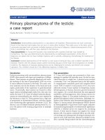
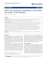
showed no evidence of malignancy or infection. Plain
radiographs demonstrated satisfactory placement of t he
cement in the verte bral body ( Figure 1). He was dis-
charged on the postoperative day si x pain free and neu-
rologically intact.
Six weeks after the initial operation, the patient com-
plained o f worsening thoracolumbar back pain (Visual
Analogue Scale (VAS) 8) requiring hospitalization. On
physical examination, incomplete sensory paraplegia
below the L1 dermatome was present without motor
impairment. The white blood cell count was 14,800 G/L
(normal range 4000-10,000 G/L) and the C-reactive pro-
tein level was 75 mg/L (normal range 0-5 mg/L) . Plain
* Correspondence:
Department of Orthopaedics, University Hospital Marburg, Baldingerstrasse,
35033 Marburg, Germany
Schofer et al. Journal of Medical Case Reports 2011, 5:101
/>JOURNAL OF MEDICAL
CASE REPORTS
© 2011 Schofer et al; licensee BioMed Central Ltd. This is an Open Access article distributed under the terms of th e Creative Commons
Attribution License (http://creativec ommons.org/licenses/by/2.0), which permits unrestricted use, distribution, and reproduction in
any medium, pro vided the original work is properly cited.
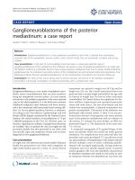
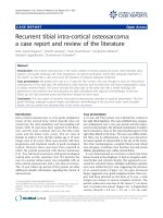
radiographs dem onstrated destruction and subtotal
resorption of the L1 vertebra, with the cement filling
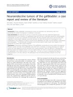
displaced and exposed (F igure 2). In addition, MRI
revealed L1 spondylitis with a right-sided psoas abscess
and compression of the lumb ar spinal cord (Figure 3).
These findings were consistent with a diagnosis of pyo-
genic spondylitis of the L1 vertebra after kyphoplasty.
Re-exploration was recommended but was refused by
the patient due to his poor general medical condition,
although he was informed about the risk of a progres-
sion to complete paralysis. The patient underwent CT-
guided aspiration and drainage of the psoas abscess.
Cultures grew group C hemolytic Streptococcus.Hewas
initially treated conservatively with a six-week course of
cefuroxime and clindamycin. The abscess cavity was irri-
gated daily with normal saline until drain removal on
post procedure day six.
The patient’ s symptoms progressed to leg paresis
without neurogenic bladder and/or bowel dysfunction.
He gave informed consent and underwent re-exploration
with dorsal spinal decompressio n, T12/L1 laminect omy
and T11 - L4 fusion using transpedicul ar fixation with a
dural rod system (Xia
®
, Stryker Howmedica
®
,Keil,
Germany). In a second procedure on postoperative day
10, ventral tran sphrenic bisegmental spondylodesis was
performed. After the removal of the residual L1 vertebra
with the cement body, adjacent discs and osteolytic end-
plates, an intracorporal stand-alone ti tanium cage (Obe-
lisc, Ulrich Medical, Ulm, Germa ny) was implanted
between T12 and L2. The patient was transferred to the
inpatient rehabilitation unit after 11 days. He made an
unevent ful recovery and his back pain impro ved signifi-
cantly (VAS 3). His neurological symptoms regressed
after six weeks, with normal biochemistry and no signs
of ongoing inflammation. At discharge, his pain was a
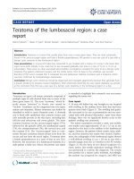
VAS 2; six months later, he was symptom free and com-
pletely a mbulatory without assistance (Figure 4). After
24 months, he had no complaints, neurologic deficit or
signs of infection. Plain radiographs demonstrated no
pseudarthrosis or disloc ation of screws, rods or the cage
(Figure 4).
Discussion
This is the first reported case of an infectious complica-
tion after kyphoplasty. Since 1998, kyphoplast y has been
Figure 1 Plain (A) and lateral (B) thoracolumbar radiographs
(T11 - L3) taken after initial kyphoplasty for treatment of an L1
compression fracture. The cement is correctly positioned in the
vertebral body.
Figure 2 Anterior posterior (A) and lateral (B) thora columb ar
radiographs (T11 - S1) six weeks after initial kyphoplasty. The
L1 vertebral body is partially resorbed. The osseous structure of the
L1 vertebral body cannot be delineated. The position of the left
cement block has shifted anteriorly and rostrally.
Figure 3 The magnetic resonance imaging T 1 gadolinium-
enhanced coronal image (A) shows spondylitis and a right-
sided psoas abscess. T1 without contrast transverse image of L1
(B) demonstrates the compressed spinal canal and inflamed right
psoas muscle. T1 sagittal image (C) shows spinal cord compression.
Schofer et al. Journal of Medical Case Reports 2011, 5:101
/>Page 2 of 4
gaining popularity for the treatment of symptomatic com-
pression fractures as outcomes have been shown to be
good [2,4]. Apart from asymptomatic cement leakage, the
morbidity is low. Complications after vertebroplasty are
also minimal, although there are 10 published cases of pri-
mary pyogenic spondylitis after vertebroplasty (Table 1)
[7-15]. Only one of these cases was without a significant
past medical history. Three were on immunosuppressive
medications, three had diabetes mellitus, three were diag-
nosed with acute urinary tract infections prior to vertebro-
plasty and one patient had Child’s A cirrhosis of the liver
secondary to prolonged alco hol abuse [8,11-13]. In addi-
tion, one patient had a grade II decubitus ulcer [12]. In
four, treatment was conservative without surgical interven-
tion [9-12]. The remaining six patients underwent re-
exploration to remove residual material and achieve
further stabilization [7,12-15]. One patient with pyogenic
spondylitis of T12 following T11 vertebroplasty was trea-
ted with drainage at T12 and subsequent vertebroplasty
using antibiotic cement [8].
There is no established evidence as to why more
infectious complications have been observed in verteb-
roplasty versus kyphoplasty. However, the incidence of
infectious complications may be attributable to comor-
bidities, suggesting that high-risk patients may need spe-
cific prophylactic antibiotic treatment in order to avoid
pyogenic spondylitis. Before our patient’s initial kypho-
plasty, preoperative imaging and blood tests did not
indicate an infectious source in the vertebral body; the
bone cylinder biopsy did not show signs of malignancy or
infection. Therefore, it is unlikely that an infection that
caused the spondylitis was already present. Although the
patient had a history of Parkinson’s disease and coronary
artery disease, these are not regarded as contraindications
to kyphoplasty. However, postoperative morbidity may be
increased with these comorbidities. One po ssible cause
Figure 4 Anterior posterior (AP) and lateral plain thoracolumbar
radiographs six and 24 months after reconstruction and
spondylodesis (T11 - L4). We performed the transpedicular fixation
with a dual rod system and vertebral replacement of the L1 vertebra
using an expandable cage. Reconstruction is stable on both AP and
lateral views at six months. Follow-up radiographs at 24 months show
no signs of pseudarthrosis or infection.
Table 1 Literature review of 10 reported cases of pyogenic spondylitis following vertebroplasty
Author Affected
vertebral
body
Side diagnosis Age Bacterium Therapy Time from
vertebroplasty
until infection
Deramond
[9]
Unstated Immunosuppressive therapy Unstated No detection Conservative Unstated
Kallmes
[10]
T12 Immunosuppressive therapy Unstated Staphylococcus epidermidis Conservative 1 month
Yu [14] T12 Urinary tract infection 78 No detection Dorsoventral
stabilization
1 month
Walker [13] T11 and T12 Urinary tract infection,
cholecystitis, meningitis,
diabetes mellitus
64 Enterobacter species Dorsoventral
stabilization
11 days
Walker [13] L3 Discectomy after
spondylodiscitis T12/L1
49 Staphylococcus aureus Dorsoventral
stabilization
8 months
Schmid
[11]
L3 - L5 Liver cirrhosis, alcohol abuse 55 No detection Conservative 2 weeks
Alfonso [7] L3 None 63 Serratia marcescens,
Stenotrophmonas maltophilia,
Burkholderia cepacia
Dorsoventral
stabilization
1 month
Vats [12] L1 Diabetes mellitus, decubital
ulcus II
73 Streptococcus agalactiae Conservative 6 months
Lin [15] T12 Immunosuppressive therapy,
urinary tract infection
65 Acinetobacter species Ventral stabilization 6 months
Chen [8] T11 Diabetes mellitus,
vertebroplasty T12
95 Proprioni acnes Drainage with
subsequent
vertebroplasty
2 months
Schofer et al. Journal of Medical Case Reports 2011, 5:101
/>Page 3 of 4
for an iatrogenic pyogenic infection could be contamina-
tion from skin flora [16]. Pyogenic spondylitis and spon-
dylodiscitis following spinal anesthesia have been
reported and this may have been the case in our patient;
if so, a single dose antibiotic prophylaxis with a first-
generation cephalosporin may have been inadequate. To
date, there are no official guidelines for antibiotic pro-
phylaxis in spinal surgery.
The cement traditionally used in kyphoplasty does not
contain antibiotics. However, the increasing use of anti-
biotic cement in endoprosthetic surgery is documented.
The use of antibiotic cement must be evaluated bearing
in mind a patient’s individual risk factors, such as age
and comorbidities. In immunocompromised patients,
the use of antibiotic cement and prolonged perioperative
antibiotic prophylaxis shouldbeconsideredinorderto
avoid infectious complications. In o ur case, we propose
thattheremaybeabenefitfromtheuseofantibiotic
cement in spine augmentation. This area requires
further investigation with controlled studies.
In addition, early and emergent s pinal cord decom-
pression of the spinal cord is the standard of care. Con-
servative treatment in this situation is not ideal but we
were limited by the patient’s refusal to proceed with our
initial recommendations. In this case, the primary pre-
senting symptom was recurrent severe back pain. There-
fore, severe back pain after a pain-free interval following
kyphoplasty must be investigated in order to rule out
pyogenic spondylitis. Another diagnosis in the differen-
tial that should be considered in such a scenario, espe-
cially without adjacent segment fractures, is vertebral
necrosis associated with cement injection.
Conclusion
Complications following kyphoplasty are rare, especially
compared with the number of surgeries performed. In
pyog enic spondylitis, treatment is laborious and extends
over a long period, often involving multiple surgeries. In
elderly patients and those with multiple comorbidities,
pyogenic spondylitis can be life-threatening. Therefore,
antibiotic prophylaxis is likely to be extremely important
for the prevention of infectious complications following
kyphoplasty in high-risk patients. In these patients, anti-
biotic cement should be considered.
Consent
Written informed consent was obtained from the patient
for publicatio n of this case report and any accompany-
ing images. A copy of the written consent is available
for review by the Editor-in-Chief of this journal.
Abbreviations
T11: eleventh thoracic vertebra; T12: twelfth thoracic vertebra; L1: first
lumbar vertebra; L2: second lumbar vertebra; L3: third lumbar vertebra; L4:
fourth lumbar vertebra; CT: computed tomography; MRI: magnetic
resonance imaging; VAS: visual analog scale.
Authors’ contributions
MDS, SL, CDP, TJH and MQ analyzed and interpreted the patient data. MDS
performed the surgery. MDS and MQ were the main authors of the
manuscript. All authors read and approved the final manuscript.
Competing interests
The authors declare that they have no competing interests.
Received: 14 March 2010 Accepted: 13 March 2011
Published: 13 March 2011
References
1. Garfin SR, Buckley RA, Ledlie J: Balloon kyphoplasty for symptomatic
vertebral body compression fractures results in rapid, significant, and
sustained improvements in back pain, function and quality of life for
elderly patients. Spine 2006, 31:2213-2220.
2. Hulme PA, Krebs J, Ferguson SJ, Berlemann U: Vertebroplasty and
kyphoplasty: a systematic review of 69 clinical studies. Spine 2006,
31:1983-2001.
3. Ledlie JT, Renfro MB: Kyphoplasty treatment of vertebral fractures: 2-year
outcomes show sustained benefits. Spine 2006, 31:57-64.
4. Schofer MD, Efe T, Timmesfeld N, Kortmann HR, Quante M: Comparison of
kyphoplasty and vertebroplasty in the treatment of fresh vertebral
compression fractures. Arch Orthop Trauma Surg 2009, 129:1391-1399.
5. Buchbinder R, Osborne RH, Ebeling PR, Wark JD, Mitchell P, Wriedt C,
Graves S, Staples MP, Murphy B: A randomized trial of vertebroplasty for
painful osteoporotic vertebral fractures. N Engl J Med 2009, 361:557-568.
6. Kallmes DF, Comstock BA, Heagerty PJ, Turner JA, Wilson DJ, Diamond TH,
Edwards R, Gray LA, Stout L, Owen S, et al: A randomized trial of
vertebroplasty for osteoporotic spinal fractures. N Engl J Med 2009,
361:569-579.
7. Alfonso Olmos M, Silva Gonzalez A, Duart Clemente J, Villas Tome C:
Infected vertebroplasty due to uncommon bacteria solved surgically: a
rare and threatening life complication of a common procedure: report
of a case and a review of the literature. Spine 2006, 31:E770-773.
8. Chen LH, Yang SC, Niu CC, Lai PL, Chen WJ: Percutaneous drainage
followed by antibiotic-impregnated cement vertebroplasty for pyogenic
vertebral osteomyelitis: a case report. J Trauma 2008, 64:E8-11.
9. Deramond H, Depriester C, Galibert P, Le Gars D: Percutaneous
vertebroplasty with polymethylmethacrylate. Technique, indications, and
results. Radiol Clin North Am 1998, 36:533-546.
10. Kallmes DF, Jensen ME: Percutaneous vertebroplasty. Radiology 2003,
229:27-36.
11. Schmid KE, Boszczyk BM, Bierschneider M, Zarfl A, Robert B, Jaksche H:
Spondylitis following vertebroplasty: a case report. Eur Spine J 2005,
14:895-899.
12. Vats HS, McKiernan FE: Infected vertebroplasty: case report and review of
literature. Spine 2006, 31:E859-862.
13. Walker DH, Mummaneni P, Rodts GE Jr: Infected vertebroplasty. Report of
two cases and review of the literature. Neurosurg Focus 2004, 17:E6.
14. Yu SW, Chen WJ, Lin WC, Chen YJ, Tu YK: Serious pyogenic spondylitis
following vertebroplasty - a case report. Spine 2004, 29:E209-211.
15. Lin WC, Lee CH, Chen SH, Lui CC: Unusual presentation of infected
vertebroplasty with delayed cement dislodgment in an
immunocompromised patient: case report and review of literature.
Cardiovasc Intervent Radiol 2008, 31(Suppl 2):S231-235.
16. Cogen AL, Nizet V, Gallo RL: Skin microbiota: a source of disease or
defence? Br J Dermatol 2008, 158:442-455.
doi:10.1186/1752-1947-5-101
Cite this article as: Schofer et al.: Primary pyogenic spondylitis following
kyphoplasty: a case report. Journal of Medical Case Reports 2011 5:101.
Schofer et al. Journal of Medical Case Reports 2011, 5:101
/>Page 4 of 4