Báo cáo y học: " Systemic lupus erythematosus associated with type 4 renal tubular acidosis: a case report and review of the literatur" pps
Bạn đang xem bản rút gọn của tài liệu. Xem và tải ngay bản đầy đủ của tài liệu tại đây (1.89 MB, 5 trang )
CASE REP O R T Open Access
Systemic lupus erythematosus associated with
type 4 renal tubular acidosis: a case report and
review of the literature
Haldane Porteous
1*
, Nadia Morgan
2
, Julio Lanfranco
1
, Monica Garcia-Buitrago
3
, Larry Young
4
and Oliver Lenz
5
Abstract
Introduction: Type 4 renal tubular acidosis is an uncommon clinical manifestation of systemic lupus
erythematosus and has been reported to portend a poor prognosis. To the best of our knowledge, this is the first
case report which highlights the successful management of a patient with systemic lupus erythematosus
complicated by type 4 renal tubular acidosis who did not do poorly.
Case presentation: A 44-year-old Hispanic woman developed a non-anion gap hyperkalemic metabolic acidosis
consistent with type 4 renal tubular acidosis while being treated in the hospital for recently diagnosed systemic
lupus erythematosus with multi-organ involvement. She responded well to treatment with corticosteroids,
hydroxychloroquine and mycophenolate mofetil. Normal renal function was achieved prior to discharge and
remained normal at the patient’s one-month follow-up examination.
Conclusion: This case increases awareness of an uncommon association between systemic lupus erythematosus
and type 4 renal tubular acidosis and suggests a positive impact of early diagnosis and appropriate
immunosuppressive treatment on the patient’s outcome.
Introduction
Inability of the kidney either to excrete sufficient net
acid or to retain sufficient bicarbonate results in a group
of disorders known as renal tubular acidoses (RTAs) [1].
These are normal anion gap hyperchloremic acidoses. In
the traditional classification, type 4 is the only variant
associated with hyperkalemia. Compared to the other
distalRTAs,intype4RTA,thecollectingductfailsto
excrete both protons and potassium. Such a scenario
arises when there is a quantitative or qualitative aldos-
terone deficiency or a genetic or acquired molecular
defect in the relevant transporters. Aldosterone activity
is necessary for adequate sodium reabsorption by the
epithelial sodium channels. These channels are located
on the luminal surface of principal cells in the terminal
portions of t he nephron. Under normal conditions, they
generate a lumen-negative potential, which is essential
for potassium and proton secretion [2].
RTA is a rare complication of systemic lupus erythe-
matosus (SLE) and can pose a diagnostic dilemma [3]. If
inappropriately treated, c hronic serum acidity ensues,
predisposing to growth retardation, nephrolithiasis, bone
disease, chronic kidney disease and even end-stage renal
disease. Type 4 RTA is less commonly associated with
SLE than type 1. Patients with type 4 RTA usually have
higher SLE disease activity index (SLEDAI) scores [4].
We report a case of a patient with a high SLEDAI score
and type 4 RTA secondary to SLE who received prompt
and appropriate treatment and did not do poorly.
Case presentation
A 44-year-old Hispanic woman presented to the hospital
with a six-month history of generalized weakness,
weight loss of 40 pounds over a four-month period a nd
a t wo-week history of progressively worsening dyspnea.
She had had hypertension for five years for which she
was being treated with lisinopril but had been noncom-
pliant for over three yea rs because of financial con-
straints. The patient denied any alcohol or illicit drug
use and was a lifelong non-smoker.
* Correspondence:
1
Department of Internal Medicine, University of Miami/Jackson Memorial
Hospital, 1611 NW 12th Avenue, Miami, FL 33136, USA
Full list of author information is available at the end of the article
Porteous et al. Journal of Medical Case Reports 2011, 5:114
/>JOURNAL OF MEDICAL
CASE REPORTS
© 2011 Porteous et al; licensee BioMed Ce ntral Ltd. This is an Open A ccess article distributed under the te rms of the Creative
Commons Attribut ion License ( which permits unrestricted use, distribution, and
reprodu ction in any medium, provided the original work is properly cited.
On admission, her blood pressure was 102/66 mm/Hg,
her temperature was 3 7°C, her respiratory rate was
24 breaths/minute, her oxygen saturation level was 92%
on room air and her pulse rate was 88 beats/minute.
A physical examination revealed an ill-looking woman
with mucosal pallor, generalized w asting and non-
tender, rubbery axillary and inguinal lymphadenopathy.
There was no evidence of cyanosis, digital clubbing, pit-
ting edema, skin rash or joint deformities. Her abdomen
was mildly distended but non-tender, with no organo-
megaly detected. Dullness to percussion and decreased
breath sounds over the left base were noted on the
respiratory system examination. The cardiovascular and
neurological examinations were unremarkable.
Initial laboratory investigations (Table 1) revealed ane-
mia, leukopenia, elevated blood urea nitrogen and ele-
vated serum creatinine. Urinalysis showed trace
protei nuria. Chest radiography revealed bilatera l pleural
effusions that were determined to be exudative in nature
on the basis of thoracocentesis. There was a high index
of suspicion for malignancy; however, the results of
chest, abdomen and pelvis computed tomography scans
and an axillary lymph node b iopsy did not confirm this.
Human immunodeficiency virus and tuberculin skin
tests were negative.
On day four of admission, she developed acute inflam-
matory arthritis of the elbows and knees. A serological
workup revealed high anti-nuclear antibody, anti-dou-
ble-stranded DNA antibody and anti-Smith antibody
titers, with low complement 3 levels. An active sediment
was noted on the basis of urinalysis (Table 2). The
patient’s renal impairment persisted, and her glomerular
filtration rate was estimated to be 53 ml/min/1.73 m
2
.
A review of blood results since admission showed evidence
of documented leukopenia on more than two occasions.
On day 5 of admission, the patient experienced dyspnea
and pleuritic chest pain. Reaccumulated pleural effusions
were noted on a chest radiograph, and an echocardiogram
showed evidence of pericardial effusion. Arterial blood gas
interpreted in conjunction with a corresponding basal
metabolic panel (Table 3) revealed the presence of a non-
anion gap hyperkalemic metabolic acidosis consistent with
type 4 RTA. Analysis of morning serum sample showed a
serum aldosterone level less than 1 ng/dl.
The renal ultra sou nd obtained showed normal kidney
morphology and no evidence of n ephrolithiasis. A renal
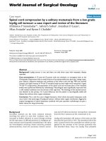
biopsy was done, which revealed diffuse global prol ifera-
tive and membranous glomerulonephritis. This was con-
sistent with lupus nephritis Renal Pathology Society/
International Society of Nephrology 2003 class IV-G(A)
and V, moderate activity index 9/24, minimal chronicity
index 1/12; minimal tubulointerstitial fibrosis and acute
tubular necrosis (Figure 1).
She was diagnosed with SLE complicated by a general-
ized lupus flare, with a SLEDAI score of 29. Overall dur-
ing this single admission, she demonstrated six of the 11
American College of Rheumatology criteria used in the
diagnosis of SLE.
The patient was treated with a course of hydroxychlor-
oquine and intravenous methylprednisone 1 g daily for
three days. Therea fter she was placed on a prednisone,
mycophenolate mofetil and hydroxychloroquine regimen.
Complete resolution of the renal impairment (Figures 2
and 3) and type 4 RTA (Figure 4) was achieved. She was
discharged after 19 days to follow-up in the Rheumatol-
ogy and Nephrology clinics. Her renal function remained
normal at the one-month follow-up clinic visit.
Discussion
The clinical manifestations of SLE are many and varied,
making it a plausible component of many differential diag-
noses [5]. It is one of several diseases known as “the great
imitators” because it often mimics other diseases. RTA is a
medical condition that involves an accumulation of acid in
the body due to a failure of the kidneys to appropriately
acidify the urine [1].
Table 1 Laboratory investigations on admission to
Jackson Memorial Hospital
Hemoglobin, 6.6 g/dl Na
+
, 131 mM/l
Hematocrit, 20.9% K
+
, 5.7 mM/l
Platelets, 544 × 10
9
/l Cl
-
, 105 mM/l
White blood cell
count,
3.5 × 10
9
/l
Neutrophils, 80.4% HCO
3
, 18 mM/l
Lymphocytes,
16.0%
Blood urea nitrogen,
60 mg/dl
Monocytes, 2.1% Creatinine, 1.69 mg/dl
Eosinophils, 0.3%
Table 2 Urinalysis results from day 4 of admission
pH 5 White blood cell count, 16 per high
power field
24-hour urinary protein,
0.81 g/day
Hyaline cast, zero to two per high-
power field
Red blood cell count, 27 per
high-power field
Squamous epithelial cells, one per
high-power field
Table 3 Arterial blood gas and basic metabolic panel
results from day 5 of admission
pH 7.34 Na
+
, 145 mM/l
HCO
3
, 14 mM/l K
+
, 5.5 mM/l
CO
2
, 26 mmHg Cl
-
, 120 mM/l
pO
2
, 96 mmHg HCO
3
, 19 mM/l
Blood urea nitrogen, 67 mg/dl
Creatinine, 1.09 mg/dl
Porteous et al. Journal of Medical Case Reports 2011, 5:114
/>Page 2 of 5
As many as 60% of adults with SLE develop overt
renal abnormalities, and 10% to 15% of patients with
lupus nephritis progress to end-stage renal failure [6].
RTA is rarely associated with SLE and, if present, is
more commonly of the type 1 than the type 4 variety
[4]. It likely represents the consequence of significant
tubulointerstitial damage, which should signal the need
for rapid treatment of the underlying lupus nephritis to
avoid future renal insufficiency [7].
In the setting of a hyperkalemic normal anion gap
metabolic acidosis and a uri ne pH less than 5.5, the
clinician should have a high index of suspicion for the
presence of type 4 R TA. Conversely, a diagnosis of
incomplete type 1 RTA may be entertained in patients
with hyperkalemia and a urine pH which is persistently
greater than 5.5 [8].
The presence of type 4 RTA can be confirmed by calcu-
lating the transtubular potassium concentr ation gradient
(TTKG) [9]. TTKG is an index of the potassium-secreting
activity of the cells in the distal tubule. In normal indivi-
duals, hyperkalemia is associated with increased aldoster-
one secretion and distal potassium excretion, leading to
a high TTKG level (usually greater than 10). A TTKG
level less than 7 is highly suggestive of hyporeninemic
hypoaldosteronism proba bly due to tubulointerstit ial
damage [10]. Our patient’ s calculated TTKG was 5.3
(Table 4).
Other causes of type 4 RTA were ruled out. Our
patient had a normal cortisol level of 14.4 μg/dl, making
adrenal insufficiency unlikely. She divulged no history of
taking non-steroidal anti-inflammatory drugs, potassium-
sparing diuretics, heparin, trimethoprim or angiotensin
receptor blocker use. A history of angiotensin-converting
enzyme inhibitor use was e licited; however, she was
completely non-compliant with the same for the past
three years, making this an unlikely cause.
Figure 1 Photomicrographs of the renal biopsy. (a) Lupus nephritis class IV-G(A) + V (hematoxylin and eosin stain; original magnification,
× 200). (b) Glomerulus showing global endocapillary proliferation, fibrinoid necrosis, neutrophilic infiltration and capillary wall thickening
(hematoxylin and eosin stain; original magnification, × 600). (c) Glomerulus showing mesangial expansion with hypercellularity and occlusion of
peripheral lumina (periodic acid-Schiff stain; original magnification, × 600). (d) Glomerulus showing thickened basement membranes with
numerous spikes and occasional dual contour (methenamine silver stain; original magnification, × 600). (e) Glomerulus showing areas of bright red
fibrinoid necrosis (trichrome stain; original magnification, × 600). (f) Minimal tubulointerstitial fibrosis (trichrome stain; original magnification, × 100).
Porteous et al. Journal of Medical Case Reports 2011, 5:114
/>Page 3 of 5
Upper Limit of Normal
Lower Limit of Normal
Figure 2 Graph showing trend of serum urea during admission.
12345678910111213141516171819
Creatinine mg/dl
1.69 1.59 1.25 1.06 1.02 1.08 1.11 1.12 1.41 1.46 1.17 1.31 1.09 1.09 0.84 0.91 0.91 0.91 0.76
0
0.2
0.4
0.6
0.8
1
1.2
1.4
1.6
1.8
CREATININE mg/dl
DAY OF ADMISSION
SERUM CREATININE TREND DURING COURSE OF ADMISSION
Creatinine mg/dl
Upper Limit of Normal
Lower Limit of Normal
Figure 3 Graph showing trend of serum creatinine during admission.
Porteous et al. Journal of Medical Case Reports 2011, 5:114
/>Page 4 of 5
Li et al. [4] noted the association between type 4 RTA
and SLE in patients with high SLEDAI scores. They
found that the degree of hyperkalemia was correlated
with a high SLEDAI score and that these patients had a
poor outcome of chronic renal insufficiency requiring
hemodialysis at admission or resulting in death. Patient s
with type 4 RTA have more extensive tubular damage
stemming from aggressive nephritis associated with
more aggressive sy stemic manifestations of SLE. Thus,
the presence of type 4 RTA is an indicator of more
aggressive SLE. In our patient, hyperkalemia and a h igh
SLEDAI score of 29 were noted; however, she did not
do poorly. Her type 4 RTA and significant renal impair-
ment resolved completely after two weeks of appropriate
therapy.
Conclusion
To the best of the authors’ knowledge this is the first
reported case of a patient with SLE who had a high
SLEDAI score and type 4 RTA secondary to lupus
nephritis, yet did not do poo rly. This case emphasizes
the fact that early diagnosis and appropriate treatment
can indeed result in the desired positive outcome.
Consent
Written informed c onsent was obtained from our
patient for publication of this case
report and any accompanying images. A copy of the
written consent is available for review by the Editor-in-
Chief of this journal.
Abbreviations
RTA: renal tubular acidosis; SLE: systemic lupus erythematosus; SLEDAI:
systemic lupus erythematosus disease activity index; TTKG: transtubular
potassium gradient.
Acknowledgements
We thank J Radkey of the Department of Pathology, Jackson Memorial
Hospital, for his help in preparing and interpreting the renal biopsy.
Author details
1
Department of Internal Medicine, University of Miami/Jackson Memorial
Hospital, 1611 NW 12th Avenue, Miami, FL 33136, USA.
2
Department of
Internal Medicine, State University of New York Downstate Medical Center,
450 Clarkson Avenue, Brooklyn, NY 11203, USA.
3
Department of Pathology,
University of Miami/Jackson Memorial Hospital, 1611 NW 12th Avenue,
Miami, FL 33136, USA.
4
Department of Rheumatology, Miami Veterans Affairs
Medical Center, 1201 NW 16th Street, Miami, FL 33125, USA.
5
Department of
Nephrology, University of Miami/Jackson Memorial Hospital, 1611 NW 12th
Avenue, Miami, FL 33136, USA.
Authors’ contributions
HP and NM collected the data and drafted the manuscript. HP and JL
contributed to the treatment of the patient. LY and OL participated in
critical revision of the report and helped draft the manuscript. All authors
read and approved the final manuscript.
Competing interests
The authors declare that they have no competing interests.
Received: 1 May 2010 Accepted: 24 March 2011
Published: 24 March 2011
References
1. Laing CM, Toye AM, Capasso G, Unwin RJ: Renal tubular acidosis:
developments in our understanding of the molecular basis. Int J Biochem
Cell Biol 2005, 37:1151-1161.
2. Karet FE: Mechanisms in hyperkalemic renal tubular acidosis. J Am Soc
Nephrol 2009, 20:251-254.
3. Caruana RJ, Barish CF, Buckalew VM Jr: Complete distal renal tubular
acidosis in systemic lupus: clinical and laboratory findings. Am J Kidney
Dis 1985, 6:59-63.
4. Li SL, Liou LB, Fang FT, Tsai WP: Symptomatic renal tubular acidosis (RTA)
in patients with systemic lupus erythematosus: an analysis of six cases
with new association of type 4 RTA. Rheumatology 2005, 44:1176-1180.
5. Rahman A, Isenberg DA: Systemic lupus erythematosus. N Engl J Med
2008, 358:929-939.
6. Cameron JS: Lupus nephritis. J Am Soc Nephrol 1999, 10:413-424.
7. Gur H, Kopolovic Y, Gross DJ: Chronic predominant interstitial nephritis in
a patient with systemic lupus erythematosus: a follow up of three years
and review of the literature. Ann Rheum Dis 1987, 46:617-623.
8. Bagga A, Sinha A: Evaluation of renal tubular acidosis. Indian J Pediatr
2007, 74:679-686.
9. Ethier JH, Kamel KS, Magner PO, Lemann J Jr, Halperin ML: The
transtubular potassium concentration in patients with hypokalemia and
hyperkalemia. Am J Kidney Dis 1990, 15:309-315.
10. West ML, Marsden PA, Richardson RM, Zettle RM, Halperin ML: New clinical
approach to evaluate disorders of potassium excretion. Miner Electrolyte
Metab 1986, 12:234-238.
doi:10.1186/1752-1947-5-114
Cite this article as: Porteous et al.: Systemic lupus erythematosus
associated with type 4 renal tubular acidosis: a case report and review
of the literature. Journal of Medical Case Reports 2011 5:114.
Figure 4 Graph depicting achievement and maintenance of
normal serum potassium, bicarbonate and urine pH in keeping
with resolution of type 4 renal tubular acidosis.
Table 4 Calculation of transtubular gradient
Urine sodium, 59 mM/l Transtubular gradient
Urine potassium, 38 mM/l Urine K/plasma K
Urine osmolality, 405 mOsm/kg Urine osmolality/plasma osmolality
Plasma potassium, 5.1 mM/l
Plasma osmolality, 288 mOsm/kg Patient’s transtubular gradient = 5.3
Porteous et al. Journal of Medical Case Reports 2011, 5:114
/>Page 5 of 5