Báo cáo y học: " Unilateral, trifocal, diaphyseal fracture of the radius with ipsilateral mid-shaft ulna fracture in an adult: a case report" doc
Bạn đang xem bản rút gọn của tài liệu. Xem và tải ngay bản đầy đủ của tài liệu tại đây (625.54 KB, 4 trang )
CAS E REP O R T Open Access
Unilateral, trifocal, diaphyseal fracture of the
radius with ipsilateral mid-shaft ulna fracture
in an adult: a case report
Mazin Ibrahim
1*
, Jenny Cwilewicz
2
, Osman H Khan
3
and Anthony Gibbon
4
Abstract
Introduction: To the best of our knowledge, a trifocal, diaphyseal fracture of the radius associated with ipsilateral
mid-shaft fracture of the ulna in an adult has not been reported in the literature to date. The AO classification
system does not include such a fracture configuration.
Case presentation: We report a case of trifocal, diaphyseal fracture of the radius with a mid-diaphyseal fracture of
the ulna in a 53-year-old Caucasian, British, right-hand dominant woman involved in a head-on collision with
another vehicle. The management of this rare fracture configuration is described and alternative treatment options
discussed.
Conclusions: We describe an unusual, complex fracture, which with prompt surgical treatment resulted in a rapid,
full and satisfactory functional recovery for our patient.
Introduction
Both bone forear m, diaphyseal fractures are commonly
encountered in clinical practice. Segmental radius shaft
fractures are, however, less commonly seen. We report a
case of trifocal, complex diaphyseal fracture of the
radius with ipsilateral mid-shaft fracture of the ulna.
Our review of the scientific literature revealed no evi-
dence of a ny previous reports relating to the surgical
treatment of such a fracture. However, the management
of a trifocal ulna fracture with bifocal radius fracture in
a child has been described previously.
Case presentation
A 53-year-old Cauc asian British, right-ha nd dominant
woman was involved in a road traffic accident while
drivi ng a car, involving a head-on co llision with another
vehicle at approxim ately 30 miles/hour. She sustained a
closed injury to the left forearm against the steering
wheel, resulting in obvious clinical def ormity. No neuro-
vascular deficit was evident.
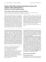
Radiographs revealed a displaced and angulated trif o-
cal fracture of the radial shaft in combination with a
displaced two-part mid-shaft ulna fracture (Figure 1).
Within 24 hours an open reduction and internal fixation
of the fracture was performed.
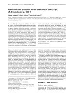
Under general anesthesia, using a direct subcutaneous
approach to the ulna, the ulna was reduced and fixed
with a seven-hole titanium dynamic compression plate
(DCP; Figure 2); 1 mm compression was applied.
The radius was exposed using Henry’ sapproach.The
distal radius fracture was fixed using a five-hole titanium
DCP while applying 1 mm compression. The proximal
three radius fragments were fixed with a nine -hole DCP
(Figure 2). Ca reful handling of the soft tissues was para-
mount and extra care was taken to avoid devascularising
any of the bone fracture segments. We also applied
1 mm compression to the proximal fracture. The middle
fracture was bridged because of inherent comminution.
After wound closure, an above-elbow back slab was
applied with the elbow held in 90 degrees of flexion.
The forearm was held elevated in a sling and our patient
was monitored for signs of compartment syndrome. Our
patient was disc harged from hospital after 48 hours of
observation in a broad arm sling; there were no immedi-
ate post-operative complications.
After two weeks, the back slab and the skin staples
were removed. There was no neurovascular deficit; only
* Correspondence:
1
24 Pinsent Court, York, UK
Full list of author information is available at the end of the article
Ibrahim et al. Journal of Medical Case Reports 2011, 5:123
/>JOURNAL OF MEDICAL
CASE REPORTS
© 2011 Ibrahim et al; licensee BioMed Central Lt d. This is an Open Acce ss articl e distribut ed under the te rms of the Creati ve Commons
Attribution License (http://creative commons.org/licenses/by/2.0), which permits unrestricted use, distribution, and reproduction in
any medium, provided the original work is properly cited.
a minor but improving subjective altered sensation over
the dorsal first web space. The range of active supina-
tion was slightly reduced, but otherwise a good range of
movement was demonstrated. Our patient was left free
of a cast and advised to mobilize her forearm.
At six weeks follow-up, our patient showed further
functional improvement. A weakened power grip was
noted and physiotherapy initiated. Results as seen on
radiographs were satisfactory.
After three months, our patient returned to work as a
cashier. She was pain free but reported a weakness in
the left forearm and occasional paresthesia over the dor-
sal first web space.
Our patient completed the Disabilities of the Arm,
Shoulder and Hand (DASH) questionnaire and scored
49.1 (measures scaled on a z ero to 100 scale: a higher
score indicates greater disability). She was finding lifting
tasks difficult and did not yet feel able to drive. She had
good and equal active and passive range of movement
of both wrist and elbow. Grip and pincer strength were
measured and values revealed an objective weakness o n
the left, although this wasconfoundedbydominant
limb strength variation.
Our patient’s final review took place six months af ter
the initial injury. She had made a complete functional
recovery with a full range of movement of elbow and
wrist joints, equal on both sides. The altered sensation
over the first dorsal web space of the left hand had con-
tinued to improve over time. She had resumed driving,
remained pain free and her grip strength had been
restored.
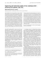
Radiographs revealed that the fractures had united
(Figure 3) and our patient was subs equently discharged.
No plan was made to remove the plates in the future.
Discussion
While diaphyseal fractures of the radius and ulna are
common, the trifocal diaphyseal fracture of the radius
with concurrent mid-shaft ulna fracture is much less
frequently encountere d. The AO classification system of
diaphysea l radius and ulna fractures has described com-
plex fractures of both bones, but only bifocal injuries
(that is, involving two points of fracture along a single
bone) [1]. No classification system as yet has described
this particular type of injury.
Our literature search did not reveal any similar cases.
However, there has been a reported case of an ipsilateral
diaphyseal fracture of the radius, ulna and radial head
[3]. This injury was similarly fixed with DCP plates but
Figure 1 Left forearm in an above-elbow back slab
(anteroposterior and lateral views from the initial injury).
Figure 2 Open reduction and internal fixati on of left radius
and ulna with dynamic compression plating (post-operative
films).
Figure 3 Left forearm open reduction and internal fixation of
radius and ulna at 6-month follow-up (anteroposterior and
lateral views).
Ibrahim et al. Journal of Medical Case Reports 2011, 5:123
/>Page 2 of 4
our patient also required a radial head replacement. Our
patient regained an almost full range of movement.
A closed ipsilateral supracondylar humerus with trifo-
cal ulna and bifocal radius fractures has also been
reported [2]. Intra-medullary pinning of diaphyseal frac-
tures of both forearm bones in adults has been found to
provide good outcomes [4], as has the use of an inter-
locking intra-medullary nail , with a mean union time of
15 weeks using an open reduction technique [5].
A retrosp ective study into 38 cases involving complex
fractures of the proximal radius and ulna in adult
patients, treated via various methods has been per-
formed. In this study there were seven early revisions
due to disassembly of the fixation system, deep infection
and insufficient fixation. A number of late complications
arose including non-union and malalignment [6]. In
cases of non-union of fractures of the radius and ulna
thereisevidencetosuggestthat plate fixation with
autologous cancellous bone grafting can resul t in high
rates of union and improved upper limb function [7].
In our case, we felt that we could achieve a satisfac-
tory outcome with open r eduction and internal fixation
using a DCP. The DCP would afford us anatomical
reduction, and rigid fixation with rotational control to
support fracture union of all segmen ts. There was con-
sideration made of using an intra-medullary nail, but
this would not provide either rigid fixation or rotational
control, and hence led to non-union, malunion and
hence a poor functional outcome. Pre-bent intra-medul-
lary nails with interlocking provide rotational stability
and reduce the risks of non-union and malunion. How-
ever, their use would be of limited value in controlling a
trifocal fracture.
Bone grafting was deemed unnecessary in our case as
we managed to reduce the fractures anatomically, under
direct compression. The radial bow and length was
achieved intra-operatively. We would advocate the use
of b one graft in such circumstances where there is
marked comminution at fracture ends a nd where ana-
tomic reduction cannot be achieved. In our patient’s
case, we also believe that careful preservation of vascu-
larity of the bone fracture segments obviated the need
for bone grafting.
Additionally, consideration was made to using a single
long DCP plate to fix the segmental radius fracture.
However, it would have been extremely diffi cult to con-
tour the plate. External fixation was deemed inappropri-
ate i n the management of this fracture pattern because
it would not allow us t o achieve anatomic reduction,
rigid fixation, and rotational control would have been
difficult to achieve.
It is well known that the use of DCP plates causes the
phenomenon of ‘stress shielding’.Inourpatient’s case,
the area of bone between the two radial DCP plates was
at potentially higher risk of fracture following another
episode of trauma, as a result of ‘ stress shielding’ .
Hence, one could advocate the removal of t he DCP
plates after fracture union. We are not planning to
remove the plates; however removal of the dist al radial
DCP plate would be easier to achieve, with a lower risk
of nerve injury, and the proximal plate could remain.
Removal of the proximal plate would be associated with
a higher risk of nerve injury.
We felt that locked compression plates were not
necessary, in view of our patient’s good bone quality.
However, they would play a useful role in older patients
who have poorer bone stock.
This report highlights a rare injury and its successful
management. Prompt surgical intervention with the
appropriate method of open reduction and internal fixa-
tion can lead to a good result.
Conclusions
The present case report highlights a rare combination of
injuries. While such injuries occur infrequently, we
should try to obtain anatomical reduction and rigid fixa-
tion to achieve the best possible functional outcome,
improve the chance of fracture union and possibly
reduce the incidence of post-operative complications.
Consent
Written informed consent was obtained from the patient
for publication of this case report and any accompany-
ing images. A copy of the writ ten consent is availabl e
for review by the Editor-in-Chief of this journal.
Author details
1
24 Pinsent Court, York, UK.
2
9 Hambleton Avenue, York, UK.
3
Orthopaedics
Department, Pinderfield Hospital, Wakefield, UK.
4
Orthopaedics Department,
York Hospital, York, UK.
Authors’ contributions
MI and JC followed up our patient, collected radiograph material, wrote the
manuscript and obtained patient consent. MI completed the initial literature
search. OK contributed to the discussion and took an editorial role. AG
performed the operation and took an editorial role. All authors read and
approved the final manuscript.
Competing interests
The authors declare that they have no competing interests.
Received: 1 April 2010 Accepted: 29 March 2011
Published: 29 March 2011
References
1. Radius/ulna diaphysis. [ />2. Ravi Mittal MS, Vijay Sharma MS: Ipsilateral supracondylar humeral and
segmental both bones forearm fracture in a child: a case report. Middle
East J Emerg Med 2005, 1:3.
3. Rafiq I, Kumar K, Sutherland AG: Ipsilateral diaphyseal fractures of radius,
ulna and radial head: case report. Internet J Orthopaedic Surg 2006, 3:1.
4. Mseddi MB, Manicom O, Filippini P, Demoura A, Pidet O, Hernigou P:
Intramedullary pinning of diaphyseal fractures of both forearm bones in
adults: 46 cases. Rev Chir Orthop Reparatrice Appar Mot 2008, 94:160-167.
Ibrahim et al. Journal of Medical Case Reports 2011, 5:123
/>Page 3 of 4
5. Gao H, Luo CF, Zhang CQ, Shi HP, Fan CY, Zen BF: Internal fixation of
diaphyseal fractures of the forearm by interlocking intramedullary nail:
short-term results in eighteen patients. J Orthop Trauma 2005, 19:384-391.
6. Chick G, Court C, Nordin JY: Complex fractures of the proximal end of the
radius and ulna in adults: a retrospective study of 38 cases. Rev Chir
Orthop Reparatrice Appar Mot 2001, 87:773-785.
7. Ring D, Allende C, Jafarnia K, Allende BT, Jupiter JB: Ununited diaphyseal
forearm fractures with segmental defects: plate fixation and autogenous
cancellous bone grafting. J Bone Joint Surg Am 2004, 86A:2440-2445.
doi:10.1186/1752-1947-5-123
Cite this article as: Ibrahim et al.: Unilateral, trifocal, diaphyseal fracture
of the radius with ipsilateral mid-shaft ulna fracture in an adult: a case
report. Journal of Medical Case Reports 2011 5:123.
Submit your next manuscript to BioMed Central
and take full advantage of:
• Convenient online submission
• Thorough peer review
• No space constraints or color figure charges
• Immediate publication on acceptance
• Inclusion in PubMed, CAS, Scopus and Google Scholar
• Research which is freely available for redistribution
Submit your manuscript at
www.biomedcentral.com/submit
Ibrahim et al. Journal of Medical Case Reports 2011, 5:123
/>Page 4 of 4