báo cáo khoa học: "Effect of iron oxide and gold nanoparticles on bacterial growth leading towards biological application" ppt
Bạn đang xem bản rút gọn của tài liệu. Xem và tải ngay bản đầy đủ của tài liệu tại đây (1.73 MB, 7 trang )
RESEARCH Open Access
Effect of iron oxide and gold nanoparticles on
bacterial growth leading towards biological
application
Saptarshi Chatterjee, Arghya Bandyopadhyay and Keka Sarkar
*
Abstract
Background: Nanoparticle-metal oxide and gold represents a new class of important materials that are
increasingly being developed for use in research and health related activities. The biological system being
extremely critical requires the fundamental understanding on the influence of inorganic nanoparticles on cellular
growth and functions. Our study was aimed to find out the effect of iron oxide (Fe
3
O
4
), gold (Au) nanoparticles on
cellular growth of Escherichia coli (E. coli) and also try to channelize the obtained result by functionalizing the Au
nanoparticle for further biological applications.
Result: Fe
3
O
4
and Au nanoparticles were prepared and characterized using Transmission electron microscopy
(TEM) and Dynamic Light Scattering (DLS). Preliminary growth analysis data suggest that the nanoparticles of iron
oxide have an inhibitory effect on E. coli in a concentration dependant manner, whereas the gold nanoparticle
directly showed no such activity. However the phase contrast microscopic study clearly demonstrated that the
effect of both Fe
3
O
4
and Au nanoparticle extended up to the level of cell division which was evident as the abrupt
increase in bacterial cell length. The incorporation of gold nanoparticle by bacterial cell was also observed during
microscopic analysis based on which glutathione functionalized gold nanoparticle was prepared and used as a
vector for plasmid DNA transport within bacterial cell.
Conclusion: Altogether the study suggests that there is metal nanoparticle-bacteria interaction at the cellular leve l
that can be utilized for beneficial biological application but significantly it also posses potential to produce
ecotoxicity, challenging the ecofriendly nature of nanoparticles.
Keywords: Bacterial Growth, magnetic nanoparticle, gold nanoparticle, Cytotoxicity
Background
Thepresenterabelongstonanotechnology.Withthe
tremendous growth in the field of science, nanobiotech-
nology has come up as a major interdisciplinary subject.
The development and application of nanotechnology has
the potential to improve greatly the quality of life. An
improved understanding of nanoparticles and biological
cell interaction can lead to the development of new sen-
sing, diagnost ic and t reatment capabilities [1- 4] such as
improved targeted drug delivery, gene therapy, magnetic
resonance imaging contrast agents and biol ogical war-
fare agent detection [5, 6]. For ins tance iron oxide nano-
particle has been widely used as carriers for targeted
drug delivery to treat several t ypes of cancer [7,8] in
biomedical research because of its biocompatibility and
magnetic properties [9,10]. Gold nanoparticle is the
other mostly appli ed nanoparticle in the field of biome-
dical sciences expanding from immunoassay [11] to in
vivo cancer targeting and imaging [12].
Though there are immense potentials of nanotechnol-
ogy, the cytotoxicity of the nanoparticles remain a major
concern. Different classes of bacteria exhibit different
susceptibil ities to nanoparticles [13] but the mechanism
controlling the toxicity is not yet understood. Moreover
different factors such as synthesis, shape, size, composi-
tion, addition of stabilizer etc can lead to different con-
clusions even for very closely related nanosuspensions
[14]. Thus the present study is aimed to investigate the
* Correspondence:
Department of Microbiology, University of Kalyani, Nadia, West Bengal, India
Chatterjee et al. Journal of Nanobiotechnology 2011, 9:34
/>© 2011 Chatterjee et al; licensee BioMed Central Ltd. This is an Open Access article distributed under the terms of the Creative
Commons Attribution License ( s/by/2.0), which permits unrestricted use, distribution, and
reproduction in any medium, provided the original work is properly cited.
effect of two widely used nanoparticles (Fe
3
O
4
& Au) on
the growth of E. coli. The growth study was followed by
microscopic study for detecting the morphological
changes. Finally, attempts were made to utilize the
results obtained for biological applications.
Result and Discussion
A) Characterization of nanoparticles
The nanoparticles (iron oxide & gold) synthesized in the
laboratory were characterized using TEM image (FEI,
Tecnai S-twin) and DLS (Malvern Zetasizer). The size of
magnetic nanoparticle was found to be 8 nm by TEM
image whereas Gold nanoparticle possessed size of 5 nm
(Figure 1, 2). The DLS data of F e
3
O
4
and Au nanoparti-
cles as shown in Figure 3, 4. indicated monodispersity.
B) Effect of Iron nanoparticle on bacterial growth
The comparative study on growth of bacteria under nor-
mal condition and under the influence of Magnetic
nanoparticle (Fe
3
O
4
) revealed the effect of Fe nanoparti-
cle on bacterial growth. The growth curve of E. coli
under normal conditions clearly depicted the lag, log,
stationary and death phase as shown in Figure 5 but
under the influence of various concentrations of iron
oxide nanoparticles (i.e 50 μg/mL, 100 μg/mL, 150 μg/
mL & 200 μg/mL) the gra dual shortening of log ph ase
was evident indicating the microbiostatic effect of iron
nanoparticle on E. col i in a concentration dependant
manner. The untreated bacterial sample at 6th hour
reached OD600 1.48 (cfu count 1.32 × 10
9
per mL)
compared to OD600 1.14 (cfu count 1.01 × 10
8
)incase
of iron oxide (200 μg/mL) treated bacterial cells (Figure
6). The reactive oxygen species (ROS) along with super-
oxide radicals (O
2-
), hy droxide radical (OH
-
) and singlet
oxygen (
1
O
2
) generated by the iron oxide nanopaticle is
thought to be the reason behind the inhibition [15].
ROS production has been found in diverse range of
metal oxide nanoparticles that may result in oxidat ive
stress, inflammation and consequent damage to pro-
teins, membranes and DNA which is one of the primary
mechanisms of nanotoxicity.
C) Effect of gold nanoparticle on bacterial growth
When E. coli was treated with various concentrations
(25 μg/mL, 50 μg/mL, 75 μg/mL & 100 μg/mL) of gold
nanoparticles no significant difference in the grow th
curve were obtained as sho wn in Figure 7. The growth
experiment under the influence of gold nanoparticle
thus reveals the nontoxic nature of the gold nanoparticle
in the bacterial system (E. coli). Hence, it can be used
for biological applications with least chances of
cytotoxicity.
D) Microscopic observation
The study was further extended at the microscopic level
using phase contrast microscope. Both the nanoparticles
were found to increase the size of the E. coli cell
abruptly. The bacterial cell size under the influence of
Fe
3
O
4
nanoparticle when compared to that of normal E.
coli cell (considering normal E. coli cell length to be
Figure 1 Transmission Electron Microscope (TEM) image of
Fe
3
O
4
nanoparticle showing the size of the nanoparticle to be
8 nm (approx).
Figure 2 Transmission Electron Microscope (TEM) image of Au
nanoparticle showing the size of the nanoparticle to be 5 nm
(approx).
Chatterjee et al. Journal of Nanobiotechnology 2011, 9:34
/>Page 2 of 7
approx. 3 μm as shown in Figure 8) showed up to a 10
fold increase in size Figure 9. The gold nanoparticle also
gave identical result where the increase of cell length
was up to 8 fold c ompared to that of normal E. coli cell
as shown in Figure 10. The E. coli cells were also found
to be clogged in between the iron oxide nanoparticles
because of the magnetic property of the nanoparticle
and the trapped cells also exhibited increased cell length
(Figure 11). Iron oxide nanoparticles due to the high
ionic strength frequently agglomerate in environmental
and biological fluids, which shield the repulsion due to
charges on the nanoparticles. Agglomeration has fre-
quently been ignored in nanotoxicity studies, even
though agglomeration would be expected to affect nano-
toxicity since it changes the size, surface area, and sedi-
mentation properties of the nanoparticles. Moreover
nanoparticles can agglomerate to some extent in the
environment or in the body before they reach their tar-
get; hence it is also desirable to study how toxicity is
affected by agglomeration [16]. Thus our study indicates
the effect of both the nanoparticles on the cellular level.
Inactivation of certain gene expression required for
‘cytokinesis’ during cell division may be considered as a
probable cause for such effect [17,18]. The result clearly
shows the involvement of the nanoparticles on the bac-
terial physiology and is a probable demonstration of
DNA nanoparticle interaction. The gold nanoparticle
showed high tendency for incorporation within bacterial
cells with the least possibility of cytotoxicity. This was
evident during microscopic study, where grain like shin-
ing spots appeared within the bacterial cells (Figure 12).
E) Biological Application of gold nanoparticle
incorporation within bacterial Cells
As t he incorporation of gold nanoparticle on E. coli cells
were evident, studies were conducted to use this phenom-
enon for bio-applications. Since glutathione has an electro-
static interaction with both gold nanoparticle and DNA,
the gold nanoparticle was surface modified using glu-
tathione followed by interaction with plasmid DNA. The
carboxyl group (COO
-
) of glycine residue electrostatically
interacts with the positivel y charged gold nan oparticle to
form glutathione functionalized gold nanoparticle. The
other free end (g-Glutamine residue) of glutathione now
posses an amine group and a carboxyl group among
which the amine group nonspecifica lly interacts with the
negatively charged phosphate group of DNA forming a
reversible electrostatic complex of gold-glutathione-DNA.
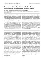
Figure 4 Size distribution intensity graph of Au nanoparticle as revealed by DLS.
Figure 3 Size distribution intensity graph of Fe
3
O
4
nanoparticle as revealed by DLS.
Chatterjee et al. Journal of Nanobiotechnology 2011, 9:34
/>Page 3 of 7
This compl ex cleaves when incorporated within the bac-
terial cell due to ionic variation liberating the intact plas-
mid DNA from go ld-glutathione complex. In our
experiment the glutathione surface functionalized gold
nanoparticles were used as a vector to insert ampicillin
resistant gene (pUC 19) in E. coli that is susceptible to
ampicillin. The result showed successful transformation of
ampicillin resistant gene in E. coli as indicated by the
growth of transformed bacteria in appropriate antibiotic
containing media. The transformation effi ciency was cal-
culated as: Transformation efficiency = (Number of trans-
formed colony/ Amount of DNA in μg ) and was found to
be 8.53 × 10
5
comparedtothatof9.55×10
3
using con-
ventional CaCl
2
mediated transformation. Thus we report
glutathione functionalized gold nanoparticle mediated
transformation as a bio-application for w hich further
research is to be carried out to make this process
generalized.
Conclusion
Finally if we consider the recent past age to be of micro
scale then the present or near future surely belongs to
nano. Since most of the natural processes also take
place in the nanometer scale therefore the associa tion of
nanotec hnolo gy and biology is expected to s olve several
biological problems. But the advanc es of the technology
in the nanoscale level also remind the possible negative
impact especially at the cellular level. From our research
the interaction of two widely used nanoparticles with
the bacterial cell was evident which opened a new
dimension of biological application in the form of Au
mediated transformation, though further research on the
mechanism of interaction can reveal the further conse-
quences which may open up a new domain of study
called ‘nanotoxicity’. However, as a cautionary note, the
results presented are not meant to be generalized
beyond the material and biological systems and condi-
tions reported here.
Moreover our study proves the effect can be modified
and channelized for human benefit.
Proper knowledge of these interactions can lead to a
safe era of nanotechnology without threat of human
health risk.
Methods
A) Preparation of Nanoparticles
i) Iron (Fe) Nanoparticle
Magnetic nanoparticles were prepared by chemical
coprecipitation of Fe
2+
and Fe
3+
ions in an alkaline solu-
tion and followed by a treatment under hydrothermal
conditions [19]. 2.7 g FeSO
4
,7H
2
O and 5.7 g FeCl
3
dis-
solved in 10 m L nanopure water (double dist ill ed water
filtered through 200 μm filter) separately. These two
solutions was thoroughly mixedandaddedtodouble
volume 10 M ammonium hydroxide with constant stir-
ring at 25°C. Then the dark blac k slurry of Fe
3
O
4
parti-
cles was heated at 80°C in a water bath f or 30 min. The
particles thus obtained exhibited a strong magnetic
response. Impurity ions such as chlorides and sulphates
were removed by washing the particles several times
with nano pure water. Then the particles are dispersed
Figure 5 Growth curve of E. coli under the influence of Fe
3
O
4
nanoparticle compared to the normal growth curve of E. coli depicting
the microbiocidal nature of the Fe
3
O
4
nanoparticle in a concentration dependant manner.
Figure 6 Comparison of colony forming unit (cfu) count of E.
coli (normal) and under the influence of Fe
3
O
4
nanoparticle at
6th hour of bacterial growth.
Chatterjee et al. Journal of Nanobiotechnology 2011, 9:34
/>Page 4 of 7
in 20 mL nanopure water and sonicated for 10 min a t
60 MHz. The yield of precipitated magnetic nanoparti-
cles was determined by removing known aliquots of the
suspension and drying to a constant mass in an oven at
60°C. The prepared magnetic nanoparticles were stable
at room temperature (25-30°C) without getting
agglomerated.
ii) Gold (Au) Nanoparticle
3mMHAuCl
4
solution was directly reduced by 10 mM
NaBH
4
solution under st irring condition. For further
and complete reduction the reaction mixture was
reduced again by 10 mg/ml solution of dextrose.
Obtained mixture was subjected to over constant
Figure 7 Growth curve of E. coli under the influence of Au nanoparticle compared to the normal growth curve of E. coli indicating the
nontoxic nature of Au nanoparticle.
Figure 8 Phase Contrast Microscopic image of E. coli grown
under normal condition.
Figure 9 Abrupt increase in E. coli cell length (up to 10 fold)
grown under the influence of iron oxide nanoparticles, as
observed under phase contrast microscope.
Figure 10 Abrupt increase in E. coli cel l length (u p to 8 fold)
grown under the influence of iron oxide nanoparticles, as
observed under phase contrast microscope.
Chatterjee et al. Journal of Nanobiotechnology 2011, 9:34
/>Page 5 of 7
stirring. Then the mixture was washed several times
with methanol using centrifugation at 65,000 rpm.
iii) Glutathione modified Gold (Au) Nanoparticle
Typically, 3.0 mM of glutathione was dissolved in 40 mL
of distilled water, and 1.0 mM of HAuCl
4
was dissolved
in 80 m L of methanol. Mixing the two solutions gener-
ates a cloudy, white suspension. Addition of 10 m M o f
NaBH
4
in 10 mL of water to this stirring suspension
results in an immediate color change to dark brown
indicating the generatio n of large cluster compounds.
After addit ional stirring, the solution was evaporated at
43°C to near dryness and excess me thanol was added to
precipitate the clusters and wash reaction byproducts
and any remaining starting material. The precipitate was
then filtered and redissolved in 10 mL of distilled water,
precipitated again with methanol, and filtered. These
steps were repeated until a fine black powder was
obtained [20].
B) Growth Experiment
Test organism Escherichia coli (E. coli)weregrown
separately in 50 mL sterilized Luria Bertani (LB) broth
medium and kept in shaker incubator at 37°C for 14
hour (overnight incubation). On the subsequent day test
organism cultures were transfe rred at the rate of 1% in
100 mL LB broth kept in 250 mL conical flasks. Various
concentrations of nanoparticles (For Fe
3
O
4
50 μg/mL to
200 μg/mL and for Au 25 μg/mL to 100 μg/mL) w ere
carefully placed into each flask, leaving one as a control
to track the normal growth of the microbial cells with-
out nanoparticles. Experiments were performed using
both a negative control (flask containing cells plus
media) and a positive control (flask containing nanopar-
ticles plus media). The flasks were shaken at 180 rpm
and 37°C in a shaker incubator. Optical density mea-
surements from each flask were taken every one hour to
record the growth of the microbes in a spectrophot-
ometer set at 600 nm. The growth ra te of microbial
cells interacting with the nanoparticles was determined
from a plot of the log of the optical density versus time.
C) Microscopic Study
The micro scopic study on the morphology i.e the shape,
size of the bacteria and interaction with the inorganic
nanoparticles were conducted using Phase contrast
microscope (Leica DM 750). 10 μLofculturewaswith-
drawn every hour and microscopic study was conducted.
The parameters were compared between normal culture
and culture under the influence of inorganic nanopar ti-
cles (Fe
3
O
4
& Au).
D) Biological Application of Au Nanoparticle
As the property of internalization of Au nanoparticle
within the E. coli cell was observed, the phenomenon
was further investigated for its potential to be used for
biological application. The insertion of Ampicillin resis-
tant gene in the form of pUC 19 (Plasmid) was tried
Figure 11 E. coli cells with abrupt cell length seen to be
clogged in between the iron oxide nanoparticle when viewed
under the phase contrast microscope.
Figure 12 Incorporation of Au nanoparticle was observed in the bacterial cell.
Chatterjee et al. Journal of Nanobiotechnology 2011, 9:34
/>Page 6 of 7
using the Au nanoparticles as vector/transport machin-
ery. E. coli cells wer e grown on LB (Luria Bertani) broth
till the O.D reaches 0.2. 10 μL of Au nanoparticles (50
μg/mL) were allowed to interact with 10 μLpUC19
DNA (Bioserve, India) taken from a stock of 0.32 ng/μL
for 2 hours at 37°C. Subsequently 1 mL of the E. coli
culture (0.2 O.D) w as centrifuged at 10,000 rcf for 1
min and 20 μL of pUC 19-Au nanoparticle mixture was
added to the pellet. 980 μLoffreshLBmediumwas
alsoaddedtoit,mixedandincubatedat37°Cfor5hrs
in shaking condition. Finally 100 μL of the culture were
withdrawn and plated on Luria Bertani agar medium
containing 50 μg/mL of ampicillin. The plates were
incubated at 37°C overnight and numbers of colon ies
were counted. The cfu (Colony Forming Unit) count
express the number of E. coli cells which posses the
ampicillin resistant property acquired due to insertion of
pUC19plasmid.Thecfucountforthenumberofbac-
terial cells in the initial stage was also noted to compare
the number of transformed cell to that of total bacterial
cell. This efficiency of this method was also compared
to that of conventional method [21] using CaCl
2
mediated transfer of plasmid DNA in competent cells.
Authors information
S. Chatterjee: M.Sc. Microbiology, Research Scholar,
University of Kalyani.
A. Bandyopadhyay: M.Sc. Microbiology, Research
Scholar, University of Kalyani.
K.Sarkar:M.Sc.,PhD, Asst. Professor, Dept. of
Microbiology, University of Kalyani.
Acknowledgements
The research work has been carried out with the financial support of Dept.
of Science & Technology, Govt. of India (Project-Nanomission: SR/NM/
NS-48/2009) and University of Kalyani, Nadia, West Bengal.
Authors’ contributions
SC carried out the growth experiments and biological application part
whereas AB was engaged in the synthesis and characterization of
nanoparticles. KS supervised in the design of the study along with critical
interpretations while drafting the manuscript. All authors read and approved
the final manuscript.
Competing interests
The authors declare that they have no competing interests.
Received: 30 June 2011 Accepted: 23 August 2011
Published: 23 August 2011
References
1. Chan WCW, Nie S: Quantum dot bioconjugates for ultrasensitive
nonisotopic detection. Science 1998, 281:2016-2018.
2. Chouly C, Pouliquen D, Lucet I, Le Jeune JJ, Jallet P: Development of
superparamagnetic nanoparticles for MRI: effect of particle size, charge
and surface nature on biodistribution. J Microencapsulation 1996,
13:245-255.
3. Couvreur P, Dubernet C, Puisieux F: Controlled drug delivery with
nanoparticles: current possibilites and future trends. Eur J Pharm
Biopharm 1994, 41:2-13.
4. Douglas SJ, Davis SS, Illum L: Nanoparticles in Drug Delivery. Crit Rev Ther
Drug Carrier Syst 1987, 3:233-261.
5. Pouliquen D, Perroud H, Calza F, Jallet P, Le Jeune JJ: Investigation of
magnetic properties of iron oxide nanoparticles used as contrast agent
for MRI. Magnetic Resonance in Medicine 1992, 24:75-84.
6. Pinto-Alphandary Andremont HA, Couvreur P: Targeted delivery of
antibiotics using liposomes and nanoparticles: research and applications.
Int J Antimicrob Agents 2000, 13:155-168.
7. Chertok B, Moffat BA, David AE, Yu F, Bergemann C, Ross BD, Yang VC: Iron
oxide nanoparticles as a drug delivery vehicle for MRI monitored
magnetic targeting of brain tumors. Biomaterials 2008, 29(4):487-496.
8. Kohler N, Sun C, Wang J, Zhang M: Methotrexate-modified
superparamagnetic nanoparticles and their intracellular uptake into
human cancer cells. Langmuir 2005, 21(19):8858-8864.
9. Gupta AK, Gupta M: Synthesis and surface engineering of iron oxide
nanoparticles for biomedical applications. Biomaterials 2005,
26(18):3995-4021.
10. Berry CC, Curtis ASG: Functionalisation of magnetic nanoparticles for
applications in biomedicine. J Phys D Appl Phys 2003, 36:R198-R206.
11. Hirsch LR, Halas NJ, West JL: Whole-blood immunoassay facilitated by
gold nanoshell-conjugate antibodies. Methods Mol Biol 2005, 303:101-11.
12. Gao X, Cui Y, Levenson RM, Chung LWK, Nie S: In vivo cancer targeting
and imaging with semiconductor quantum dots. Nat Biotechnol 2004,
22:969-76.
13. Fu G, Vary PS, Lin CT: Anatase TiO2 nanocomposites for antimicrobial
coatings. J Phys Chem B 2005, 109:8889-8898.
14. Warheit DB: How meaningful are the results of nanotoxicity studies in
the absence adequate material characterization? Toxicol Sci 2008,
101:183-185.
15. Sies H: Oxidative stress: oxidants and antioxidants. Exp Physiol
1997,
82(2):291-295.
16. Zook JM, Maccuspie RI, Locascio LE, Halter MD, Elliott JT: Stable
nanoparticle aggregates/agglomerates of different sizes and the effect
of their size on hemolytic cytotoxicity. Nanotoxicology 2010, 1-14, Early
online Edition, 13th Dec.
17. Osborn MJ, Rothfield L: Cell shape determination in Escherichia coli. Curr
Opin Microbiol 2007, 10(6):606-10.
18. Trun NJ, Gottesman S: On the bacterial cell cycle: Escherichia coli mutants
with altered ploidy. Genes Dev 1990, 4:2036-2047.
19. Bandyopadhyay A, Chatterjee S, Sarkar K: Rapid isolation of genomic DNA
from E. coli XL1 Blue strain approaching bare magnetic nanoparticles.
Current Science 2011, 101(2):210-214.
20. Schaaff TG, Whetten RL: Giant Gold-Glutathione Cluster Compounds:
Intense Optical Activity in Metal-Based Transitions. J Phys Chem B 2000,
104:2630-2641.
21. Sambrook J, Russell DW: Preparation and transformation of competent E.
coli using calcium chloride. In Molecular Cloning: A Laboratory Manual.
Volume 1 3 edition. New York, Cold Spring Harbor Laboratory Press;
2001:116, Volume 1
doi:10.1186/1477-3155-9-34
Cite this article as: Chatterjee et al.: Effect of iron oxide and gold
nanoparticles on bacterial growth leading towards biological
application. Journal of Nanobiotechnology 2011 9:34.
Chatterjee et al. Journal of Nanobiotechnology 2011, 9:34
/>Page 7 of 7