báo cáo khoa học: "Split-Inteins for Simultaneous, site-specific conjugation of Quantum Dots to multiple protein targets In vivo" pptx
Bạn đang xem bản rút gọn của tài liệu. Xem và tải ngay bản đầy đủ của tài liệu tại đây (5.59 MB, 14 trang )
RESEA R C H Open Access
Split-Inteins for Simultaneous, site-specific
conjugation of Quantum Dots to multiple protein
targets In vivo
Anna Charalambous
†
, Ioanna Antoniades
†
, Neophytos Christodoulou and Paris A Skourides
*
Abstract
Background: Proteins labelled with Quantum Dots (QDs) can be imaged over long periods of time with ultrahigh
spatial and temporal resolution, yielding important information on the spatiotemporal dynamics of proteins within
live cells or in vivo. However one of the major problems regarding the use of QDs for biological imaging is the
difficulty of targeting QDs onto proteins. We have recently developed a DnaE split intein-based method to
conjugate Quantum Dots (QDs) to the C-terminus of target proteins in vivo. In this study, we expand this approach
to achieve site-specific conjugation of QDs to two or more proteins simultaneously with spectrally distinguishable
QDs for multiparameter imaging of cellular functions.
Results: Using the DnaE split intein we target QDs to the C-terminus of paxillin and show that paxillin-QD
conjugates become localized at focal adhesions allowing imaging of the formation and dissolution of these
complexes. We go on to utilize a different split intein, namely Ssp DnaB mini-intein, to demonstrate N-terminal
protein tagging with QDs. Combination of these two intein systems allowed us to simultaneously target two
distinct proteins with spectrally distinguishable QDs, in vivo, without any cross talk between the two intein systems.
Conclusions: Multiple target labeling is a unique feature of the intein based methodology which sets it apart from
existing tagging methodologies in that, given the large number of characterized split inteins, the number of
individual targets that can be simultaneously tagged is only limited by the number of QDs that can be spectrally
distinguished within the cell. Therefore, the intein-mediated approach for simultaneous, in vivo, site-specific (N- and
C-terminus) conjugation of Quantum Dots to multiple protein targets opens up new possibilities for bioimaging
applications and offers an effective system to target QDs and other nanostructures to intracellular compartments as
well as specific molecular complexes.
Background
Visualizing protein localization, activity-dependent
translocation and protein-protein interactions in vivo,in
real time has become vital for unraveling the complexity
and dynamics of biological interactions [1,2]. Organic
fluorophores have been widely used for these purposes
but are subject to various limitations, most notably a
lack of photostability and relatively low emission inten-
sity, limiting study of long and short term dynamics
respecti vely, especially when imaging takes place in vivo
and in highly auto-fluorescent embryos [3]. QDs, such
as CdSe-ZnS core-shell nanoparticles, are inorganic
fluorophores that circumvent these limitations due to
their superior optical properties and are thus a promis-
ing alternative bioimaging tool. In contrast to organic
fluorophores, QDs act as robust, broadly tunable man-
ometers that can be excited by a single light source,
offer extremely high fluorescence intensity, wide excita-
tion spectra, narrow and tunable emission spectra, large
stokes shift and resistance to photobleaching [4-9].
However QDs have a number of limitations which
need to be resolved before their full potential can be
realized including i) lack of versatile techniques for
selective and site-specific targeting of QDs to biomole-
cules within specific cell compartments or within mole-
cular complexes in vivo (ii) lack of QDs that can be
* Correspondence:
† Contributed equally
Department of Biological Sciences, University of Cyprus, P.O. Box 20537 1678
Nicosia, Cyprus
Charalambous et al. Journal of Nanobiotechnology 2011, 9:37
/>© 2011 Charalambous et al; licensee BioMed Central Ltd. This is an Ope n Access article distributed un der the terms of the Creative
Commons Attribution License ( /by/2.0), which permits unrestricted use, distribution, and
reproduction in any me dium , provided the original work is properly cited.
targeted to biomolecules with controllable stoichiometry
(iii) lack of compact QDs with small hydrodynamic dia-
meters, close to those of biological macromolecules (iv)
lack of methodologies f or the eff icient del ivery of QDs
into cells [9,10]. Although some of the above issues are
gradually being resolved, site specifi c targeting of QDs to
proteins in vivo, still remains a major problem [11,12].
One promising approach is based on the use of polyhisti-
dine peptides (His-tags) fused to proteins of interest. His-
tags can bind with high affinity and specificity to bivalent
metal atoms such as Ni
2+
or Zn
2+
and can therefore effi-
ciently assemble on the QD surface w ith a well-defined
orientation [13]. Another approach exploits the highly
specific yet non-covalen t interaction between the bacter-
ial streptavidin protein and the small molecule vitamin
biotin. QDs conjugated to streptavidin can bind with
high affinity and specificity to proteins biotinylated under
physiological conditions [14]. Furthermore, the use of
HaloTag proteins, which are haloalkane dehalogenase
bacter ial proteins that have been mutated to readily form
a covalent bond with chloroalkanes has also been
explored [15]. Because chloroalkanes are very rare func-
tional groups in biology, one can label a HaloTag fusion
protein with QDs that display chloroalkane groups.
Even though these strategies afford stable QD-protein
conjugates capable of withstanding complex biological
environments for prolonged periods of time without sig-
nificant dissociation, they are restrictive in that they do
not allow labelling of different proteins simultaneously
for multiparameter imaging of cellular functions. To
address this challenge, we decided to take advantage of
an intein-mediated ligation system. Inteins are polypep-
tide sequences that are able to self-excise during a pro-
cess termed protein splicing, rejoining the two flanking
extein sequences by a native pept ide bond [16-21].
Molecular mechanisms of protein splicing have been
studied and they involve N ® S(or®O) acyl shift at
the splice sites [18,22,23], formation of a branched inter-
mediate [24,25] and cyclization of an invariant Asn resi-
due at the C-terminus of the intein to form succinimide
[26], leading to excision of the intein and ligatio n of the
exteins. Inteins have been widely used for in vit ro pro-
tein semi-synthesis [20,27], segmental isotopic labelling
[28], QD nanosensor synthesis [29-31]in vivo protein
cyclization [32,33] and in vivo conjugation of QDs to
biomolecular t argets [34]. Nearly 200 intein and intein-
like sequences have been found in a wide variety of ho st
proteins and in microorganisms belonging to bacteria,
archaea and eukaryotes [35]. Inteins share only low
levels of sequence similarity but they share striking simi-
larities in structure, reaction mechanism and evolution
[36]. It is thought that inteins first originated with just
the splicing domain and then acquired the endonuclease
domain, with the latter conferring genetic mobility to
the intein [35]. During intein evolution however, some
inteins lost sequence continuity, such as the DnaE split
intein, and as a result they exist in two fragments cap-
able of protein trans splicing [37].
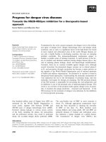
We have recently used the DnaE split intein to site-
specifically conjugate QDs to the C-terminus of the PH
domain of Akt and Btk, in vivo (Figure 1A)[34]. We
have now utilized a new split intein to allow conjugation
of QDs to the N-terminus of target proteins. This
expands the possibilities of the intein-based system
allowing for the first time in vivo site specific conjuga-
tion of QDs and other nanostructures to the N terminus
of target proteins. W e selected the Ssp DnaB mini-
intein, to achieve N-terminal protein QD labelling, given
that the N-terminal part was small enough to be synthe-
tically produced and shown to be capable of trans-spli-
cing and protein modification [38,39]. This mini-intein
lost its endonuclease domain during evolution and cur-
rently consists of just the 130aa protein splicing domain
plus a 24aa linker sequence in place of the endonuclease
domain [35,40]. Recent work by Sun W. et al. demon-
strated that the Ssp DnaB mini-intein remained profi-
cient in protein trans-splicing when artificially split in
the loop region between the b-strands, b2andb3, pro-
ducing an N-terminal part of 11 aa and a C-terminal
part of 143aa (Figure 1B) [41].
We go on to show that inteins can be used to target
QDs to specific molecular complexes within living cells
and embryos. Specifically through the targeting of QDs
to the C-terminus of paxillin, we generated a full length
protein-QD complex. Paxillin-QD conjugates localized
efficiently to focal adhesion complexes within the cells
of the developing embryo. Imaging of these complexes
in real time revealed that QDs would associate with
newly formed focal adhesions and would be released
once the complexes were disassembled. Finally, split
intein based QD conjugation may be extended to simul-
taneous and multiple protein tagging as long as func-
tionally orthogonal split inteins are used, in order to
prevent undesired side products due to cross-reactivity
[42]. Through the combination of the C and N termin al
intein systems we were able for the first time to simulta-
neously target two distinct proteins with spectrally resol-
vable QDs in vivo.Thisistoourknowledgetheonly
methodology that will allow conjugation of mu ltiple tar-
gets with QDs without cross reactivity and should serve
as an important addition to existing labeling methods.
Results and Discussion
Quantum Dots targeted to Focal Adhesion Complexes
following in vivo, intein-mediated conjugation to the C-
terminus of paxillin
We have recently used intein based conjugation to cova-
lently conjugate QDs to the C-terminus of the Plekstrin
Charalambous et al. Journal of Nanobiotechnology 2011, 9:37
/>Page 2 of 14
homology domain of Akt. Using this methodology we
were able to site-specifically tag a protein domain with
QDs in vivo for the first time, effectively generating QD
biosensors that could respond to PI3K activation by
translocating to the cell membrane [34]. We now
wanted to examine whether this methodology could be
used i) to tag a full length protein and more importantly
ii) to target QDs to sp ecific molecular complexes within
the cell. We decided to target paxillin, a focal-adhesion
associated protein implicated in the regulation of actin
cytoskeletal organization and cell motility [43]. To inves-
tigate whether we could target QDs to focal adhesion
complexes via paxillin in vivo, we injected both blasto-
meres of 2-cell stage Xenopus embryos with the probe
(DnaE I
C
-QDot
585
) and RNA encoding the target pro-
tein (in this case, Paxillin-EGFP-DnaE I
N
). The presence
of EGFP on the paxillin was required as it would allow
us to monitor and compare the distribution of the QDs
vs paxillin. Embryos were allowed to develop to stage 8,
at which point animal cap cells were dissociated,
induced with activin, plated onto fibronectin coated
slides and observ ed by time-lapse microscopy. We first
examined the localization of Paxillin-EGFP and found
that, as previously reported, it localized at focal-adhe-
sions, especiall y at the filopodia and lamelipodia, gener-
ated by mesodermal cells during migration on
fibronectin substrates (Figure 2A) [44]. Furthermore,
QDs translocated to focal adhesions in cells derived
from embryos injected with both DnaE I
C
-QDot
585
and
RNA, where they colocalized with Paxillin-EGFP (Figure
2A). On the other hand, in cells that did not express the
Paxillin-EGFP-DnaE I
N
, QDs remained in the cytosol
(Figure 2B).
To confirm formation of QD-protein conjugates we
used a biochemical approach. Xenopus embryos were
injected as follows: i) Uninjected ii) DnaE I
C
-QDot
585
ii)
RNA encoding Paxillin-EGFP-DnaE I
N
,iii)DnaEI
C
-
QDot
585
+ RNA encoding Paxillin-EGFP -DnaE I
N
.
Embryos were lysed when they reached stage 10 and
loaded onto an agarose gel. QDot
585
were visuali zed
with the ethidium bromide emission filter under UV
excitation and EGFP was imaged with a band pass 500/
50 filter set on UVP iBox Imagi ng System. As shown in
Figure 2C a smeary band of the expect ed molecular
weight for the Paxillin-EGFP appeared in lysates of
Xenopus embryos injected with the RNA encoding the
corresponding target protein. This band could not be
detected in lysates of uninjected Xenopus embryos or
Xenopus embryos injected with the probe (I
C
peptide
conjugated QD
585
) only. Higher MW bands correspond-
ing to the semi-synthetic products appeared only in
lysates of Xenopus embryos inject ed with both the RNA
encoding the target protein (Paxillin-EGFP) and the
probe (I
C
peptide conjugated QD
585
). Importantly, this
new band overlaps with the QD signal. Commercially
available streptavidin-coated QDs bear 4-10 streptavidin
Figure 1 In vivo conjugation of QDs to the C- or N-terminus of target proteins via intein mediated protein splicing. (A) Schematic
representation of site-specific Ssp DnaE split intein-mediated conjugation of QDs to the C-terminus of the PH domain of Akt. (B) Schematic
representation of site specific Ssp DnaB mini intein-mediated conjugation of QDs to the N-terminus of mem-EGFP.
Charalambous et al. Journal of Nanobiotechnology 2011, 9:37
/>Page 3 of 14
molecules (53 kD each)/QD giving 16-40 biotin binding
sites implying 16-40 conjugated Paxillin-EGFP protein
molecules per QD, resulting in a significant increase in
size that results in trapping of the conjugates in the gel
wells and preventing their migration. Video microscopy
revealed that foc al adhesion formation and disassembly
is very rapid in these highly migratory cells. In addition
and as shown in time lapse images, QDs would associate
with newly formed focal adhesion complexes (Figure
3A) and would be released once the complexes were
disassembled (Figure 3B).
We repeated the above described experiment using
commercially available QDs from Invitrogen (15-20 nm
in diameter) from all the emission wavelengths (525,
565, 585, 655) coupled to streptavidin. Conjugates of
paxillin-EGFP with QDs from all the emission wave-
lengths tested were successfully targeted to the focal
adhesions. However, there was a definitive size depen-
dence in their ability to target focal adhesions, with
longer wavelength emitting QDs showing a diminished
capacity to do so (Figure 4). These results emphasize
the need for the generation of biocompatible and col-
loidally stable long wavelength QDs with smaller effec-
tive hydrodynamic radii.
Quantum Dots targeted to the cell membrane, following
in vivo intein-mediated conjugation to the N-terminus of
a membrane targeted variant of EGFP (memEGFP)
Although intein-based C-terminal conjugation of QDs to
proteins is a valuable tool, it is often necessary to tag a
protein at the N-terminus in order to achieve a func-
tional conjugate. Thus, we wanted t o implement an
intein based strategy that would enable site-specific N-
terminal conjugation in vivo,tocomplementtheC-
terminal tagging system we have already described [34].
In addition we wanted to test a shorter synthetic peptide
that would make this approach more affordable as well
as easy. The value of using short synthetic intein
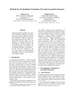
Figure 2 In vivo conjugation of QDs to the C-terminus of Paxillin-EGFP via intein mediated protein splicing. Co-localization of QDot
585
with Paxillin-EGFP on focal-adhesions of mesodermal cells during migration. Stage 2 Xenopus embryos were injected with (A) probe (DnaE I
C
-
QDot
585
)(in red) and RNA encoding Paxillin-EGFP-DnaE I
N
(in green) or (B) probe (DnaE I
C
-QDot
585
) alone. Fluorescence images of animal cap
cells dissociated from stage 8 Xenopus embryos, induced with activin, and plated onto fibronectin coated slides. (A) Yellow shows overlap
between red QDot
585
and green EGFP indicating successful QD-protein conjugation in vivo. (B) In the absence of Paxillin-EGFP, QDs do not
target focal adhesions but remain diffusely localized in the cytosol. (C &D) Biochemical characterization of protein-QD conjugates. Xenopus
embryos were injected as follows, C: i) Uninjected ii) DnaE I
C
-QDot
585
ii) Paxillin-EGFP-DnaE IN RNA iii) DnaE IC-QDot
585
+ Paxillin-EGFP-DnaE IN
RNA, D: i) Uninjected ii) QDot
585
-DnaB I
N
iii) DnaB I
C
-memEGFP RNA iv) QDot
585
-DnaB I
N
+ DnaB I
C
-memEGFP RNA, lysed at stage 10 and loaded
onto a 0.5% agarose gel in this order, from left to right. QDot
585
were visualized with the ethidium bromide emission filter under UV excitation
and EGFP was imaged with a band pass 500/50 filter set on UVP iBox
Imaging System. The ligation products appear as a single band under the
GFP and QD filters, only in lysates of Xenopus embryos injected with RNA + QD probe (vertical white arrows). Bands corresponding to Paxillin-
EGFP and memEGFP proteins not conjugated to QDs are detectable under the GFP filter, in lysates of Xenopus embryos injected with RNA only
and QD probes + RNA, but not QDs only (horizontal arrows). Bands corresponding to QD probes are detectable under the QD filter, in lysates of
Xenopus embryos injected with the QD probes only or the QD probes + RNA, but not RNA only, (horizontal arrows).
Charalambous et al. Journal of Nanobiotechnology 2011, 9:37
/>Page 4 of 14
Figure 3 Paxillin-QD conjugates a ssociate with newly formed focal adhesion complexes and are released once the complexes are
disassembled. Xenopus embryos were injected at the 2-cell stage with the probe (DnaE I
C
-QDot
525 or 585
) and RNA encoding Paxillin-EGFP-DnaE
I
N
. Animal cap cells were dissociated from stage 8 Xenopus embryos, induced with activin, and plated onto fibronectin coated slides. (A) Time
lapse images (time-interval: 30 sec) show paxillin-QD conjugates associating with newly formed focal adhesion complexes at the filopodia and
lamellipodia of mesodermal cells during their migration on fibronectin substrates (see arrows). (B) Time lapse images (time-interval: 10 sec) show
paxillin-QD conjugates being released from focal adhesion complexes as they disassemble during migration of mesodermal cells on fibronectin
substrates (see arrows).
Charalambous et al. Journal of Nanobiotechnology 2011, 9:37
/>Page 5 of 14
peptides capable of trans-splicing was initially demon-
strated for C-terminus-specific modifications of recom-
binant proteins using artificially split Npu DnaE [45]
and Ssp Gy rB inteins [42] and more recently for N-ter-
minus-specific modifications using Ssp DnaB mini-
intein [41]. B y taking advantage of the latter we imple-
mentedthestrategyshowninFigure1B.Wewenton
to examine whether this approach could be used suc-
cessfully for in vivo conjugation of QDs to the N-ter-
minus of target proteins using a membrane-targeted
variant of EGFP as a target. This construct, generated
by the genetic fusion of the enhanced GFP to the far-
nesylation sequence of p21(Ras) (memEGFP) was
selected due to its ability to constitutively localize to
the cell membrane as it would provide clear visual
confirmation of successful conjugation in the intact
embryo [46]. In addition, it is a good example of a tar-
get protein that cannot be QD-tagged at the C termi-
nus as that would interfere with the membrane
tethering function of the farnesylated residues and
would lead to elimination of membrane anchoring.
To demonstrate in vivo N-terminal labelling of mem-
EGFP with QDs, we injected both blastomeres of two-
cell stage Xenopus embryos with the probe (QDot
605
-
DnaB I
N
) and with RNA encoding the target protein
(in this case, DnaB I
C
-memEGFP). As shown in Figure
Figure 4 Increased QD size imposes constraints on the translocation efficiency of Paxillin-EGFP-QD conjugates to the focal adhesion
complexes. Co-localization of QDots
525
, QDots
565
and QDots
655
with Paxillin-EGFP on focal adhesion complexes. Note that unlike QDot
525
, the
QDot
655
are not recruited as effectively to the focal adhesion complexes.
Charalambous et al. Journal of Nanobiotechnology 2011, 9:37
/>Page 6 of 14
5A, QDs translocated to the cell membrane in cells
derived from the embryo injected with both QDot
605
-
DnaB I
N
and RNA, where they colocalized with mem-
EGFP. On the other hand, in cells that do not express
the DnaB I
C
-memEGFP, QDs were not targeted to the
membrane but remained in the cytosol (Figure 5B).
Despite the fact that most QDs colocalize with the tar-
get protein to the plasma membrane, a significant
amount of QDs remain in the cytosol. This is due to
the fact that the initial streptavidin QD solution con-
tains a mixture of streptavidin-conjugated and uncon-
jugatedQDsasshowninFigure6,aswellasdueto
gradual loss of both the intein peptide and the target
protein from the QD surface, as a result of proteolytic
degradation, as discussed in the Conclusions section.
This problem will be significantly ameliorated when
QDs with more stable surface modifications become
commercially available.
In order to confirm conjugation of QDs to the N-ter-
minus of memEGFP biochemically, we prepared lysates
from injected embryos, which were run on an agarose
gel, in a similar fashion to what has already been
described above for the C-terminal conjugation of QDs
to paxillin. As shown in Figure 2D, conjugation of QDs
to the N-terminus of memEGFP was succe ssful leading,
to a higher molecular weight product, absent from the
QD only lane.
Ssp DnaE and DnaB inteins do not cross splice and
therefore facilitate simultaneous targeting of Quantum
Dots to two different proteins in vivo
Several naturally occurring and artificially split inteins
have been examined for their orthogonality and it was
found that inteins can cross-splice when s haring a high
degr ee of sequence identity and similarity. In fact it has
been shown that the natural DnaE split inteins from
Figure 5 In vivo conjugation of QDs to the N-terminus of mem-EGFP via intein mediated protein splicing. (A) Co-localization of QDot
605
with mem-EGFP on the cell membrane. Fluorescence images of stage 10 Xenopus embryos microinjected with the probe (QDot
605
-DnaB I
N
)
shown in red, in one blastomere at the two-cell stage, and then injected with RNA encoding the target protein (DnaB I
C
-memEGFP) shown in
green, in three of four blastomeres. Yellow shows the overlap between red QDot
605
and green EGFP indicating successful QD-protein
conjugation in a live embryo. (B) In embryos injected with the probe (QDot
605
-DnaB I
N
) alone, in the absence of RNA encoding the target
protein (DnaB I
C
-memEGFP), QDs do not target the cell membrane but remain diffusely localized in the cytosol.
Charalambous et al. Journal of Nanobiotechnology 2011, 9:37
/>Page 7 of 14
Nostoc Punctiforme and Synech ocystis sp. PCC 6803
cross-splice [47] as do the DnaE split inteins from three
other cyanobacteria (Nostoc sp.PCC7120, Oscillatoria
Limnetica and Thermosynechococcus vulcanus) [48].
Given that the naturally occurring Ssp DnaE split intein
and the artificially split mini intein, Ssp DnaB, do not
sha re any sequence similarity as indicated by a protein-
protein BLAST and can afford effective conjugation of
QDs to the C- and N-terminus of target proteins
respectively we decided to exploit this combination for
QD-targeting to multiple proteins in vivo,simulta-
neously. To demonstrate that memEGFP and Akt-EGFP
fusion proteins can be simultaneously and specifically
targeted by spectrally resolvable QDs, without cross
reactivity we performed in vivo injections with a mix-
ture of complementary QD-intein peptide probes and
targ et protein RNAs. More specifically we injected both
blastomeres of two-cell stage Xenopus embryos with the
probes QDot
585
-DnaB I
N
and DnaE I
C
-QDot
705
and the
corresponding RNAs encoding DnaB I
C
-memEGFP and
Akt-EGFP-DnaE I
N
.AsshowninFigure7A,both
QDot585 and QDot705 translocated to the cell mem-
brane in cells derive d from the embryo injected with
the complementary probes where they colocalized with
memEGFP and Akt-EGFP. We predicted that the N-ter-
minus of the DnaE intein would not react with the C-
terminus of the DnaB intein and vice versa, as the spe-
cific interactions that facilitate the splicing reaction,
notably recognition of the complementary N- or C-
intein and consequent non-covalent association for for-
mation of an active-intein intermediate, could not be
formed given that there is no sequence similarity. To
examine if cross splicing between Ssp DnaE and Ssp
DnaB inteins occurs we injected both blastomeres of
two-cell stage Xenopus embryos with the probe
QDot
655
-DnaB I
N
and RNA encoding Akt-EGFP-DnaE
I
N
.AsshowninFigure7B,Akt-EGFPclearlytargetto
the cell membrane whereas QDot
655
remain diffuse in
the cytoplasm. Clearly, Akt-EGFP-QD conjugates do
not form, implying that the two inteins cannot cross
splice. Similar results were obtained when we e xam-
ined the reverse combination, that is when we injected
two-cell stage Xenopus embryos with the probe DnaE
I
C
-QDot
655
and RNA encoding DnaB I
C
-memEGFP
(Figure 7B).
This experiment thus demonstrates that intein-
mediated trans splicing facilitates simultaneous and spe-
cific tagging of two protein targets within the sa me
embryo with spectrally resolvable QDs without cross
splicing. Given the large number of orthogonal inteins it
is possible that more than two targets can be simulta-
neously tagged with different QDs or different
nanostructures.
Conclusions
Herein, we describe an intein-based system for conjuga-
tion of QDs to target proteins in vivo.Thisapproach
has several advantages over existing methodologies that
make it truly unique, including i) site-specificity (N- or
C-terminus), ii) low-intrinsic reactivity towards endo-
genous proteins which do not contain the intein motif
required for splicing, thus eliminating mis-targeting of
the QDs, iii) versatility conferred by the ability to target
QDs to a single protein within any cellular compartment
or molecular complex and iv) the ability to target spec-
trally resolvable QDs to multiple protein targets simulta-
neously without cross reactivity.
We have previously shown site-specific conjugation of
QDs to the C-terminus of target proteins by using the
naturally-split DnaE intein [34]. However, C-terminal
protein labelling with QDs can in some c ases, interfere
with protein localization and/or biological function, as
Figure 6 Evaluation of commercially available streptavidin
coated QDs. Commercially available streptavidin coated QDot
605
(from Invitrogen) were incubated with biotinylated DNA (lane 1)
and non biotinylated DNA (lane 2) at a molar ratio of 1:100, for 30
minutes at room temperature. Following the conjugation reaction
the DNA-QD mixtures were run on a 1% agarose gel to assess the
percentage of QDs capable of efficient biotin-streptavidin
conjugation. QDot
605
were imaged using the ethidium bromide
filter set on the UVP iBoxImaging System. As shown, the QDs used
in our experiments exhibit great variability in terms of their biotin
binding ability (see arrows). Arrow 1 indicates QDs that are unable
to bind biotin.
Charalambous et al. Journal of Nanobiotechnology 2011, 9:37
/>Page 8 of 14
Figure 7 Simultaneou s targeting of QDs to two different proteins via Ssp DnaE and Ssp DnaB intein mediated splicing without cross
reactivity. Fluorescence images of stage 10 Xenopus embryos injected with (A) the probes QDot
585
-DnaB I
N
and DnaE I
C
-QDot
705
and the
corresponding RNAs encoding DnaB I
C
-memEGFP and Akt-EGFP-DnaE I
N
, (B) the probe QDot
655
-DnaB I
N
and RNA encoding Akt-EGFP-DnaE I
N
or
the probe DnaE I
C
-QDot
655
and RNA encoding DnaB I
C
-memEGFP. Both QDot585 and QDot705 translocated to the cell membrane in cells
derived from the embryo injected with the complementary probes where they colocalized with memEGFP and Akt-EGFP. In contrast, Akt-EGFP
and mem-EGFP clearly target to the cell membrane whereas the non-complementary probes, remain diffuse in the cytoplasm, implying that the
two inteins do not cross react.
Charalambous et al. Journal of Nanobiotechnology 2011, 9:37
/>Page 9 of 14
can C-terminal fusion of fluorescent proteins [49-51].
This is due to interference with protein sorting or tar-
geting signals located at the C-terminus of proteins,
such as two common ER retrieval signals, the dilysine
motifandthetetrapeptideKDEL,aswellasthetype1
peroxisomal targeting signal peptide SKL [50]. A C-
terminal tag or marker could also disrupt signals for the
incorporation of lipid anchors. For example, many mem-
bers of the Ras superfamily carry sequences that signal
the attachment of lipid anchors at their C-termini [51].
A class of plasma membrane proteins, including cell
adhesion molecules or receptors have a glycosylpho-
sphatidylinositol (GPI) linker [49]. The molecular signals
engaging the lipid modification enzyme complexes
reside at the C-terminus of these proteins and would
definitely be disrupted by the addition of a fluorescent
protein or QD. We therefore took advantage of the arti-
ficially split Ssp DnaB intein originally described by Sun,
W. et al. [41], for site-specific conjugation of QDs to the
N-terminus of target proteins. Ssp DnaB intein has been
split artificially at a site (S1) proximal to the N- term-
inal, producing an N-termin al piece of only 11 aa in
length and a C-terminal piece of 144 aa in length [41].
This novel artificially split intein is quite useful due to
the ease of chemical peptide synthesis and due to the
fact that such short peptides are not prone to misfold-
ing. We used the S1 split intein for site-specific conjuga-
tion of QDs to the N-terminus of a model target protein
in vivo, n amely mem-EGFP, and have shown that QD-
memEGFP conjugates localize to the cell membrane and
can be monitored in real time within the developing
Xenopus embryo (Figure 5). Thus, the ability to target
QDs to the N-terminus of proteins is very helpful for
bioimaging studies aiming at determining protein locali-
zation and function, given that there are numerous pro-
teins bearing C-terminal post-translational modifications
or a C-terminal critical domain whose function would
be impeded if a bulky QD was conjugated at the C-
terminus.
We have also demonstrated, using this methodology,
that Quantum Dots can be targeted via paxillin to focal
adhesions, a specific molecular complex, for t he first
time. Focal Adhesions (FAs) are c omprised of a and b
integrin heterodimers that form a br idge between the
intracellular actin cytoskeleton and the extracellular
matrix (ECM) [52]. While the extracellular domain of
integrins binds directly to ECM proteins, the cytoplas-
mic tail is linked to the actin cytoskeleton via signalling
and adapter proteins, such as focal adhesion kinase
(FAK), vinculin, talin and paxillin [52]. FAs play a cru-
cial role in cell adhesion, spreading and motility by reg-
ulating various signal transduction pathways leading to
rearrangement of the actin cytoskeleton [53,54]. We
have demonstrated that QDs can be efficiently targeted
to focal adhesions via paxillin without altering protein
localization and/or function. In fact Paxillin-QD conju-
gates retained full functionality as indicated by their
ability to i) translocate to focal adhesions at the cell
membrane (Figure 2A) and ii) associate with newly
formed focal adhesion complexes and be released once
the complexes were disassembled (Figure 3). This is an
inherent advantage of QDs over fluorescent proteins
since the former are conjugated to target protei ns post-
translationa lly and do not therefore interfere with pro-
tein folding and tertiary structure.
A useful additional application of this intein-based
methodology is t he simultaneous and specific conjuga-
tion of Q Ds to multiple proteins target s in vivo.
Although fluorescent proteins already provide a straight-
forward solution to this problem [3]. Q D-conjugation
methods are attractive complements given the superior
optical properties of QDs over fluorescent proteins [55].
Double in vi vo labeling becomes possible with our sys-
tem due to the existence of orthogonal pairs of split
inteins that do not cross splice and therefore allow dif-
ferent protein targets to be simultaneously and specifi-
cally tagged with spectrally resolvable QDs within the
cell. Such orthogonal split-intein combinations include
Ssp DnaE and Sce VMA, Ssp DnaB and Sce VMA, Ssp
DnaB and Mxe GyrA [42] to mention but a few and
now Ssp DnaE and Ssp DnaB. I n fact, given the large
number of characterized split inteins, the number of
individual targets that can be simultaneously tagged is
only limited by the number of QDs that can be spec-
trally distinguished. Moreover, the fact that the trans-
splicing reactions proceed with an identical molecular
mechanism ensures similar reaction rates for QD-conju-
gation that would aid the comparison of the properties
of the two proteins-otherwise the first protein of interest
is already redistributing while the second protein is not
yet sufficiently labelled. We have shown in this work
that Ssp DnaE and Ssp DnaB inteins do not cross splice
and may therefore b e used to specifically target spec-
trally r esolvable QDs to different proteins simulta-
neously in vivo (Figure 7).
Despit e the successful conjugation of QDs to both the
N and C terminus of target proteins, the current metho-
dology and the materials used have certain limitations
that need to be noted. We have observed that a pool of
QDs remains in the cytosol, even when the target pro-
tein is in excess. This was expected in the case of paxil-
lin, a cytosolic protein occasionally localized to th e focal
adhesion compl exes on the cell membrane, but came as
a surprise in the case of memEGFP, a protein expected
to be exclusively localized on the cell membrane. An
unwanted result of the presence of free QDs in the cyto-
sol was the reduction of signal to noise ratio. These
QDs are most likely not conjugated to the target protein
Charalambous et al. Journal of Nanobiotechnology 2011, 9:37
/>Page 10 of 14
due to the following two reasons. Firstly the commer-
cially available solution of Streptavidin-coated QDs used
in these experiments, contains both streptavidin-conju-
gated and free QDs (see Figure 6). This implies that
even if the splicing reaction i s 100% efficient, a portion
of free QDs is still present in the cell. Secondly, in the
Xenopus model, translation begins after the Midblastula
Transition (~12 hours post injection). By that time, a
portion of the streptavidin-conjugated QDs may have
lost the streptavidin or the intein peptide (due to pro-
teolytic degradation). This, in effect, generates additional
free QDs, which will remain in the cytosol, thus redu-
cing the apparent conjugation efficiency. Given that as
the embryo develops, the amount of conjugated QDs is
progressively reduced and given the target proteins’
degradation rate, it is importanttonotethatthetime
frame for imaging can be quite small. In addition, the
presence of free QDs in the cytosol greatly impedes
visualization of target proteins that do not localize to a
specific organelle or structure in the cell, even early on.
These limitations raise the need for i) commercially
available QDs capable of retaining their conjugated bio-
molecule longer and ii) improved methodologies to
ensure that the starting material consist of 100% conju-
gated QDs.
Our present results indicat e efficient, covalent and
site-specific in vivo-fusion of QDs to either the N- or C-
terminus of a target protein within any cellular compart-
ment or molecular complex. This methodology is nota-
ble due to its potential diagnostic and therapeutic
applications , as it make s the targeti ng of nanostructures
and nanodev ices to different intracel lular compartments
and signalling complexes a viable possibility. Further-
more, this method is unique in that it facilitates QD
conjugation to multiple target proteins, as long as ortho-
gonal intein pairs are used. The number of potential
applications for double (or multiple) in vivo labelling i s
quite large. Most obv iously, protein localizations of two
or more species can be followed simultaneously and
protein-protein interacti ons may be explored using QDs
suited for FRET experiments. In conclusion the intein-
mediated approach for simultaneous, in vivo, site-speci-
fic (N- and C-terminus) conjugation of Quantum Dots
to multiple protein targets, should serve as a powerful
tool for bioimaging applications.
Methods
Embryos and explants
Xenopus laevis embryos from induced spawning [56]
were staged ac cording to Nieuwkoop and Faber (1967).
Operation techniques and buffer (MMR, Ubbels, 1983)
have been described [56]. Xenopus embryos were ferti-
lized in vitro and dejellied using 2% cysteine-HCl, pH
7.8, then maintained in 0.1× Marc’s Modified Ringer’s
(0.1× MMR). Microinjections were performed in 4%
Ficoll in 0.33× MMR. The embryos were injected with
RNA and QDs conjugated to either the C-terminal part
of DnaE Intein (DnaE-I
C
) or the N-terminal part of Ssp
DnaB mini-intein (DnaB-I
N
) through a bio tin-streptavi-
din bond, at the 2 and 4-cell stage according to estab-
lished prot ocols [57]. After injection s the embryos were
cultured in 4% Ficoll in 0.33× MMR until stage 8 and
then cultured in 0.1× MMR at room temperature. For
in vivo assays, the embryos were transferred to slides for
time lapse movies using Zeiss Axiocam MR3 and the
Axiovision software 4.6 to monitor GFP-Q D co-
localization.
Electrophoretic evaluation of streptavidin-coated QDs
Commercially available streptavidin coated QDot
605
(from Invitrogen) were incubated with biotinylated DNA
and non biotinylated DNA at a molar ratio of 1:100, for
30 minutes at room temperature. Following the conjuga-
tion reaction the DNA-QD mixtures were run on a 1%
agarose gel to assess the percentage of QDs capable of
efficient biotin-streptavidin conjugation. QDot
605
were
imaged using the ethidium bromide filter set on the
UVP iBox Imaging System.
Chemical Synthesis of biotinylated C-terminus DnaE intein
peptide (DnaE I
C
-Biotin) and biotinylated N-terminus
DnaB mini-intein peptide (Biotin-DnaB I
N
)
The 47 amino acid peptide sequence of the C-terminus
DnaE intein peptide (DnaE I
C
-Biotin):
MVKVIGRRSLGVQRIFDIGLPQDHNFLLAN-
GAIAANCFDYKDDDDK(Ahx-Biotin)G
The 11 amino acid peptide sequence of the N-termi-
nus DnaB intein peptide (Biotin-DnaB I
N
):
Biotin-KKK-Ahx-CISGDSLISLA
Biotin was conjugated to a C-terminal Lysine (K) on
DnaE I
C
via an Ahx linker (6 carbon inert linker) and to
a N-terminal Cysteine (C) on DnaB I
N
via a three lysine
linker and Ahx. Both peptides were synthesized on a 0.5
mmol scale on a 4-methylbenzhydrylamine (MBHA)
resin according to the in-situ neutralization/HBTU acti-
vation protocol for Boc SPPS [58]. In order to put a bio-
tin at the C-terminus of DnaE intein, it was necessary to
add an ext ra amino acid, Lys, at the C-t erminus. In
order to put a biotin at the N-terminus of DnaB intein,
it was necessary to add three extra Lys, at the N-termi-
nus. Lysines serve as a linking point for biotin as well as
a spacer between the peptide and biotin. The DnaE I
C
peptide contains a cysteine protected with the NPyS
group which was added as the last amino acid in the
synthesis. Following chain assembly, global de-protec-
tion and cleavage from the support was achieved by
treatment with HF containing 4% v/v pcresol, for 1 hour
at 0°C. Following removal of the HF, the crude peptide
Charalambous et al. Journal of Nanobiotechnology 2011, 9:37
/>Page 11 of 14
products were precipitated and washed with anhydrous
cold Et
2
O before being dissolved in aqueous acetonitrile
(50% B) and lyophilized. The crude peptides were puri-
fied by preparative HPLC using a linear gradient of 25-
45% B over 60 minutes. The purified peptides were
characterized as the desired product by ESMS. The lyo-
philized biotinylated DnaE I
C
peptide was dissolved in
60% DMSO at a concentration of 1 mg/ml. The lyophi-
lized biotinylated DnaB I
N
peptide was dissolved in PBS
at a concentration of 1 mg/ml.
In vitro conjugation of DnaE I
C
-Biotin and Biotin-DnaB I
N
to streptavidin-coated QDs
The biotinylated peptides were diluted to 50 μMand
used at 1:1 vol ume ratio with streptavidin-coated QDs
(1 μM) (from Invitro gen or eBiosciences). To allow for-
mation of the biotin-streptavidin bond we incubate at
24°C for 30 min. To remove any excess unbound pep-
tide the conjugate was filtered through microcon centri-
fugal filter units (YM100) [59].
Analysis of QD-peptide conjugates
Analysis of QD-peptide conjugati on was performed by
electrophoresis at 60 V for 4 h at 4°C using a 0.5% agar-
osegel.Noloadingbufferwasaddedtothesamples
before loading. Gels were visualized under the ethidium
bromide filter (515-570 nm) with a UVP Imager (data
not shown).
Alternatively analysis of QD peptide conjugation was
performed by spotting nitrocellulose membranes (What-
man). Biotinylated peptides and peptides that did n ot
contain the biotin modification were spotted on nitro-
cellulose membrane and blocked in PBS containing 1%
BSA for 30 min at room temperature. The nitrocellulose
membrane was then soaked in PBS containing streptavi-
din-coated QDs (1:500 dilution) for 30 min at room
temperature. The membrane was washed with PBS-
Tween 20 (1%) twice and visualized under the ethidium
bromide filter (515-570 nm) with a UVP Imager (data
not shown).
Plasmids and Cloning
All plasmids were constructed using standard molecular
biology techniques and they were sequenced to verify
correct coding.
pCS2++-Paxillin-EGFP- I
N
A PCR fragment amplified with F
pax
(5’ AAATCGA-
TATGGACGACCTCGAT 3’ )andR
egfp
(5’
CCGAATTCCTTGTACAGCTCGTC 3’)encodingpax-
illin-EGFP, using the pEGFP-N3 plasmid (from
Addgene) as template was inserted into the multiple
cloning site of the pCS2++ plasmid by restriction
enzyme digest wit h ClaI-EcoRI. A PCR fragment ampli-
fied with IGpr61 (AAGGAATTCAAGTTTGC
GGAATATTGCCTCAGTTTTGG) and IGpr63
(AAGCTCGAGTTATTTAATTGTCCCAGCG) encod-
ing I
N
with 5 N-terminal extein residues (KFAEY), using
the pJJDuet30 plasmid (from Addgene) as template was
inserted at the C-terminus of Paxillin-EGFP on pCS2++
between the EcoRI-XhoI restriction sites.
pCS2++-DnaB I
C
-memEGFP
The membrane targeted EGFP variant was constructed
by genetically engineering a membrane targeting
sequence, namely c-HaRas at the C-terminus of EGFP,
via sequential PCR. Initially, a PCR fragment was encod-
ing EGFP and half of the cHaRas membrane targeting
sequence was amplified using the pEGFP-N3 plasmid
(from Addgene) as template. The primers used for the
first PCR were: F
EGFP
(5’ AGCGAATTCATGGTGAG-
CAAGGGCGAGGAG 3’ )andRA
EGFP-cHaras
(5’
gggccactctcatcaggagggttcagcttCTTGTACAGCTCGTC-
CATGCCG 3’). A second PCR followed using this PCR
product and the following primers: F
EGFP
(5’ AGC-
GAATTCATGGTGAGCAAGGGCGAGGAG 3’)and
RB
EGFP-cHaras
(5’ GCCTCGAGtcaggagagcacacacttgcagct-
catgcagccggggccactctc 3’). This PCR fragment encoded
the fusion EGFP-cHaRas and was inserted into the mul-
tiple cloning site of the pCS2++ plasmid by restriction
enzyme digest with EcoRI-XhoI. A PCR fragment ampli-
fied with F
IC
(ACATCGATatgttatcaccagaaata-
gaaaagttgtctcag) and R
IC
(CTGAATTCgttatggacaat
gatgtcattggcgac) encoding DnaB I
C
using the pMAL
plasmid (kind gift from Dr. Xiang-Qin Liu) as template,
was inserted upstream and in frame with the EGFP-
cHaRas on pCS2++ between the ClaI-EcoRI restriction
sites.
All plasmids were transcribed into RNA using mMes-
sage mMachine Sp6 kit (Ambion) and the mRNAs were
purified using the Mega Clear kit (Ambion). Microinjec-
tions performed in Ficoll as mentioned above.
Electrophoretic analysis of protein trans-splicing
Biochemical analysis of protein-trans splicing was per-
formed by lysis of injected Xenopus embr yos at stage 10.
Lysis was performed by pipetting up and down in the
presence of proteinase inhibitors (Sigma) and DNAse
(Roche). Lysates were then loaded onto agarose gels run
at 100 V for 2 h, at 4°C. Gels were visualized with a
UVP Imager.
Activin-induced Cell migration assays
Animal cap explants were prepared from stage 8
embryos. Cells were dissociated in CMFM (Ca
2+
and
Mg
2+
free medium) and then treated with activin pro-
tein (1 U/ml in 1×CMFM) for 1 hour. The dissociated
cells were subsequently plated in Modified Barth’s Solu-
tion [60] into fibronectin-coated chambered coverslips
(VWR). Coverslips were coated with 0.1 mg/ml
Charalambous et al. Journal of Nanobiotechnology 2011, 9:37
/>Page 12 of 14
fibronectin (Sigma, diluted to the appropriate concentra-
tion with MBS) for 2 hours at room temperature, and
then blocked with bovine serum albumin (BSA; 50 mg/
ml in MBS).
Image analysis
Timelapse analysis of dissociated cells was performed
using a Zeiss Axiocam MR3 camera attached to a Zeiss
Axiovert 135. Images were acquired and timelapse files
assembled using Axiovision software 4.6.
Acknowledgements
Funding was provided by the Cyprus Research Promotion Foundation
(ΑΝΑΒΑΘΜΙΣΗ/0609/28). It is acknowledged that the published research
work is co-funded by the European Regional Development Fund.
Authors’ contributions
PS conceived of the study, participated in its design and coordi nation and
helped to draft the manuscript. AC participated in the design and
coordination of the study and drafted the manuscript. IA carried out carried
out the molecular and biochemical studies and the in vivo assays. NC
helped to carry out some of the in vivo experiments. All authors read and
approved the final manuscript.
Competing interests
The authors declare that they have no competing interests.
Received: 6 April 2011 Accepted: 15 September 2011
Published: 15 September 2011
References
1. Finley KR, Davidson AE, Ekker SC: Three-color imaging using fluorescent
proteins in living zebrafish embryos. Biotechniques 2001, 31:66.
2. Giuliano KA, Post PL, Hahn KM, Taylor DL: Fluorescent protein biosensors:
measurement of molecular dynamics in living cells. Annu Rev Biophys
Biomol Struct 1995, 24:405.
3. Giepmans BN, Adams SR, Ellisman MH, Tsien RY: The fluorescent toolbox
for assessing protein location and function. Science 2006, 312:217.
4. Bruchez M, Moronne M, Gin P, Weiss S, Alivisatos AP: Semiconductor
nanocrystals as fluorescent biological labels. Science 1998, 281:2013.
5. Chan WC, Nie S: Quantum dot bioconjugates for ultrasensitive
nonisotopic detection. Science 1998, 281:2016.
6. Dahan M, et al: Diffusion dynamics of glycine receptors revealed by
single-quantum dot tracking. Science 2003, 302:442.
7. Han M, Gao X, Su JZ, Nie S: Quantum-dot-tagged microbeads for
multiplexed optical coding of biomolecules. Nat Biotechnol 2001, 19:631.
8. Larson DR, et al: Water-soluble quantum dots for multiphoton
fluorescence imaging in vivo. Science 2003, 300:1434.
9. Michalet X, et al: Quantum dots for live cells, in vivo imaging, and
diagnostics. Science 2005, 307:538.
10. Courty S, Luccardini C, Bellaiche Y, Cappello G, Dahan M: Tracking
individual kinesin motors in living cells using single quantum-dot
imaging. Nano Lett 2006, 6:1491.
11. Howarth M, et al: Monovalent, reduced-size quantum dots for imaging
receptors on living cells. Nat Methods 2008, 5:397.
12. Smith AM, Nie S: Minimizing the hydrodynamic size of quantum dots
with multifunctional multidentate polymer ligands. J Am Chem Soc 2008,
130:11278.
13. Medintz IL, et al: Self-assembled nanoscale biosensors based on
quantum dot FRET donors. Nat Mater 2003, 2
:630.
14. Howarth M, Takao K, Hayashi Y, Ting AY: Targeting quantum dots to
surface proteins in living cells with biotin ligase. Proc Natl Acad Sci USA
2005, 102:7583.
15. So MK, Yao H, Rao J: HaloTag protein-mediated specific labeling of living
cells with quantum dots. Biochem Biophys Res Commun 2008, 374:419.
16. Colston MJ, Davis EO: The ins and outs of protein splicing elements. Mol
Microbiol 1994, 12:359.
17. Cooper AA, Stevens TH: Protein splicing: self-splicing of genetically
mobile elements at the protein level. Trends Biochem Sci 1995, 20:351.
18. Xu MQ, Perler FB: The mechanism of protein splicing and its modulation
by mutation. Embo J 1996, 15:5146.
19. Evans TJT, Xu MQ: Mechanistic and kinetic considerations of protein
splicing. Chem Rev 2002, 102:4869.
20. Giriat I, Muir TW: Protein semi-synthesis in living cells. J Am Chem Soc
2003, 125:7180.
21. Muralidharan V, Muir TW: Protein ligation: an enabling technology for the
biophysical analysis of proteins. Nat Methods 2006, 3:429.
22. Chong S, et al: Protein splicing involving the Saccharomyces cerevisiae
VMA intein. The steps in the splicing pathway, side reactions leading to
protein cleavage, and establishment of an in vitro splicing system. J Biol
Chem 1996, 271:22159.
23. Shao Y, Xu MQ, Paulus H: Protein splicing: evidence for an N-O acyl
rearrangement as the initial step in the splicing process. Biochemistry
1996, 35:3810.
24. Xu MQ, et al: Protein splicing: an analysis of the branched intermediate
and its resolution by succinimide formation. Embo J 1994, 13:5517.
25. Xu MQ, Southworth MW, Mersha FB, Hornstra LJ, Perler FB: In vitro protein
splicing of purified precursor and the identification of a branched
intermediate. Cell 1993, 75:1371.
26. Shao Y, Xu MQ, Paulus H: Protein splicing: characterization of the
aminosuccinimide residue at the carboxyl terminus of the excised
intervening sequence. Biochemistry 1995, 34:10844.
27. Lew BM, Mills KV, Paulus H: Protein splicing in vitro with a semisynthetic
two-component minimal intein.
J Biol Chem 1998, 273:15887.
28. Busche AE, et al: Segmental isotopic labeling of a central domain in a
multidomain protein by protein trans-splicing using only one robust
DnaE intein. Angew Chem Int Ed Engl 2009, 48:6128.
29. Cowburn D, Shekhtman A, Xu R, Ottesen JJ, Muir TW: Segmental isotopic
labeling for structural biological applications of NMR. Methods Mol Biol
2004, 278:47.
30. Zuger S, Iwai H: Intein-based biosynthetic incorporation of unlabeled
protein tags into isotopically labeled proteins for NMR studies. Nat
Biotechnol 2005, 23:736.
31. Xia Z, et al: Multiplex detection of protease activity with quantum dot
nanosensors prepared by intein-mediated specific bioconjugation. Anal
Chem 2008, 80:8649.
32. Evans TC Jr, et al: Protein trans-splicing and cyclization by a naturally
split intein from the dnaE gene of Synechocystis species PCC6803. J Biol
Chem 2000, 275:9091.
33. Scott CP, Abel-Santos E, Wall M, Wahnon DC, Benkovic SJ: Production of
cyclic peptides and proteins in vivo. Proc Natl Acad Sci USA 1999,
96:13638.
34. Charalambous A, Andreou M, Skourides PA: Intein-mediated site-specific
conjugation of Quantum Dots to proteins in vivo. J Nanobiotechnology
2009, 7:9.
35. Perler FB: InBase: the Intein Database. Nucleic Acids Res 2002, 30:383.
36. Saleh L, Perler FB: Protein splicing in cis and in trans. Chem Rec 2006,
6:183.
37. Wu H, Hu Z, Liu XQ: Protein trans-splicing by a split intein encoded in a
split DnaE gene of Synechocystis sp. PCC6803. Proc Natl Acad Sci USA
1998, 95:9226.
38. Ludwig C, Pfeiff M, Linne U, Mootz HD: Ligation of a synthetic peptide to
the N terminus of a recombinant protein using semisynthetic protein
trans-splicing. Angew Chem Int Ed Engl 2006, 45:5218.
39. Ludwig C, Schwarzer D, Mootz HD: Interaction studies and alanine
scanning analysis of a semi-synthetic split intein reveal thiazoline ring
formation from an intermediate of the protein splicing reaction. J Biol
Chem 2008, 283:25264.
40. Telenti A, et al: The Mycobacterium xenopi GyrA protein splicing
element: characterization of a minimal intein. J Bacteriol 1997, 179:6378.
41. Sun W, Yang J, Liu XQ: Synthetic two-piece and three-piece split inteins
for protein trans-splicing. J Biol Chem 2004, 279:35281.
42. Volkmann G, Iwai H: Protein trans-splicing and its use in structural
biology: opportunities and limitations. Mol Biosyst 6:2110.
43. Schaller MD: Paxillin: a focal adhesion-associated adaptor protein.
Oncogene 2001, 20:6459.
44. Stylianou P, Skourides PA: Imaging morphogenesis, in Xenopus with
Quantum Dot nanocrystals. Mech Dev 2009.
Charalambous et al. Journal of Nanobiotechnology 2011, 9:37
/>Page 13 of 14
45. Aranko AS, Zuger S, Buchinger E, Iwai H: In vivo and in vitro protein
ligation by naturally occurring and engineered split DnaE inteins. PLoS
One 2009, 4:e5185.
46. Jiang W, Hunter T: Analysis of cell-cycle profiles in transfected cells using
a membrane-targeted GFP. Biotechniques 1998, 24:349.
47. Iwai H, Zuger S, Jin J, Tam PH: Highly efficient protein trans-splicing by a
naturally split DnaE intein from Nostoc punctiforme. FEBS Lett 2006,
580:1853.
48. Dassa B, Amitai G, Caspi J, Schueler-Furman O, Pietrokovski S: Trans protein
splicing of cyanobacterial split inteins in endogenous and exogenous
combinations. Biochemistry 2007, 46:322.
49. Mayor S, Riezman H: Sorting GPI-anchored proteins. Nat Rev Mol Cell Biol
2004, 5:110.
50. Purdue PE, Lazarow PB: Peroxisome biogenesis. Annu Rev Cell Dev Biol
2001, 17:701.
51. Zhang FL, Casey PJ: Protein prenylation: molecular mechanisms and
functional consequences. Annu Rev Biochem 1996, 65:241.
52. Lo SH: Focal adhesions: what’s new inside. Dev Biol 2006, 294:280.
53. Hynes RO: Integrins: bidirectional, allosteric signaling machines. Cell 2002,
110:673.
54. Ridley AJ, et al: Cell migration: integrating signals from front to back.
Science 2003, 302:1704.
55. Smith AM, Gao X, Nie S: Quantum dot nanocrystals for in vivo molecular
and cellular imaging. Photochem Photobiol 2004, 80:377.
56. Winklbauer R: Mesodermal cell migration during Xenopus gastrulation.
Dev Biol 1990, 142:155.
57. Smith WC, Harland RM: Injected Xwnt-8 RNA acts early in Xenopus
embryos to promote formation of a vegetal dorsalizing center. Cell 1991,
67:753.
58. Schnolzer M, Alewood P, Jones A, Alewood D, Kent SB: In situ
neutralization in Boc-chemistry solid phase peptide synthesis. Rapid,
high yield assembly of difficult sequences. Int J Pept Protein Res 1992,
40:180.
59. Stronati A, et al: Relationships between sperm DNA fragmentation, sperm
apoptotic markers and serum levels of CB-153 and p,p’-DDE in
European and Inuit populations. Reproduction 2006, 132:949.
60. Leibovitz A, et al: Classification of human colorectal adenocarcinoma cell
lines. Cancer Res 1976, 36:4562.
doi:10.1186/1477-3155-9-37
Cite this article as: Charalambous et al.: Split-Inteins for Simultaneous,
site-specific conjugation of Quantum Dots to multiple protein targets In
vivo. Journal of Nanobiotechnology 2011 9:37.
Submit your next manuscript to BioMed Central
and take full advantage of:
• Convenient online submission
• Thorough peer review
• No space constraints or color figure charges
• Immediate publication on acceptance
• Inclusion in PubMed, CAS, Scopus and Google Scholar
• Research which is freely available for redistribution
Submit your manuscript at
www.biomedcentral.com/submit
Charalambous et al. Journal of Nanobiotechnology 2011, 9:37
/>Page 14 of 14