Vaginal Surgery for Incontinence and Prolapse - part 3 pptx
Bạn đang xem bản rút gọn của tài liệu. Xem và tải ngay bản đầy đủ của tài liệu tại đây (608.56 KB, 30 trang )
50 Vaginal Surgery for Incontinence and Prolapse
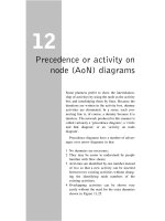
Figure 4.19. A: The pubococcygeal line (arrow) used as a reference
point radiographically is drawn from the inferior pubic symphysis to the
sacrococcygeal junction. B: Compared to the normal exam in A, this
image shows prolapse of the bladder (b) and vaginal vault (long arrow)
below the pubococcygeal line, compatible with a cystocele and vaginal
vault prolapse. A rectocele is also seen as an anterior bulge (arrowhead)
in relation to the anal canal (asterisk). (From Pannu HK. Dynamic MR
imaging of female organ prolapse. Radiol Clin North Am 2003;41(2):409–
423. © 2003, with permission from Elsevier.)
Figure 4.20. A: Typical H configuration of the vagina (long arrows) is
seen in this MRI image. B: A paravaginal detachment (arrow). (From
Pannu HK. Dynamic MR imaging of female organ prolapse. Radiol Clin
North Am 2003;41(2):409–423. © 2003, with permission from Elsevier.)
Prolapse 51
Other Modalities
Transperineal ultrasound has been described to
assess dynamic function of the pelvic fl oor (81).
Dynamic anorectal endosonography has also
been described and may detect the presence of
enteroceles (82). The role of these alternate
modalities has not been fully elucidated and
needs further study.
Conclusion
A thorough pelvic assessment is necessary prior
to any planning regarding surgical or nonsurgi-
cal intervention for pelvic organ prolapse.
Patient history will direct the physician to look
for appropriate fi ndings on physical examina-
tion. The Pelvic Organ Prolapse Quantifi cation
system is gaining wider acceptance with physi-
cians involved in the care of women with pelvic
fl oor disorders as it has been shown to be valid
and reproducible, and it facilitates effective
communication of treatment outcomes among
clinicians and researchers. Several studies have
shown that physical examination may not be
accurate in diagnosing certain pelvic fl oor
defects such as paravaginal defects whose clini-
cal relevance has yet to be fully elucidated. The
use of pelvic fl oor imaging may complement the
clinical assessment of the pelvic fl oor, but its use
needs to be further studied and defi ned prior to
advocating its routine use. Ultimately the goal of
the evaluation is to fully appreciate the extent of
the prolapse and to relate that to any visceral
or sexual dysfunction that may coexist.
References
1. Cardozo LD, Stanton SL. Genuine stress incontinence
and detrusor instability—a review of 200 patients. Br J
Obstet Gynaecol 1980;87:184–188.
2. Summitt RL, Stovall TG, Bent AE, et al. Urinary incon-
tinence: correlation of history and brief offi ce evalua-
tion with multichannel urodynamic testing. Am J
Obstet Gynecol 1992;166:1835–1844.
3. Walters MD, Shields LE. The diagnostic value of history,
physical examination and the Q-tip cotton swab test in
women with urinary incontinence. Am J Obstet Gynecol
1989;159:145–149.
4. Rosenzweig BA, Pushkin S, Blumenfeld D, et al. Preva-
lence of abnormal urodynamic test results in continent
women with severe genitourinary prolapse. Obstet
Gynecol 1992;79:539–542.
5. American College of Obstetricians and Gynecologists.
Pelvic organ prolapse. ACOG Technical Bulletin No.
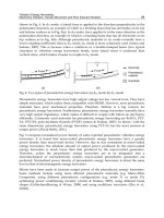
Figure 4.21. Cystocele, cervical prolapse with enterocele, and perineal
descent. A: Normal resting image. B: Compared to A, this study was per-
formed during defecation and shows a cystocele (B), a prolapsed cervix
(long arrow), a widened rectovaginal space (thick arrow), and a low-lying
rectum (R). (From Pannu HK. Dynamic MR imaging of female organ pro-
lapse. Radiol Clin North Am 2003;41(2):409–423. © 2003, with permis-
sion from Elsevier.)
52 Vaginal Surgery for Incontinence and Prolapse
214. Washington, DC: American College of Obstetri-
cians and Gynecologists, 1995.
6. Rosenzweig BA. Genital prolapse and lower urinary
tract dysfunction. Int Urogynecol J 1993;4:278–281.
7. Enhorning GE. Simultaneous recording of intravesical
and intraurethral pressure: a study of urethral closure
in normal and stress incontinent women. Acta Clin
Scand 1961;176:1.
8. Richardson DA, Bent AE, Ostergard DR. The effect
of uterovaginal prolapse on urethrovesical pressure
dynamics. Am J Obstet Gynecol 1983;146:901–905.
9. Bergman A, Koonings PP, Ballard CA. Predicting post-
operative urinary incontinence development in women
undergoing operation for genitourinary prolapse. Am
J Obstet Gynecol 1998;158:1171–1175.
10. Versi E, Lyell DJ, Griffi ths DJ. Videourodynamic diag-
nosis of occult genuine stress incontinence in patients
with anterior vaginal wall relaxation. J Soc Gynecol
Invest 1998;5:327.
11. Bump RC, Fantl AJ, Hurt WG. The mechanism of
urinary continence in women with severe uterovaginal
prolapse: results of barrier studies. Obstet Gynecol
1988;72:291.
12. Myers DL, Lasala CA, Hogan JW, et al. The effect of
posterior wall support defects on urodynamic indices
in stress urinary incontinence. Obstet Gynecol
1988;91:710.
13. Grady M, Kozminski M, DeLancey J, et al. Stress incon-
tinence and cystoceles. J Urol 1991;145:1211–1213.
14. Weil A, Gianoni A, Rottenberg RD, et al. The risk of
postoperative urinary incontinence after surgical
treatment of genital prolapse. Int Urogynecol J 1993;4:
74–79.
15. Rosenzweig BA, Soffi ci AR, Thaomas S, et al. Urody-
namic evaluation of voiding in women with cystocele.
J Reprod Med 1992;37:162–166.
16. Jackson SL, Weber AM, Hull TL, et al. Fecal inconti-
nence in women with urinary incontinence and pelvic
organ prolapse. Obstet Gynecol 1997;89:423–427.
17. Leigh RJ, Tumberg LA. Fecal incontinence: the unvoiced
symptom. Lancet 1982;1:1349–1351.
18. Thomas TM, Egan M, Walgrove A, et al. The prevalence
of fecal and double incontinence. Commun Med
1984;6:216–220.
19. Field SM, Hilton P. The prevalence of sexual problems
in women attending for urodynamic investigation. Int
Urogynecol J 1993;4:212–215.
20. Haase P, Skibsted L. Infl uence of operations for stress
incontinence and/or genital descensus on sexual life.
Acta Obstet Gynecol Scand 1988;67(7):659–661.
21. Diokno AC, Brown MB, Herzog AR. Sexual function in
the elderly. Arch Intern Med 1990;150(1):197–200.
22. Weber AM, Walters MD, Schover LR, et al. Sexual func-
tion in women with uterovaginal prolapse and urinary
incontinence. Obstet Gynecol 1995;85(4):483–487.
23. Mouritsen L, Larsen JP. Symptoms, bother and
POPQ in women referred with pelvic organ prolapse.
Int Urogynecol J Pelvic Floor Dysfunct 2003;14(2):
122–127.
24. Ellerkmann RM, Cundiff GW, Melick CF, et al. Correla-
tion of symptoms with location and severity of pelvic
organ prolapse. Am J Obstet Gynecol 2001;185(6):
1332–1337.
25. Barber MD, Visco AG, Wyman JF, et al. Continence
Program for Women Research Group. Sexual function
in women with urinary incontinence and pelvic organ
prolapse. Obstet Gynecol 2002;99(2):281–289.
26. Weber AM, Walters MD, Piedmonte MR, et al.
Anterior colporrhaphy: a randomized trial of three
surgical techniques. Am J Obstet Gynecol 2001;185(6):
1299–1304.
27. Spence-Jones C, Kamm MA, Henry MM, et al. Bowel
dysfunction: a pathogenic factor in uterovaginal pro-
lapse and urinary stress incontinence. Br J Obstet Gyn-
aecol 1994;101(2):147–152.
28. Sze EH, Karram MM. Transvaginal repair of vault pro-
lapse: a review. Obstet Gynecol 1997;89(3):466–475.
29. Paraiso MF, Ballard LA, Walters MD, et al. Pelvic
support defects and visceral and sexual function in
women treated with sacrospinous ligament suspension
and pelvic reconstruction. Am J Obstet Gynecol
1996;175(6):1423–1430.
30. Holley RL, Varner RE, Gleason BP, et al. Recurrent
pelvic support defects after sacrospinous ligament
fi xation for vaginal vault prolapse. J Am Coll Surg
1995;180(4):444–448.
31. Shull BL, Capen CV, Riggs MW, et al. Preoperative and
postoperative analysis of site-specifi c pelvic support
defects in 81 women treated with sacrospinous liga-
ment suspension and pelvic reconstruction. Am J
Obstet Gynecol 1992;166(6 pt 1):1764–1768.
32. Swift SE, Pound T, Dias JK. Case-control study of etio-
logic factors in the development of severe pelvic organ
prolapse. Int Urogynecol J Pelvic Floor Dysfunct
2001;12(3):187–192.
33. Samuelsson EC, Victor FTA, Tibblin G, et al. Signs of
genital prolapse in a Swedish population of women 20
to 59 years of age and possible related factors. Am J
Obstet Gynecol 1999;180:299–305.
34. Mant J, Painter R, Vessey M. Epidemiology of genital
prolapse: observations from the Oxford Family Plan-
ning Association Study. Br J Obstet Gynaecol 1997;
104(5):579–585.
35. Olsen AL, Smith VJ, Bergstrom JO, et al. Epidemiology
of surgically managed pelvic organ prolapse and
urinary incontinence. Obstet Gynecol 1997;89(4):501–
506.
36. Gurel H, Gurel SA. Pelvic relaxation and associated risk
factors: the results of logistic regression analysis. Acta
Obstet Gynecol Scand 1999;78(4):290–293.
37. Strohbehn K, Jakary JA, Delancey JO. Pelvic organ
prolapse in young women. Obstet Gynecol 1997;90(1):
33–36.
38. Bruskewitz R. Female incontinence: signs and symp-
toms. In: Raz S, ed. Female Urology. Philadelphia: WB
Saunders, 1983:45–50.
39. Porges RF. A practical system of diagnosis and classifi -
cation of pelvic relaxations. Surg Gynecol Obstet
1963;117:769–773.
40. Beecham CT. Classifi cation of vaginal relaxation. Am J
Obstet Gynecol 1980;1:136(7):957–958.
41. Baden WF, Walker TA. Genesis of the vaginal profi le:
a correlated classifi cation of vaginal relaxation. Clin
Obstet Gynecol 1992;15(4):1048–1054.
42. Bump RC, Mattiasson A, Bo K, et al. The standardiza-
tion of terminology of female pelvic organ prolapse
and pelvic fl oor dysfunction. Am J Obstet Gynecol
1996;175:10–17.
43. Thiede HA. Urogynecology: comments and caveats.
Am J Obstet Gynecol 1987;157:536.
Prolapse 53
44. Barber MD, Cundiff GW, Weidner AC, et al. Accuracy
of clinical assessment of paravaginal defects in women
with anterior vaginal wall prolapse. Am J Obstet
Gynecol 1999;181(1):87–90.
45. Segal JL, Vassallo BJ, Kleeman SD, et al. Paravaginal
defects: prevalence and accuracy of preoperative detec-
tion. Int Urogynecol J Pelvic Floor Dysfunct 2004 (Jul
1) [Epub ahead of print] 15(6):378–383.
46. Richardson AC. The rectovaginal septum revisited: its
relationship to rectocele and its importance in rectocele
repair. Clin Obstet Gynecol 1993;36(4):976–983.
47. Cundiff GW, Weidner AC, Visco AG, et al. An anatomic
and functional assessment of the discrete defect
rectocele repair. Am J Obstet Gynecol 1998;179(6 pt
1):1451–1456.
48. Burrows LJ, Sewell C, Leffl er KS, et al. The accuracy of
clinical evaluation of posterior vaginal wall defects.
Int Urogynecol J Pelvic Floor Dysfunct 2003;14(3):
160–163.
49. Toglia MR, DeLancey JO. Anal incontinence and the
obstetrician-gynecologist. Obstet Gynecol 1994;84(4 pt
2):731–740.
50 Barber MD, Lambers A, Visco AG, et al. Effect of patient
position on clinical evaluation of pelvic organ prolapse.
Obstet Gynecol 2000;96:18–22.
51. Visco AG, Wei JT, McClure LA, et al. Effects of exami-
nation technique modifi cations on pelvic organ pro-
lapse quantifi cation (POP-Q) results. Int Urogynecol J
Pelvic Floor Dysfunct 2003;14(2):136–140.
52. Swift SE, Herring M. Comparison of pelvic organ
prolapse in the dorsal lithotomy compared with
the standing position. Obstet Gynecol 1998;91:961–
964.
53. Silva WA, Kleeman S, Segal J, et al. Effects of a full
bladder and patient positioning on pelvic organ
prolapse assessment. Obstet Gynecol 2004;104(1):
37–41.
54. Hall AF, Theofrastous JP, Cundiff GW, et al. Interob-
server and intraobserver reliability of the proposed
International Continence Society, Society of Gyneco-
logic Surgeons, and American Urogynecologic Society
pelvic organ prolapse classifi cation system. Am J Obstet
Gynecol 1996;175:1467–1470.
55. Kobak WH, Rosenberger K, Walters MD. Interobserver
variation in the assessment of pelvic organ prolapse.
Int Urogynecol J Pelvic Floor Dysfunct 1996;7:121–
124.
56. Scotti RJ, Flora R, Greston WM, et al. Characterizing
and reporting pelvic fl oor defects: the revised New York
classifi cation system. Int Urogynecol J Pelvic Floor
Dysfunct 2000;11(1):48–60.
57. Swift SE, Freeman R, Petri E, et al. Proposal for a world-
wide, user-friendly classifi cation system for pelvic
organ prolapse (abstract). 26
th
annual meeting of the
International Urogynecologic Association, Melbourne,
Australia, December 5–7, 2001.
58. Steele A, Mallipeddi P, Welgoss J, et al. Teaching the
pelvic organ prolapse quantitation system. Am J Obstet
Gynecol 1998;179(6 pt 1):1458–1463.
59. Maglinte DD, Kelvin FM, Hale DS, et al. Dynamic
cystoproctography: a unifying diagnostic approach to
pelvic fl oor and anorectal dysfunction. AJR 1997;
169(3):759–767.
60. Kelvin FM, Maglinte DD. Radiologic investigation of
prolapse. J Pelv Surg 2000;6:218–220.
61. Yang A, Mostwin JL, Rosenshein NB, et al. Pelvic fl oor
descent in women: dynamic evaluation with fast MR
imaging and cinematic display. Radiology 1991;179(1):
25–33.
62. Pannu HK, Kaufman HS, Cundiff GW, et al. Dynamic
MR imaging of pelvic organ prolapse: spectrum of
abnormalities. Radiographics 2000;20(6):1567–1582.
63. Kelvin FM, Maglinte DD, Hornback JA, et al. Pelvic
prolapse: assessment with evacuation proctography
(defecography). Radiology 1992;184(2):547–551.
64. Altringer WE, Saclarides TJ, Dominguez JM, et al. Four-
contrast defecography: pelvic “fl uoroscopy.” Dis Colon
Rectum 1995;38(7):695–699.
65. Halligan S. Commentary: imaging of anorectal func-
tion. Br J Radiol 1996;69(827):985–988.
66. Stoker J, Halligan S, Bartram CI. Pelvic fl oor imaging.
Radiology 2001;218(3):621–641.
67. Bartram CI, Turnbull GK, Lennard-Jones JE. Evacua-
tion proctography: an investigation of rectal expulsion
in 20 subjects without defecatory disturbance. Gastro-
intest Radiol 1988;13(1):72–80.
68. Shorvon PJ, McHugh S, Diamant NE, et al. Defecogra-
phy in normal volunteers: results and implications. Gut
1989;30(12):1737–1749.
69. van Dam JH, Ginai AZ, Gosselink MJ, et al. Role
of defecography in predicting clinical outcome of
rectocele repair. Dis Colon Rectum 1997;40(2):201–
207.
70. Halligan S, Bartram CI, Park HJ, et al. Proctographic
features of anismus. Radiology 1995;197(3):679–682.
71. Kelvin FM, Hale DS, Maglinte DD, et al. Female pelvic
organ prolapse: diagnostic contribution of dynamic
cystoproctography and comparison with physical
examination. AJR 1999;173(1):31–37.
72. Vanbeckevoort D, Van Hoe L, Oyen R, et al. Pelvic fl oor
descent in females: comparative study of colpocystode-
fecography and dynamic fast MR imaging. J Magn
Reson Imaging 1999;9(3):373–377.
73. Kelvin FM, Maglinte DD, Hale DS, et al. Female pelvic
organ prolapse: a comparison of triphasic dynamic MR
imaging and triphasic fl uoroscopic cystocolpoproctog-
raphy. AJR 2000;174(1):81–88.
74. Kaufman HS, Buller JL, Thompson JR, et al. Dynamic
pelvic magnetic resonance imaging and cystocolpo-
proctography alter surgical management of pelvic
fl oor disorders. Dis Colon Rectum 2001;44(11):
1575–1583.
75. Tunn R, Paris S, Taupitz M, et al. MR imaging in
posthysterectomy vaginal prolapse. Int Urogynecol J
Pelvic Floor Dysfunct 2000;11(2):87–92.
76. Lienemann A, Anthuber C, Baron A, et al. Dynamic MR
colpocystorectography assessing pelvic-fl oor descent.
Eur Radiol 1997;7(8):1309–1317.
77. Fielding JR, Dumanli H, Schreyer AG, et al. MR-based
three-dimensional modeling of the normal pelvic fl oor
in women: quantifi cation of muscle mass. AJR
2000;174(3):657–660.
78. Klutke C, Golomb J, Barbaric Z, et al. The anatomy of
stress incontinence: magnetic resonance imaging of the
female bladder neck and urethra. J Urol 1990;143(3):
563–566.
79. Huddleston HT, Dunnihoo DR, Huddleston PM 3rd,
et al. Magnetic resonance imaging of defects in
DeLancey’s vaginal support levels I, II, and III. Am
J Obstet Gynecol 1995;172(6):1778–1782.
54 Vaginal Surgery for Incontinence and Prolapse
80. Tunn R, Paris S, Fischer W, et al. Static magnetic reso-
nance imaging of the pelvic fl oor muscle morphology
in women with stress urinary incontinence and pelvic
prolapse. Neurourol Urodyn 1998;17(6):579–589.
81. Beer-Gabel M, Teshler M, Barzilai N, et al. Dynamic
transperineal ultrasound in the diagnosis of pelvic fl oor
disorders: pilot study. Dis Colon Rectum 2002;45(2):
239–245.
82. Karaus M, Neuhaus P, Wiedenmann TB. Diagnosis of
enteroceles by dynamic anorectal endosonography. Dis
Colon Rectum 2000;43(12):1683–1688.
Fecal continence is a complex function with
multiple factors contributing to normal conti-
nence: anatomic integrity, function, innerva-
tion, compliance, capacity, sensation, and stool
characteristics. The evaluation of fecal inconti-
nence can also be complex, with a variety of
investigations aimed at the different compo-
nents of continence. A thorough evaluation is
necessary to identify the type of incontinence
and its etiology so that the correct treatment
can be selected.
History
A directed history and physical examination
are essential in evaluating a patient with fecal
incontinence and help guide the selection of
studies to be performed. As this is a sensitive
topic, very pointed questions must be asked,
as the patient may not volunteer specifi cs. The
history starts with defi ning the patient’s incon-
5
Fecal Incontinence
Sharon G. Gregorcyk
55
History . . . . . . . . . . . . . . . . . . . . . . . . . . . . . . . . . 55
Physical Examination . . . . . . . . . . . . . . . . . . . . . 56
Special Physiologic Testing . . . . . . . . . . . . . . . . 56
Anal Manometry . . . . . . . . . . . . . . . . . . . . . . . . . 57
Electromyography . . . . . . . . . . . . . . . . . . . . . 57
Pudendal Nerve Terminal Motor
Latency . . . . . . . . . . . . . . . . . . . . . . . . . . . . . 58
Endoanal Ultrasound . . . . . . . . . . . . . . . . . . . 59
Magnetic Resonance Imaging . . . . . . . . . . . . 59
Cinedefecography . . . . . . . . . . . . . . . . . . . . . . . . 60
Conclusion . . . . . . . . . . . . . . . . . . . . . . . . . . . . . . 60
tinence and its severity. The physician must
determine if the patient’s incontinence is to gas,
liquid, and/or solid stool, and the volume of
stool lost. The patient who has minor seepage
and otherwise full control over her stool is
approached differently than the patient with
complete incontinence. While recording the
number of episodes of incontinence will assist
with determining the severity of the patient’s
incontinence, one must keep in mind that some
patients adapt their entire life to being near a
bathroom so that they may avoid an episode of
incontinence. Other changes in lifestyle may
include the use of pads and carrying a change
of underwear. Factors such as these must be
taken into account.
A variety of scoring systems exist and are
aimed at objectively quantifying a patient’s
incontinence. Most scoring systems include the
type of incontinence (solid, liquid, gas), fre-
quency of episodes, lifestyle alteration, and
use of pads. Table 5.1 demonstrates a common
scoring system utilized. All of the scoring systems
have limitations. Adding a quality of life assess-
ment questionnaire improves upon these limita-
tions as it takes more into account the effect the
patient’s incontinence has on her daily life (see
appendix). Since these two tools do not change
one’s management of the patient, they are not
routinely used by all physicians. However, these
tools bring an objectivity to the evaluation,
which is important in comparing results of
procedures in the literature.
The patient should be questioned about
urgency or any change in her bowel habits, which
56 Vaginal Surgery for Incontinence and Prolapse
might indicate a problem such as a colitis or irri-
table bowel syndrome. Even with an intact func-
tioning sphincter mechanism, continence may be
diffi cult when a large watery stool is presented
with extreme urgency. Dietary and medication
history should be recorded as well as any past
medical history. Some systemic disorders such as
diabetes, alcoholism, and connective tissue dis-
eases can predispose a woman to incontinence
with or without mitigating factors. An obstetric
history is very important. The number of vaginal
deliveries, episiotomies, obstetric tears, and use
of forceps with delivery have all been associated
with fecal incontinence. A history of pelvic or
anal surgery should be documented as well. The
patient should also be asked if she has urinary
incontinence, as a signifi cant number of patients
are affl icted with this problem as well.
Physical Examination
Although a complete physical examination
should be performed, the emphasis is placed on
the perineum and digital rectal examination.
For the experienced surgeon, the history and
physical exam alone may be all that is necessary
to develop a therapeutic plan in some patients.
Inspection of the perineum is fi rst performed.
Important fi ndings to note include a patulous
anus, loss of perineal body, scarring (Figure
5.1), perineal soiling, muscular defect, dermati-
tis, or a mucosa ectropion. Asking the patient to
bear down might help the physician identify
a prolapsing hemorrhoid or complete rectal
prolapse. Straining also is necessary in order
to evaluate the presence of perineal descent, an
enterocele, or a cystocele.
Sensation can be assessed by touch or with a
Q-Tip, and the presence or absence of the anocu-
taneous refl ex, also known as an anal wink,
should be noted. This refl ex is a transient con-
traction of the external sphincter in response to
the stimulation of the perianal skin and suggests
an intact innervation via the pudendal nerve. The
digital rectal examination should start within the
anal canal where one can assess both the resting
tone and the patient’s squeeze. A more aggressive
digital examination proximally can then be per-
formed to rule out a mass within the rectum or a
fecal impaction. Inserting a fi nger into the vagina
during the rectal examination is helpful in evalu-
ating the rectovaginal septum, as well as the ante-
rior sphincter. In the offi ce, a proctosigmoidoscopy
can be performed to evaluate for infl ammatory
or neoplastic conditions.
Special Physiologic Testing
Once the history and physical examination have
been completed, the physician may have suffi -
cient information to plan treatment. In 11% to
51% of cases (1), the history and physical exam
alone are adequate for the evaluation of fecal
incontinence. An example is the patient with
incontinence who has suffered an obstetric
injury and who upon examination has good
Table 5.1. Incontinence scoring system (30)
Type of
incontinence Never Rarely Sometimes Usually Always
Solid 0 1 2 3 4
Liquid 0 1 2 3 4
Gas 0 1 2 3 4
Pad usage 0 1 2 3 4
Lifestyle 0 1 2 3 4
alteration
The score may range from 0 (perfect continence) to 20 (complete
incontinence).
Rarely, less than once per month; sometimes, less than once per
week, once or more per month; usually, less than once per day, once
or more per week; always, once per day or more.
Figure 5.1. Gapping anus and scarred perineum on physical exam.
Fecal Incontinence 57
tone and squeeze pressures with a palpable
anterior defect. This patient can be directly
counseled with regard to a surgical repair versus
attempts at biofeedback. Although further
physiologic testing for this patient may be of
benefi t for objective documentation, it is not
necessary to plan the patient’s treatment. Other
patients are not so easily diagnosed and further
information is necessary. A variety of investiga-
tive tools exist to evaluate fecal incontinence,
with no one testing modality providing all the
information needed with regard to all of the
components of continence.
Anal Manometry
Anal manometry is typically performed by placing
a four- or eight-channel catheter with radial ports
into the anal canal and measuring the pressures
at rest and with the patient squeezing. The rectal-
anal inhibitory refl ex (RAIR), rectal sensation,
compliance, and capacity are also measured.
Normal values are listed in Table 5.2.
Although digital examination assesses the
resting and squeeze pressures, anal manometry
is a reliable and reproducible way to quantify the
pressures (2–5). This information can be useful
for documentation purposes and may be used
for comparison after treatment. For the patient
with an isolated external sphincter injury,
one would expect a normal resting tone with
decreased squeeze pressure, whereas a patient
with an isolated internal sphincter injury such as
from a sphincterotomy would have a decreased
resting pressure and normal squeeze. A decreased
resting pressure and squeeze pressure (Figure
5.2) may be seen in a combined sphincter injury
or with a neurogenic etiology.
The RAIR is the relaxation of the proximal
internal anal sphincter in response to rectal dis-
tention such as when a substance is presented to
the rectum. This refl ex allows for sampling of the
substance to discern if it is gas, liquid, or solid.
The RAIR is measured by infl ating a balloon into
the rectum with 10 cc or more of air and observ-
ing for a decrease in the pressure to 15% below
the baseline. The RAIR is absent in Hirschsprung’s
and Chagas’ disease and is commonly absent
with rectal prolapse.
Rectal sensation can be measured at the time
of manometry or separately, as it simply involves
infl ating a balloon placed in the rectum. Resec-
tion of the rectum, infl ammation, or radiation
proctitis may result in a lower compliance with
less volume required to cause a rise in the rectal
pressure. As the pressure rises above that of the
sphincters, incontinence may result. Compli-
ance is calculated by taking the difference in
pressure between the initial rectal sensation and
rectal fullness and dividing that into the volume
of fl uid necessary to achieve that difference (2).
Although the measurements from anal
manometry can be helpful, they do not by them-
selves determine the etiology of a patient’s
incontinence. The measurements do not even
indicate if a patient is incontinent or to what
degree. A patient can have abnormal values and
be continent or normal values and be inconti-
nent. In a study by McHugh and Diamant (6),
almost 40% of patients with fecal incontinence
had normal resting and squeeze pressures on
anal manometry. Thus, it is important to balance
the results from anal manometry with the history
and physical exam.
Electromyography
Anal sphincter electromyography (EMG)
records the electrical activity of the striated
Table 5.2. Normal parameters for anal manometry
Parameters Normal
Resting pressure 40–70 mmHg
Squeeze pressure 100–180 mmHg
Rectal-anal inhibitory reflex Present
Sensory threshold 10–30 cc
Rectal capacity 100–250 cc
Rectal compliance 3–15 ccH
2
O/mmHg
Figure 5.2. Anal manometry with low resting and squeeze pressures.
58 Vaginal Surgery for Incontinence and Prolapse
muscles of the anorectum (7). This electrical
activity may be recorded with surface elec-
trodes, concentric needle electrodes, or single-
fi ber needle electrodes. Measurements from the
EMG provides information about the innerva-
tion and functional state of the motor units
within a muscle. Pudendal nerve terminal motor
latency (PNTML) is a type of surface EMG that
is addressed separately in the next section.
Besides PNTML, surface EMG is utilized with
biofeedback therapy. It is a simple, well-
tolerated method of EMG but it is imprecise
and limited in value.
Concentric needle EMG and single-fi ber
needle EMG are much more precise than surface
EMG. In general, the measurements from needle
EMG can delineate muscle that has undergone
denervation and reinnervation. Thus it can be
used to map injuries to the muscle as well as
evaluate for neurogenic conditions. The single-
fi ber needle EMG is the most accurate and mea-
sures action potentials from individual muscle
fi bers from which the fi ber density is calculated.
Fiber density is a sensitive way to detect and
quantitate rearrangement of the muscle fi ber in
the motor unit. Needle EMG has signifi cant
drawbacks including the expense of the equip-
ment, pain associated with inserting the needles
(8), and the diffi culty of doing the exam itself,
which is quite time-consuming. The utility of
needle EMG with fecal incontinence is contro-
versial, and its routine use is not advocated
owing to poor patient compliance and limited
additional value provided.
Pudendal Nerve Terminal Motor Latency
The pudendal nerve innervates the external
anal sphincter and puborectalis. Injury to this
nerve is one of the possible etiologies of incon-
tinence. Pudendal nerve terminal motor latency
(PNTML) is the measurement of the nerve con-
duction velocity in the terminal part of the
pudendal nerve (9). The device for measuring
the PNTML consists of a stimulating electrode
that is positioned at the tip of the index fi nger
and a recording electrode located at the base of
the fi nger (Figure 5.3). The pudendal nerve is
stimulated at Alcock’s canal, resulting in con-
traction of the sphincter muscles. The technique
requires extensive practice and may not be pos-
sible in the obese or muscular patient owing to
anatomic factors. The time from the stimula-
tion to movement of the muscle is measured. A
normal PNTML value is 2.0 ± 0.2 ms. Prolonged
PNTML may be seen in patients with neuro-
genic fecal incontinence, perineal descent, and
rectal prolapse. Of note, PNTML also increases
with age.
Pudendal nerve terminal motor latency is pri-
marily used in fecal incontinence to predict out-
comes of surgical therapy. Its use, however, in
predicting outcomes is controversial, with some
studies supporting poorer outcomes in patients
with prolonged latency and other studies
showing no difference (Table 5.3). Even patients
with bilateral pudendal neuropathy may benefi t
from surgical repair, with Nikiteas et al (10)
demonstrating a 60% success rate for overlap-
ping sphincteroplasty in patients with bilateral
prolonged PNTML. Patient selection is impor-
tant, as a success rate that high would not be
expected in the patient with a gapping anus and
minimal muscle movement. As with all the
testing modalities, PNTML, when used, should
Figure 5.3. Pudendal nerve stimulating device.
Table 5.3. Results of sphincteroplasty based on pudendal nerve
function
Patients Patients
without with
neuropathy neuropathy
First author Year n (% success) (% success) p value
Londono- 1994 94 55 30 <.001
Schimmer
(31)
Sitzler (32) 1996 31 67 70 NS
Gilliland (33) 1997 100 63 10 <.01
Young (34) 1998 56 90 78 NS
Karoui (35) 2000 28 32 56 NS
NS, nonsignificant.
Fecal Incontinence 59
be only one piece of the puzzle and not a sole
deciding factor.
Endoanal Ultrasound
Endoanal ultrasound provides direct imaging
of the internal and external anal sphincters as
well as the puborectalis. A radial probe with a
high-frequency transducer such as a 10-mHz
device is used to obtain 360-degree images of
the anal canal. Endoanal ultrasound is very
accurate at assessing the structural integrity of
the sphincters (11–13). Defects, scarring, thin-
ning of sphincters, and other local pathology
can be visualized. The procedure is very well
tolerated and is more accurate than EMG or
anal manometry (8,14,15). In fact, Sultan et al
(16) compared the accuracy of detecting anal
sphincter defects using clinical exam, anal
manometry, EMG, and endoanal ultrasound.
The results were 50%, 75%, 75%, and 100%,
respectively. The accuracy, however, does
depend on the experience of the sonographer.
One must have intimate knowledge of the
anatomy to accurately interpret the ultrasound.
The external sphincter has mixed echogenicity
and extends further distally than the hypoechoic
band of internal sphincter. Proximally, one
sees the horseshoe-shaped puborectalis (Figure
5.4A), which can be mistaken for an anterior
sphincter defect. As the probe is withdrawn into
the mid-anal canal, both the internal and exter-
nal sphincters are best visualized and should be
intact rings (Figure 5.4B). By inserting a fi nger
into the vagina, the distance between the probe
and fi nger is measured, with a normal value
being 1.0 to 1.5 cm. A thinner muscle implies a
defect or scar. Defects in the external sphincter
muscle are seen as an interruption in the parallel
mixed echogenic layer (Figure 5.5). The inter-
vening scar tissue appears as an amorphous
texture usually with low refl ectiveness.
Endoanal ultrasound is safe, inexpensive, and
well tolerated. These factors combined with its
accuracy make it the procedure of choice in
defi ning the anatomy of the internal and exter-
nal anal sphincters. Although a sphincter defect
may be present, it does not necessarily mean
that the patient is incontinent, or if the patient
is incontinent, it does not necessarily mean that
the defect is the principal cause of the patient’s
incontinence. Karoui et al (17) demonstrated
sphincter defects in 335 incontinent patients and
in 43% of 115 continent patients. Hence, clinical
correlation is essential.
Magnetic Resonance Imaging
Magnetic resonance imaging (MRI) with an
endoanal coil is a radiographic technique that
can be used to image the sphincter muscles. The
external sphincter muscle and pelvic fl oor
muscles are well demonstrated on MRI. Even
IAS
PBR
IAS
EAS
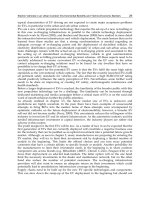
Figure 5.4. A: Normal upper anal canal. Top of picture is anterior and
shows the open horseshoe shape that can be misdiagnosed as a defect.
IAS, internal anal sphincter; PBR, puborectalis muscle. B: Normal anal
sphincters at mid-anal canal on endoanal ultrasound. IAS, internal anal
sphincter; EAS, external anal sphincter.
60 Vaginal Surgery for Incontinence and Prolapse
external sphincter muscle atrophy can be
detected with MRI, whereas this is diffi cult to
do on endoanal ultrasound (18,19). The ability
of MRI to detect fat gives it this advantage. As
the external sphincter atrophies, the striated
muscle is replaced with fat. Magnetic resonance
imaging is less effective in evaluating the inter-
nal sphincter. Endoanal ultrasound provides
superior imaging of the internal anal sphincter
with regard to defects and atrophy (20).
Multiple studies have compared endoanal
MRI to endoanal ultrasound, with some fi nding
that MRI is superior (21), others fi nding that
ultrasound is superior (22), and still others
fi nding that the two techniques are equivalent
(23). All the studies, however, agree that endo-
anal ultrasound is less expensive, more widely
available, and faster than MRI. Thus endoanal
ultrasound should be the initial imaging modal-
ity for fecal incontinence, reserving MRI for
cases where one might need to assess for atrophy
of the external sphincter or weakness of the
pelvic fl oor.
Cinedefecography
Cinedefecography is a radiographic procedure
that images the dynamics of defecation (24–28).
The patient’s rectum is fi lled with a barium
mixture that has the consistency of stool. The
vagina and small bowel are opacifi ed as well.
With the patient on a radiolucent commode, a
fl uoroscopic videotape is made capturing the
patient during rest, squeeze, push, evacuation,
and postevacuation. This exam is able to
demonstrate rectoceles, perineal descent,
spastic pelvic fl oor, intussusception, rectal pro-
lapse, enteroceles, and leakage of contrast. Most
of these fi ndings, however, are more benefi cial
in evaluating the patient with constipation sus-
pected of having obstructed defecation (28).
Overall cinedefecography is of limited value in
studying the patient with fecal incontinence
and thus is not routinely used (29).
Conclusion
The evaluation of fecal incontinence is a
complex process and should be tailored to the
individual patient. The history and physical
exam alone may be adequate in some patients,
but many patients require more extensive
investigation. Some institutions use all the
investigations at their disposal for every patient,
but the utility of that approach is mainly for
purposes of documentation and publication.
Still anorectal physiological testing and endo-
anal ultrasound are valuable tools that can help
in guiding one’s management of a patient with
fecal incontinence. In addition to being used in
the patient’s initial evaluation, these tests may
also be used in monitoring the patient’s prog-
ress and determining in an objective manner
what has been altered in the course of treat-
ment. The decision of which tests to utilize
and when is based on the physician’s clinical
Figure 5.5. Anterior sphincter defect
(top of picture and marked with dotted
lines) demonstrated on endoanal
ultrasound.
Fecal Incontinence 61
evaluation and judgment. One must remember
that these tests are only tools, and correlation
to the patient’s history and physical exam is
always necessary.
References
1. Vaizey CJ, Kamm MA. Prospective assessment of
the clinical value of anorectal investigations. Digestion
2000;61:207–214.
2. Jorge JMN, Wexner SD. Anorectal manometry: tech-
niques and clinical applications. South Med J 1993;80:
924–931.
3. Morgado P Jr, Wexner SD, Jorge JMN. Discrepancies in
anal manometric pressure measurement—important or
inconsequential? Dis Colon Rectum 1994;37:820–830.
4. Pfeifer J, Oliveira L, Park UC, et al. The relation of mano-
metry to age and gender. Tech Proctol 1996;1:10–13.
5. Yang Y-K, Wexner SD. Anal pressure vectography no
apparent benefi t for sphincter evaluation. Colorectal
Dis 1994;9:92–95.
6. McHugh SM, Diamant NE. Effects of age, gender and
parity on anal canal pressures. Dig Dis Sci 1987;32:
726–736.
7. Wexner SD, Marchetti F, Salanga VD, et al. Neuro-
physiologic assessment of the anal sphincters. Dis
Colon Rectum 1991;34:606–612.
8. Tjandra JJ, Milsom JW, Schroeder T, et al. Endoluminal
ultrasound is preferable to electromyography in
mapping anal sphincteric defects. Dis Colon Rectum
1993;36:689–692.
9. Jorge JMN, Wexner SD, Ehrenpreis ED, et al. Does peri-
neal descent correlate with pudendal neuropoathy? Dis
Colon Rectum 1993;36:475–483.
10. Nikiteas N, Korsgen S, Kuman D, et al. Audit of sphinc-
ter repair. Factors associated with poor outcome. Dis
Colon Rectum 1996;39:1164–1170.
11. Bartram CI, Sultan AH. Anal endosonography in faecal
incontinence. Gut 1995;37:4–6.
12. Law PJ, Kamm MA, Bartram CI, et al. Anal endosonog-
raphy in the investigation of faecal incontinence. Br J
Surg 1991;78(3):312–314.
13. Yang YK, Wexner SD, Nogueras JJ, et al. The role of
anal ultrasound in the assessment of benign anorectal
diseases. Coloproctology 1993;5:260–264.
14. Law PJ, Kamm MA, Bartram CI, et al. A comparison
between electromyography and anal endosonography
in mapping external anal sphincter defects. Dis Colon
Rectum 1990;78(4):448–450.
15. Sultan AH, Kamm MA, Hudson CN, et al. Anal endo-
sonography for identifying external sphincter defects
confi rmed histologically. Br J Surg 1994;81:463–465.
16. Sultan AH, Kamm MA, Hudson CN, et al. Endosonogra-
phy of the anal sphincter: normal anatomy and com-
parison with manometry. Clin Radiol 1994;49:368–374.
17. Karoui S, Sevoue-Collet C, Koning E, et al. Prevalence
of anal defects revealed by sonography in 335 incon-
tinent patients and 115 continent patients. AJR 1999;
173:389–392.
18. deSouza NM, Puni R, Zbar A, et al. MR imaging of the
anal sphincter in multiparious women using an endo-
anal coil: correlation with the in vitro anatomy and
appearances in fecal incontinence. AJR 1996;167(6):
1465–1471.
19. Rociu E, Stoker J, Eijkemans MJ, et al. Fecal inconti-
nence: endoanal US versus endoanal MR imaging.
Radiology 1999;212:453–458.
20. Malouf AJ, Williams AB, Halligan S, et al. Prospective
assessment of accuracy of endoanal MR imaging and
endosonography in patients with fecal incontinence.
AJR 2000;175:741–745.
21. Maier AG, Funovics MA, Kruezer SH, et al. Evaluation
of perinal sepsis: comparison of anal endosonography
and magnetic resonance imaging. J Magn Reson Imaging
2001;14:254–260.
22. Orsoni P, Barthet M, Portier F, et al. Prospective com-
parison of endosonography, magnetic resonance
imaging and surgical fi ndings in anorectal fi stula and
abscess complicating Crohn’s disease. Br J Surg 1999;
86:360–364.
23. Schwartz DA, Wiersema MJ, Dudiak KM, et al. A com-
parison of endoscopic ultrasound, magnetic resonance
imaging and exam under anesthesia for evaluation of
Crohn’s perianal fi stulas. Gastroenterology 2001;121:
1064–1072.
24. Agachan F, Pfeifer J, Wexner SD, et al. Defecography
or proctography. Results of 744 patients. Dis Colon
Rectum 1996;39:899–905.
25. Jorge JMN, Wexner SD, Ger GC, et al. Cinedefecogra-
phy and electromyography in the diagnosis of relaxing
puborectalis syndrome. Dis Colon Rectum 1993;36:
668–676.
26. Jorge JMN, Wexner SD, Marshetti F, et al. How reliable
are currently available methods of measuring anorectal
angle? Dis Colon Rectum 1992;35:332–338.
27. Jorge JMN, Yang Y-K, Wexner SD. Incidence and sig-
nifi cance of sigmoidoceles as determined to new
classifi cation system. Dis Colon Rectum 1994;37(11):
1112–1117.
28. Kelvin FM, Maglinte DDT, Benton JT. Evacuation proc-
tography (defecography) an aid to the investigation
of pelvic fl oor disorders. Obstet Gynecol 1994;82(2):
307–314.
29. Soffer EE, Hull T. Fecal incontinence: a practical
approach to evaluation and treatment. Am J Gastroin-
test 2000;95:1873–1880.
30. Jorge JMN, Wexner SD. Etiology and management
of fecal incontinence. Dis Colon Rectum 1993;36:
77–97.
31. Londono-Schimmer EE, Garcia-Duperly R, Nicholls RJ,
et al. Pudendal nerve latencies are predictive of outcome
following functional results. Int J Colorectal Dis 1994;
9:110–113.
32. Sitzler PJ, Thomson JPS. Overlap repair of damaged
anal sphincter. Dis Colon Rectum 1996;39:1356–1360.
33. Gilliland R, Altomare DF, Moreira H Jr, et al. Pudendal
nerve latencies are predictive of outcome following
anterior sphincteroplasty. Dis Colon Rectum 1997;40:
A13(abstr).
34. Young CJ, Mathur MN, Eyers AA, et al. Successful over-
lapping anal sphincter repair: relationship to patient
age, neuropathy and colostomy formation. Dis Colon
Rectum 1998;41:344–349.
35. Karoui S, Leroi A-M, Koning E, et al. Results of sphinc-
teroplasty in 86 patients with anal incontinence. Dis
Colon Rectum 2000;43:813–820.
36. Rockwood TH, Church JM, Fleshman JW, et al. Fecal
incontinence quality of life scale: quality of life instru-
ment for patients with fecal incontinence. Dis Colon
Rectum 2000;43:9–17.
62 Vaginal Surgery for Incontinence and Prolapse
Appendix: Fecal Incontinence
Quality of Life Scale (36)
Q1: In general, would you say your health is:
1 Excellent
2 Very Good
3 Good
4 Fair
5 Poor
Q2: For each of the items, please indicate how
much of the time the issue is a concern for
you due to accidental bowel leakage. (If it
is a concern for you for reasons other than
accidental bowel leakage, then check the
box “Not Apply.”)
Q3: Due to accidental bowel leakage, indicate
the extent to which you AGREE or DISAGREE
with each of the following items. (If it is a
Most of Some of A little of None of
the time the time the time the time Not apply
Q2. Due to accidental bowel leakage:
a. I am afraid to go out.
b. I avoid visiting friends.
c. I avoid staying overnight away from home.
d. It is difficult for me to get out and do things like going
to a movie or to church.
e. I cut down on how much I eat before I go out.
f. Whenever I am away from home, I try to stay near a
restroom as much as possible.
g. It is important to plan my schedule (daily activities)
around my bowel pattern.
h. I avoid traveling.
i. I worry about not being able to get to the toilet in time.
j. I feel I have no control over my bowels.
k. I can’t hold my bowel movement long enough to get to
the bathroom.
l. I leak stool without even knowing it.
m. I try to prevent bowel accidents by staying very near a
bathroom.
concern for you for reasons other than acci-
dental bowel leakage, then check the box
“Not apply.”)
Strongly Somewhat Somewhat Strongly
agree agree disagree disagree Not apply
Q3. Due to accidental bowel leakage:
a. I feel ashamed.
b. I cannot do many things I want to do.
c. I worry about bowel accidents.
d. I feel depressed.
e. I worry about others smelling stool on me.
f. I feel like I am not a healthy person.
g. I enjoy life less.
h. I have sex less often than I would like to.
i. I feel different from other people.
j. The possibility of bowel accidents is always
on my mind.
k. I am afraid to have sex.
l. I avoid traveling by plane or train.
m. I avoid going out to eat.
n. Whenever I go someplace new, I specifically
locate where the bathrooms are.
Fecal Incontinence 63
Q4: During the past month, have you felt so
sad, discouraged, hopeless, or had so many
problems that you wondered if anything
was worthwhile?
1. Extremely so, to the point that I have just
about given up
Scales range from 1 to 5, with a 1 indicating a lower functional status of quality of life. Scale scores are
the average (mean) response to all items in the scale (e.g., add the responses to all questions in a scale
together and then divide by the number of items in the scale. “Not apply” is coded as a missing value in
the analysis for all questions.)
Scale 1. Lifestyle (ten items): Q2a, Q2b, Q2c, Q2d, Q2e, Q2fg, Q2h, Q3b, Q3l, Q3m
Scale 2. Coping/behavior (nine items): Q2f, Q2i, Q2j, Q2k, Q2m, Q3d, Q3h, Q3j, Q3n
Scale 3. Depression/self-perception (seven items): Q1, Q3d, Q3f, Q3g, Q3i, Q3k, Q4 (question 1 is reverse
coded.)
Scale 4. Embarrassment (three items): Q2l, Q3a, Q3e
2. Very much so
3. Quite a bit
4. Some—enough to bother me
5. A little bit
6. Not at all
Electrodiagnostic testing of the pelvic fl oor is
becoming increasingly common in clinical
pelvic medicine and pelvic fl oor research.
Along with history, physical exam, and urody-
namics, neurophysiologic testing can help in
the diagnosis of certain pelvic fl oor disorders
and to determine if a central or peripheral
neurologic problems exists. Electrodiagnostic
testing is also emerging in studies investigating
the etiology of pelvic fl oor disorders. There-
fore, a basic understanding of the principles
and techniques used in electrodiagnostic medi-
cine are essential for reconstructive pelvic
surgeons.
6
Neurophysiologic Testing
Kimberly Kenton
65
Electrophysiologic Testing . . . . . . . . . . . . . . . . 65
Nerve Conduction Studies . . . . . . . . . . . . . . . . . 66
Stimulating . . . . . . . . . . . . . . . . . . . . . . . . . . . . . . 66
Recording . . . . . . . . . . . . . . . . . . . . . . . . . . . . . . . 66
Compound Muscle Action Potential . . . . . . . . 66
Pudendal Nerve Conduction Studies . . . . . . . . 68
Perineal Nerve Conduction Studies . . . . . . . . . 68
Clinical Applications . . . . . . . . . . . . . . . . . . . 69
Sacral Refl ex Testing . . . . . . . . . . . . . . . . . . . . . 69
Urethral Anal Refl ex . . . . . . . . . . . . . . . . . . . . . . 69
Bladder Anal Refl ex . . . . . . . . . . . . . . . . . . . . . . 69
Clitoral Anal Refl ex . . . . . . . . . . . . . . . . . . . . . . 69
Clinical Applications . . . . . . . . . . . . . . . . . . . 70
Electromyography . . . . . . . . . . . . . . . . . . . . . . . . 70
Surface electrodes . . . . . . . . . . . . . . . . . . . . . . 70
Concentric Needle Electrodes . . . . . . . . . . . . 70
Urethral EMG . . . . . . . . . . . . . . . . . . . . . . . . . 72
External Anal Sphincter . . . . . . . . . . . . . . . . 72
Clinical Applications . . . . . . . . . . . . . . . . . . . 72
Conclusion . . . . . . . . . . . . . . . . . . . . . . . . . . . . . . 72
This chapter introduces the most common
electrodiagnostic techniques used in women with
pelvic fl oor disorders, including pudendal and
perineal nerve conduction studies, sacral refl ex
testing, and surface and concentric needle elec-
tromyography. It describes the technique, advan-
tages, and disadvantages of the technique, and
how the technique can be applied clinically. A
brief review of neurophysiology is presented to
provide a basis for understanding the pathophys-
iology that leads to nerve and muscle disorders
and how the electrodiagnostic studies work.
Electrophysiologic Testing
Skeletal muscles are activated by electrical
impulses (action potentials), which can be gener-
ated along myelinated and unmyelinated axons.
In unmyelinated axons, action potential propa-
gation occurs by each small area of nerve under-
going depolarization activating its neighbor in a
continuous fashion. Many nerve cell axons are
covered myelin, which improves impulse con-
duction by increasing speed of current move-
ment down the inside of the membrane. The
myelin acts as an insulator, allowing the action
potential generated at one node to “jump” to the
next node signifi cantly increasing the velocity of
action potential propagation. The amount of
myelin surrounding a nerve and the distance
between the nodes of Ranvier are directly pro-
portional to the nerves diameter and conduction
velocity. A large myelinated nerve conducts more
quickly than small unmyelinated nerves. Large,
66 Vaginal Surgery for Incontinence and Prolapse
myelinated motor axons conduct action poten-
tials, and then branch out into a few terminal
branches, which attach to a single muscle fi ber.
Axonal and demyelinating neuropathies can
decrease the distance between nodes and slow
nerve conduction velocity, which can be recorded
clinically during electrodiagnostic testing. Table
6.1 lists nerve fi ber classifi cations.
Nerve Conduction Studies
A nerve conduction study is the introduction of
an action potential in the peripheral nervous
system and the subsequent recording of the
neural impulse at some location distant to the
site of stimulation. Nerve conduction studies
measure the velocity of action potential propa-
gation and the magnitude of the response,
thereby allowing one to make clinical judg-
ments about the health of a particular nerve.
Depending on the type of nerve stimulated and
where the recording electrodes are placed, one
can measure three different types of responses:
pure sensory nerve action potentials, compound
nerve action potentials from mixed sensory and
motor nerves, and pure motor nerve evalua-
tions by measuring compound muscle action
potentials (CMAPs).
Compound muscle action potentials have
been traditionally used to evaluate neuropathies
in women with pelvic fl oor disorders. Nerve
conduction studies facilitate identifying precise
neural injuries or more generalized neuropathic
injuries along portions of the peripheral nervous
system. As with all electrodiagnostic tests, it is
important to have a thorough understanding of
peripheral neuromuscular anatomy prior to per-
forming nerve conduction studies.
Stimulating
When performing nerve conduction studies, a
stimulus is given at a predefi ned site using a
surface or monopolar needle electrode. Most
pelvic fl oor electromyographers use surface
electrodes to stimulate, reserving needle elec-
trodes for nerves that are hard to stimulate
with surface electrodes because of excess fat or
edema. The magnitude of stimulus used in
routine nerve conduction studies is referred to
as the supramaximal stimulus. The supramaxi-
mal stimulus is approximately 20% to 30%
above the stimulus, which does not produce
any further increase in CMAP response because
all the nerve fi bers to the muscle are being
depolarized. The larger, myelinated axons are
depolarized fi rst, and then at supramaximal
stimulation, the smaller, myelinated axons are
depolarized.
Recording
After stimulating the nerve, it is necessary to
record the response. Surface or monopolar
needle electrodes can be used. When recording
a muscle response, three electrodes are neces-
sary: an active, a reference, and a ground. The
active electrode should be placed directly over
the muscle being studied, and the reference
electrode should be placed some distance from
the muscle.
Compound Muscle Action Potential
A CMAP is the biphasic waveform obtained
from stimulating a nerve proximal to a muscle
Table 6.1. Classification of nerve fibers
Sensory and Sensory Diameter Velocity
motor fibers fibers (nm) (m/s) Function
A α Ia 10–20 50–120 Motor: alpha motor neurons
Sensory: muscle spindle afferents
A α Ib 10–20 50–120 Sensory: Golgi tendon organs, touch and pressure
A-β II 4–12 25–70 Motor: neurons to intra/extrafusal muscle fibers
Sensory: secondary muscle spindle afferents, touch, vibratory, and pressure receptors
A-γ 2–8 10–50 Motor: gamma motor neurons to intrafusal muscle fibers
A-δ III 1–5 3–30 Sensory: small, lightly myelinated; touch, pain, and temperature
B 1–3 3–15 Motor: small, unmyelinated preganglionic autonomic fibers
C IV 1 2 Motor: all postganglionic autonomic fibers
Sensory: pain and temperature
Neurophysiologic Testing 67
and recording the potential directly over the
muscle. A CMAP, or M response, is a summa-
tion of all the muscle fi bers that are depo-
larized by a single stimulated nerve. Several
parameters recorded from the CMAP are
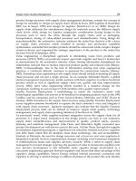
useful in electrodiagnostic testing (Figure 6.1).
Onset latency is the time from nerve stimula-
tion to the initial upward defl ection of the
CMAP. Onset latency refl ects neural activation
at the cathode, propagation of the action poten-
tial along the nerve, and transmission at the
neuromuscular junction. Therefore, an abnor-
mality at any of these sites can result in pro-
longed latency. Latency measures only the
large, heavily myelinated, fastest conducting
axons in a nerve. If a nerve has lost many
axons, but a few myelinated axons remain
intact, the onset latency will be normal. When
the latency is prolonged, one can assume
signifi cant loss of neuromuscular function.
However, if only a few axons conduct the nerve
impulse at a normal velocity, the latency can
be normal despite signifi cant neural injury.
Therefore, latency is not a sensitive measure of
nerve injury. Amplitude is measured from
baseline to the maximum point of the wave-
form. Amplitude refl ects the total number of
axons and muscle fi bers being tested and pro-
vides an estimate of the amount of functioning
tissue. It is less reliable than latency because
the distance between the electrode and muscle
infl uences it. Area is the space under the
portion of the waveform above baseline and
provides the most direct estimate of function-
ing tissue. Compound muscle action potential
duration is typically measured from the onset
latency to where it crosses the baseline. Dura-
tion and shape of the waveform measure the
temporal dispersion of all the individual fi bers.
Nerve conduction velocity is the rate an action
potential propagates along the stimulated
nerve. It is calculated by dividing the length of
nerve over which the action potential travels
by the time required to travel the distance.
However, in motor nerve conduction studies
the latencies between two different sites of
stimulation are subtracted from one another
to account for the delay at the neuromuscular
junction. Nerve conduction velocities are dif-
fi cult to obtain on the pudendal nerve owing
to the nerve’s anatomic course and the inabil-
ity to stimulate at two well-defi ned sites.
Nerve conduction velocities are affected by
the diameter of the nerve (large nerves conduct
more quickly), temperature (cooler tempera-
tures increase latency and amplitude), and age
(age greater than 60 years decreases nerve
conduction velocity and amplitude). Therefore,
delayed nerve conduction velocities in these
instances may not be abnormal.
Stimulus
Onset
Latency
Duration
Area
Amplitude
Figure 6.1. Compound muscle action potential (CMAP).
68 Vaginal Surgery for Incontinence and Prolapse
Pudendal Nerve Conduction Studies
The pudendal nerve branches, after exiting
Alcock’s canal, form three terminal branches:
the inferior hemorrhoidal, the perineal, and
the dorsal nerve to the clitoris. The inferior
hemorrhoidal and perineal branches contain
efferent fi bers to the external anal sphincter
and urethral sphincter, respectively, which
can be measured using nerve conduction
techniques.
Pudendal nerve conduction studies are the
most commonly reported electrodiagnostic
tests done on the pelvic fl oor. First described
by Kiff and Swash (1) in 1984 to study patients
with fecal incontinence, they have been used to
investigate the role of pudendal neuropathy in
stress urinary incontinence and pelvic organ
prolapse (2–6). A St. Mark’s electrode (Figure
6.2) consists of a stimulating cathode and anode
and two recording electrodes, which can be
attached to a gloved index fi nger. The stimulat-
ing electrodes are located at the tip of the index
fi nger and the recording electrodes at the base.
The pudendal nerve is then stimulated at the
level of the ischial spine. If stimulation is
applied transrectally, the recording electrodes
are located at the external anal sphincter. In
women, it is preferable to stimulate the puden-
dal nerve using a transvaginal approach with
surface electrodes placed over the external anal
sphincter at the 3 and 9 o’clock positions with
the patient in dorsal lithotomy. Normative data
using this technique in 42 continent women
have been established (Table 6.2). Older age,
more vaginal deliveries, and a wide genital
hiatus were associated with longer pudendal
and perineal nerve terminal motor latencies
(7). These data are consistent with normative
data determined in other electrodiagnostic
laboratories (8).
Perineal Nerve Conduction Studies
The amplitude and latency of fi bers to the ure-
thral sphincter can be measured at the same
time that pudendal nerve conduction studies
are being done. Ring electrodes consisting of
two pieces of platinum wire wound onto a small
cylinder are available, which slip onto the end
of a Foley catheter (Figure 6.3). When the elec-
trode is placed 1 cm distal to the Foley balloon
and the balloon is secured at the level of
the urethrovesical junction, the electrode can
record neuromuscular activity from the striated
urethral sphincter. Stimulating the pudendal
nerve at the ischial spine and recording from
the urethral sphincter and external anal sphinc-
ter simultaneously facilitate recording the
CMAP from the pudendal (inferior hemor-
rhoidal) and perineal branches. Normal values
Figure 6.2. St. Mark’s electrode.
Table 6.2. Normative pudendal and perineal nerve conduction
parameters
Latency (ms) Amplitude (μV)
Pudendal 1.94 (1.55–2.54) 101 (20–260)
Perineal 2.18 (1.84–3.33) 63 (14–199)
From Olsen et al. (7).
Figure 6.3. Ring electrode on Foley catheter.
Neurophysiologic Testing 69
for perineal latencies and amplitudes are listed
in Table 6.2.
Clinical Applications
Pudendal and perineal nerve conduction studies
established the link between pudendal neuro-
pathy and stress urinary incontinence and
fecal incontinence (2–6). Prolonged terminal
motor latencies have also been shown after
vaginal incontinence and prolapse surgery
(26,27), suggesting that some anterior vaginal
wall dissection leads to distal pudendal nerve
injury.
Pudendal nerve terminal motor latencies are
most frequently reported in case series of women
undergoing anal sphincteroplasty. Authors have
attempted to predict surgical outcomes based on
normal versus abnormal pudendal nerve func-
tion, with varying results (9–11). One hundred
subjects underwent anterior overlapping anal
sphincteroplasty after pudendal nerve testing.
Sixty-two percent of subjects with normal
pudendal nerve terminal motor latencies had
“successful” outcomes versus only 17% of sub-
jects with unilateral or bilateral pudendal nerve
terminal motor latencies (10). Other authors
have reported good postoperative success in
patients with prolonged pudendal nerve termi-
nal motor latencies (9).
The clinical usefulness of pudendal and peri-
neal nerve terminal motor latencies is hotly
debated. They should not be used in isolation
from other electrodiagnostic tests when evaluat-
ing pelvic fl oor injuries. Generally, EMG follows
nerve conduction studies since EMG is more
sensitive for detecting neuropathic injury.
Sacral Reflex Testing
Stimulation of certain pelvic fl oor structures
results in refl ex contractions of pelvic fl oor
skeletal muscles. Techniques to test these
refl exes in the pelvic fl oor are used to measure
efferent and afferent nerve activity, as well as
neurotransmission through the pelvic plexus
and sacral nerve routes. Refl exes from the
urethra and bladder travel through visceral
afferent pathways through autonomic nerves
to the sacral cord and are then carried through
the pudendal nerve to the external anal
sphincter.
Urethral Anal Reflex
Urethral anal refl exes were fi rst described by
Bradley et al (12), who used the ring electrode
mounted on a Foley catheter to stimulate while
recording from electrodes over the anal sphinc-
ter (3 and 9 o’clock positions—dorsal lithot-
omy). This refl ex involves afferent fi bers from
the urethra, which synapse in the conus medul-
laris and travel through pudendal efferents to
the external anal sphincter. Injuries to the pelvic
plexus or cauda equina frequently result in
absence of the urethral anal refl ex. If the patient
is unable to sense the stimulus, but the refl ex is
intact, she likely has an injury in the sensory
cortex or ascending spinal cord. Abnormal ure-
thral anal refl exes with preserved bladder anal
refl exes are common after multiple urethral
surgeries. Responses with latencies greater than
100 ms are voluntary and not considered refl ex
responses. Amplitudes of the responses are not
used clinically, but are currently being assessed
in research protocols. Normal values for women
can be found in Table 6.3 (13).
Bladder Anal Reflex
This refl ex is performed similarly to the ure-
thral anal refl ex, except the site of stimulation
is the bladder. Abnormal bladder anal refl exes
with preserved urethral anal refl ex latencies
suggest denervation injury to the bladder wall.
This can be seen in women with urinary reten-
tion or voiding problems owing to overdisten-
tion injuries.
Clitoral Anal Reflex
Surface electrodes are placed paraclitorally to
stimulate while recording is done at the surface
electrodes placed over the external anal sphinc-
ter. This refl ex passes through the pudendal
afferents to the spinal cord back through puden-
dal efferent fi bers to the anal sphincter. These
roots are often affected in cauda equina disease
Table 6.3. Normative sacral reflex latencies cutoffs
Reflex Sensory threshold (mA) Latency (ms)
Urethral anal 8 82
Bladder anal 37 85
Clitoral anal 9 55
70 Vaginal Surgery for Incontinence and Prolapse
but are not affected in conditions that disrupt
the pelvic plexus.
Clinical Applications
Anything that affects the pelvic plexus can
potentially disrupt the urethral and bladder anal
refl exes. This can be seen in peripheral neuropa-
thies with signifi cant autonomic components
and after radical pelvic surgery or radiation. The
clitoral anal refl ex should be preserved because
the course of this branch is not involved.
Pudendal neuropathy typically results in pro-
longed or absent clitoral anal refl ex with preser-
vation of the urethral and bladder anal refl exes.
The afferent limb of the pathway through the
pelvic plexus is less affected and is a temporally
longer portion of the pathway. Lesions in the
conus medullaris and cauda equina frequently
produce abnormalities in all sacral refl exes.
Suppression of the urethral anal refl ex by
actively trying to void is a measure of upper
motor neuron function (14). If a patient is unable
to suppress the response during voiding, she may
have a lesion in the suprasacral spinal cord.
Electromyography
Electromyography (EMG) is the recording and
study of electrical activity from striated muscles
and can be used to distinguish between normal,
denervated, denervated and reinnervated, and
myopathic muscle. The electrical activity can be
recorded using surface or needle electrodes and
then displayed on the oscilloscope screen of an
electrodiagnostic instrument. Voluntary elec-
trical activity is recorded as motor unit action
potentials (MUAPs), which represent the sum-
mation of activity from multiple motor units.
Motor units are composed of a single anterior
horn cell, its axon, and all the skeletal muscle
fi bers it serves.
A variety of electrode types are used for EMG.
Each has different properties and capabilities.
The most common electrodes used in the pelvic
fl oor are surface and concentric needle
electrodes (CNE).
Surface Electrodes
Surface electrodes are placed on the skin over
the muscle being evaluated and can be used to
evaluate patterns of muscle activity. Surface
electrodes record a summation of electrical
activity from the muscle, but cannot distinguish
individual MUAPs, and therefore cannot be
used to diagnose or quantify neuropathy or
myopathy. They are easier to use and less painful
than needle electrodes, but provide less reliable
information owing to signal distortion by inter-
vening skin, subcutaneous tissue, and volume
conduction from other muscles.
Surface electrodes are commonly used during
urodynamic studies to assess striated urethral
sphincter activity. Electrodes are placed on
either side of the perineal body or anal sphinc-
ter, and neuromuscular activity is recorded
during the cystometry and voiding portions of
the study. An increase in activity is normally
seen during fi lling with an absence activity
during voiding or episodes of detrusor overac-
tivity. This setup records neuromuscular activity
of multiple pelvic fl oor muscles, not just the stri-
ated urethral sphincter, making it diffi cult to dif-
ferentiate which muscle is contributing to the
signal. A recent study comparing perineal surface
to urethral CNE during urodynamics demon-
strated that needle tracings were consistently
more interpretable than surface recordings (15).
Needle tracings demonstrated urethral relax-
ation with voiding 79% of the time, whereas
surface recordings demonstrated urethral relax-
ation only 28% of the time.
Concentric Needle Electrodes (CNE)
Electromyographers consider EMG the gold
standard for studying peripheral striated neuro-
muscular disease. Needle electrodes are inserted
directly into the muscle, providing an accurate
portrayal of the electrical signals to diagnose
neuropathy or myopathy. The main advantage of
CNE is the ability to quantify neuromuscular
function. The small recording area at the beveled
tip of a CNE differentiates activity from approxi-
mately 20 nearby muscle fi bers. The wire or
active electrode is referenced to the needle shaft,
reducing the activity recorded from nearby
muscles. Concentric needle electrodes have the
advantages of being able to record EMG activity
with little interference from other muscles, a
predictable recording surface, and the absence
of a separate reference electrode.
Three types of activity can be recorded with
CNE: insertional, spontaneous, and MUAPs.
Neurophysiologic Testing 71
Insertional activity is the electrical activity
detected by the CNE as it passes through the
muscle at rest. When the electrode is in healthy
muscle, the insertional activity returns to base-
line in 300 ms. Decreased insertional activity
indicates that the electrode is not in muscle or
the muscle has undergone severe atrophy and
replacement by electrically inactive tissue. This
is commonly seen in the anal sphincter at the 12
o’clock position in women with long-standing
anal sphincter disruptions.
Spontaneous activity is persistent electrical
activity after the CNE is inserted and results
from marked membrane instability of the muscle
or neuron innervating it. Unlike most skeletal
muscles, which are electrically silent at rest, the
pelvic fl oor muscles have baseline tonic electri-
cal activity, making it more diffi cult to detect
spontaneous activity. The most common form of
spontaneous activity is the presence of positive
sharp waves or fi brillation potentials. Fibrilla-
tion potentials are action potentials of single
muscle fi bers that have been denervated. The
density of the fi brillation potentials is a rough
estimate of the number of denervated muscle
fi bers. Fibrillation potentials develop within 1 to
3 weeks after the loss of innervation. The fi nal
type of spontaneous activity reported in pelvic
fl oor muscles is complex repetitive discharges.
Complex repetitive discharges are high-
frequency, abrupt onset and offset waveforms
associated with neuropathy and voiding dys-
function in women. Fowler’s syndrome, fi rst
described in 2003, is the triad of urinary reten-
tion, urethral complex repetitive discharges, and
polycystic ovaries in young women (16). Reten-
tion in this group of patients is thought to be due
to “overactivity” of the striated urethral sphinc-
ter resulting from direct spread of electrical exci-
tation from muscle fi ber to muscle fi ber.
Motor unit action potential analysis in the
sphincters can be done at rest and with volun-
tary activity. Figure 6.4 shows a normal MUAP.
Nerve injury results in characteristic changes in
MUAP parameters of duration, amplitude, and
polyphasia. After nerve injury, a muscle fi ber
can be reinnervated by regrowth of the original
axon or a nearby axon. If a nearby axon reaches
the denervated muscle fi ber, it will supply more
muscle fi bers, creating a more complex MUAP.
The new complex waveform tends to be poly-
phasic (number of times a MUAP crosses the
baseline). New axons are initially not well
myelinated and conduct impulses more slowly;
as a result, newly reinnervated muscle has long
duration MUAPs. The MUAPs have larger ampli-
tudes because one motor unit is supplying more
muscle fi bers.
Motor unit action potential analysis can be
done using one of three techniques: manual
MUAP, individual MUAP, and computerized
multi-MUAP programs. A study comparing the
techniques in the anal sphincter demonstrated
that individual MUAP and multi-MUAP analy-
ses were most sensitive for differentiating
neuropathic from normal muscle (17). Mean
quantitative parameters from the three tech-
niques are different, so normative data from one
technique cannot be used for another (17).
Motor unit recruitment refers to the pattern
in which motor units are recruited by the spinal
cord. Muscles increase force by increasing the
frequency and number of individual motor units
are fi ring. Therefore, as voluntary effort is
Duration
Rise
Time
Phase
Amplitude
Baseline Crossing
Satellite
Turns
Figure 6.4. Motor unit action
potential.
72 Vaginal Surgery for Incontinence and Prolapse
increased, an increased number and frequency
of MUAPs should be seen. At maximum effort,
so many motor units are fi ring that individual
MUAPs cannot be distinguished, resulting in
an interference pattern. Computerized software
programs are available to measure interference
patterns as well.
Urethral EMG
Concentric needle EMG of the striated urethral
sphincter is done with the woman in the dorsal
lithotomy position. Anesthetic cream can be
applied to the external urethral meatus 10
minutes prior to the study to optimize patient
comfort. The CNE is inserted 5 mm above the
external urethral meatus. The striated urethral
sphincter is located at the mid-urethra, 1.5 cm
from the external urethral meatus.
External Anal Sphincter
Needle exam of the anal sphincter is also done
with the patient in dorsal lithotomy after appli-
cation of anesthetic cream. The external anal
sphincter can be located with a digital rectal
exam and the CNE inserted parallel to the
muscle at 3 and 9 o’clock. Twenty MUAPs should
be examined from each site.
Clinical Applications
Concentric needle EMG has been used in pelvic
fl oor muscles to confi rm the association between
pelvic nerve injury, and vaginal delivery, stress
incontinence, and fecal incontinence. Signifi -
cant changes in MUAP morphology have been
reported after vaginal childbirth by multiple
authors (18–20). Needle EMG of the levator ani
and external anal sphincter muscles has shown
electromyographic evidence of denervation
with reinnervation in women with stress urinary
incontinence and pelvic organ prolapse (20,21).
Two studies have used quantitative CNE of the
urethral sphincter in women undergoing incon-
tinence surgery (22–24). Fisher et al (23) dem-
onstrated more advanced neuropathic changes
in women with persistent stress urinary incon-
tinence. Kenton et al (24) studied 89 women
undergoing Burch urethropexy with CNE and
found signifi cant differences in EMG parame-
ters of women with successful incontinence
surgery, suggesting that these women had better
innervation of their urethral sphincters. Spe-
cifi c EMG criteria were established, which could
predict surgical success 100% of the time.
Gregory et al (19) recently reported quantita-
tive CNE data from the anal sphincter of 23 nul-
liparous and 28 vaginally parous women. Motor
unit action potentials from the 23 nulliparas had
signifi cantly higher amplitudes, longer dura-
tions, and more phases, providing further evi-
dence that vaginal childbirth results in pudendal
neuropathy.
Concentric needle EMG of the anal sphincter
can also be used to map the anal sphincter prior
to surgical repair. The exam should include the
anterior quadrant in addition to the 3 and 9
o’clock positions. This will provide information
about where the muscle is intact/disrupted and
about whether the muscle has sustained dener-
vation or denervation/reinnervation injury.
Some normative CNE MUAP parameters have
been reported for the external anal sphincter
and levator ani (20,25). No normative data exist
for the striated urethral sphincter.
Conclusions
Electrodiagnostic testing has both clinical and
research applications in pelvic fl oor disorders.
Clinical evidence suggests that certain types of
reconstructive surgery may impact pelvic fl oor
innervation. Zivkovic et al (26) measured peri-
neal nerve terminal motor latencies before and
after vaginal reconstructive surgery and found
signifi cantly prolonged terminal motor latencies
in women who underwent vaginal needle sus-
pension procedures. Similarly, Benson and
McClellan (27) found signifi cantly prolonged
pudendal and perineal nerve terminal motor
latencies in 27 women undergoing vaginal
prolapse repair, while the terminal motor laten-
cies of 21 women undergoing abdominal pro-
lapse repair were not different. The authors
then compared postoperative perineal terminal
motor latencies of the women with optimal
and suboptimal prolapse repairs. Pudendal
neuro pathy was signifi cantly more common in
the women with suboptimal repairs. In a well-
done randomized controlled trial of abdominal
versus vaginal reconstructive surgery, Benson
(13) found superior anatomic results of prolapse
repair in the abdominal group. Another ran-
Neurophysiologic Testing 73
domized controlled trial also demonstrated ana-
tomic superiority of the abdominal approach
(29). These data suggest that vaginal reconstruc-
tive surgery results in denervation of the pelvic
fl oor musculature, which may impact anatomic
success of the surgery. There is also increasing
data that preoperative pelvic fl oor denervation
may impact surgical outcomes, particularly for
continence procedures. Two studies demon-
strated a relationship between urethral sphinc-
ter neuropathy and outcome of continence
surgery (23,24).
Much of our current understanding of the eti-
ology of pelvic fl oor disorders has come from
both nerve conduction studies and EMG of the
pelvic fl oor muscles in women with stress incon-
tinence, fecal incontinence, and pelvic organ
prolapse. We understand that surgery can impact
pelvic innervation, and electrodiagnosis has
also confi rmed the relationship between vaginal
childbirth and pudendal neuropathy. The degree
of denervation and pelvic fl oor injury can be
measured and therefore studied. Such measure-
ments have some correlation with clinical out-
comes, but further research refi ning techniques
and establishing normative electrodiagnostic
parameters for the urethral sphincter anal
sphincter, and levator ani are imperative
(25,30,31). No normative data exist for the stri-
ated urethral sphincter.
Pelvic fl oor electrodiagnostic studies may aid
in the clinical diagnosis of some pelvic fl oor
disorders and help to predict outcomes of
incontinence surgery. However, confi rmatory
studies are necessary. Clinicians who wish to
add electrodiagnosis to their clinical evaluation
of patients with pelvic fl oor disorders should
have proper training in nerve conduction
studies and EMG or work in a multidisciplinary
setting with a neurologist or physiatrist trained
in electrodiagnosis.
References
1. Kiff ES, Swash M. Normal proximal and delayed distal
conduction in the pudendal nerves of patients with
idiopathic (neurogenic) faecal incontinence. J Neurol
Neurosurg Psychiatry 1984;47:820–823.
2. Snooks SJ, Badenoch DF, Tiptaft RC, Swash M. Perineal
nerve damage in genuine stress urinary incontinence.
An electrophysiological study. Br J Urol 1985;57:
422–426.
3. Snooks SJ, Barnes PR, Swash M. Damage to the inner-
vation of the voluntary anal and periurethral sphincter
musculature in incontinence: an electrophysiological
study. J Neurol Neurosurg Psychiatry 1984;47:1269–
1273.
4. Snooks SJ, Henry MM, Swash M. Faecal incontinence
due to external anal sphincter division in childbirth is
associated with damage to the innervation of the pelvic
fl oor musculature: a double pathology. Br J Obstet
Gynaecol 1985;92:824–828.
5. Snooks SJ, Swash M, Henry MM, Setchell M. Risk
factors in childbirth causing damage to the pelvic fl oor
innervation. Int J Colorect Dis 1986;1:20–24.
6. Snooks SJ, Swash M, Mathers SE, Henry MM. Effect of
vaginal delivery on the pelvic fl oor: a 5-year follow-up.
Br J Surg 1990;77:1358–1360.
7. Olsen AL, Ross M, Stansfi eld RB, Kreiter C. Pelvic fl oor
nerve conduction studies: establishing clinically rele-
vant normative data. Am J Obstet Gynecol 2003;189:
1114–1119.
8. Benson JT. Pelvic fl oor neurophysiology: an AAEM
Workshop. American Association of Electrodiagnostic
Medicine Workshop: American Association of Electro-
diagnostic Medicine, 1998.
9. Chen AS, Luchtefeld MA, Senagore AJ, Mackeigan JM,
Hoyt C. Pudendal nerve latency. Does it predict
outcome of anal sphincter repair? Dis Colon Rectum
1998;41:1005–1009.
10. Gilliland R, Altomare DF, Moreira H, Jr., Oliveira L,
Gilliland JE, Wexner SD. Pudendal neuropathy is pre-
dictive of failure following anterior overlapping sphinc-
teroplasty. Dis Colon Rectum 1998;41:1516–1522.
11. Sangwan YP, Coller JA, Barrett RC, Murray JJ, Roberts
PL, Schoetz DJ Jr. Prospective comparative study of
abnormal distal rectoanal excitatory refl ex, pudendal
nerve terminal motor latency, and single fi ber density
as markers of pudendal neuropathy. Dis Colon Rectum
1996;39:794–798.
12. Bradley WE, Timm GW, Rockswold GL, Scott FB.
Detrusor and urethral electromyelography. J Urol 1975;
114:891–894.
13. Benson JT, Lucente V, McClellan E. Vaginal versus
abdominal reconstrucitve surgery for the treatment of
pelvic support defects: a prospective randomized study
with long-term outcome evaluation. Am J Obstet
Gynecol 1996;175:1418–1422.
14. Sethi RK, Bauer SB, Dyro FM, Krarup C. Modulation of
the bulbocavernosus refl ex during voiding: loss of inhi-
bition in upper motor neuron lesions. Muscle Nerve
1989;12:892–897.
15. Mahajan ST, FitzGerald MP, Kenton K, Shott S, Bru-
baker L. Concentric needle electrodes are superior to
perineal surface patch electrodes for EMG documenta-
tion of urethral sphincter relaxation during voiding.
Pelvic Med Surg 2004;10:56.
16. Fowler CJ. Urinary retention in women. BJU Int 2003;
91:463–464.
17. Podnar S, Vodusek DB. Standardisation of anal sphinc-
ter EMG: high and low threshold motor units. Clin
Neurophysiol 1999;110:1488–1491.
18. Allen R. Pelvic fl oor damage and childbirth: a neuro-
physiological study. Br J Obstet Gynaecol 1990;97:770–
779.
19. Gregory WT, Lou JS, Stuyvesant A, Clark AL. Quantita-
tive electromyography of the anal sphincter after
uncomplicated vaginal delivery. Obstet Gynecol 2004;
104:327–335.
20. Weidner AC, Sanders DB, Nandedkar SD, Bump RC.
Quantitative electromyographic analysis of levator ani
74 Vaginal Surgery for Incontinence and Prolapse
and external anal sphincter muscles of nulliparous
women. Am J Obstet Gynecol 2000;183:1249–1256.
21. Aanestad O, Flink R. Urinary stress incontinence. A
urodynamic and quantitative electromyographic study
of the perineal muscles. Acta Obstet Gynecol Scand
1999;78:245–253.
22. Incontinence. In: Abrams P, Cardozo L, Khoury S,
Wein A, eds. 2nd International Consultation on Incon-
tinence, July 1–3, Plymbridge Distributers Ltd, Plym-
outh, UK, 2001. Health Publication Ltd, 2002.
23. Fisher JR, Hale DS, McClellan E, Benson JT. The use of
urethral electrodiagnosis to select the method of
surgery in women with intrinsic sphincter defi ciency.
Int Urogynecol 2001;12:33.
24. Kenton K, FitzGerald MP, Shott S, Brubaker L. Role of
urethral electromyography in predicting outcome of
Burch retropubic urethropexy. Am J Obstet Gynecol
2001;185:51–55.
25. European urinalysis guidelines—Summary. Scand J
Clinic Lab Invest 2000;60:1–96.
26. Zivkovic F, Tamussino K, Ralph G, Schied G,
Auer-Grumbach M. Long-term effects of vaginal dissec-
tion on the innervation of the striated urethral sphinc-
ter. Obstet Gynecol 1996;87:257–260.
27. Benson J, McClellan E. The effect of vaginal dissection
on the pudendal nerve. Obstet Gynecol 1993;82:387–
389.
28. Welgoss JA, Vogt VY, McClellan EJ, Benson JT. Rela-
tionship between surgically induced neuropathy and
outcome of pelvic organ prolapse surgery. Int Urogy-
necol J 1999;10:11–14.
29. Maher CF, Qatawneh AM, Dwyer PL, Carey MP, Cornish
A, Schluter PJ. Abdominal sacral colpopexy or vaginal
sacrospinous colpopexy for vaginal vault prolapse: a
prospective randomized study. [see comment]. Am J
Obstet Gynecol 2004;190:20–26.
30. Podnar S, Vodusek DB, Stalberg E. Comparison of
quantitative techniques in anal sphincter electromyog-
raphy. Muscle Nerve 2002;25:83–92.
31. Weidner AC, Barber MD, Visco AG, Bump RC, Sanders
DB. Pelvic muscle electromyography of levator ani and
external anal sphincter in nulliparous women and
women with pelvic fl oor dysfunction. Am J Obstet
Gynecol 2000;183:1390–1399; discussion 1399–1401.
Standard treatment for stress urinary inconti-
nence (SUI) in women has evolved over the last
few decades. The development of effective sur-
gical modalities and the recent explosion in the
availability of minimally invasive treatment
options have altered the playing fi eld, and have
provided a wider range of treatment options for
women with SUI. With these alternatives come
the opportunity and the responsibility to assess
how successful these treatments are. The area
7
Outcome Measures for Assessing Efficacy of
Incontinence Procedures
Adam G. Baseman and Gary E. Lemack
75
Diffi culties in Assessing Treatment
Outcome . . . . . . . . . . . . . . . . . . . . . . . . . . . . 75
Symptom Assessment . . . . . . . . . . . . . . . . . . . . 76
Effect of Study Methodology . . . . . . . . . . . . 76
Incontinence Questionnaires . . . . . . . . . . . . 76
Objective Measures to Assess
Volume/Frequency of Urine Loss . . . . . . 77
Pad Tests . . . . . . . . . . . . . . . . . . . . . . . . . . . . . 77
Voiding Diaries . . . . . . . . . . . . . . . . . . . . . . . . 80
Physiologic Assessment: Urodynamic
Testing . . . . . . . . . . . . . . . . . . . . . . . . . . . . . 81
Uses of Urodynamic Testing . . . . . . . . . . . . 81
Accuracy of Urodynamic Testing . . . . . . . . 81
Assessing Treatment Success . . . . . . . . . . . . 82
Assessing Adverse Outcomes . . . . . . . . . . . . 82
Objective Measures to Assess Anatomic
Changes Following Treatment . . . . . . . . . 82
Radiologic Studies . . . . . . . . . . . . . . . . . . . . . 82
Q-Tip Tests . . . . . . . . . . . . . . . . . . . . . . . . . . . 83
Stress Tests . . . . . . . . . . . . . . . . . . . . . . . . . . . 83
Recent Trends . . . . . . . . . . . . . . . . . . . . . . . . . . . 83
Conclusion . . . . . . . . . . . . . . . . . . . . . . . . . . . . . . 84
of outcome assessment itself has evolved over
the last several years, becoming more struc-
tured in the approach to defi ning treatment
success. This process is particularly important
for treatments aimed at SUI, where an improve-
ment in the quality of life is the ultimate goal,
and for which success can be defi ned in a
number of different ways. Indeed, it may be
impossible to identify a single parameter that
can be used to defi ne success in every patient
undergoing treatment for SUI. This chapter
explores current methods to analyze outcome
of SUI treatment.
Difficulties in Assessing
Treatment Outcome
Defi ning treatment success for SUI remains a
contentious issue. One of the fi rst consider-
ations is whether to rely on purely objective
outcome measures confi rming the absence of
SUI, for example the absence of urodynamically
demonstrable SUI following treatment of SUI.
Advocates of this argument would contend that
since one is treating a particular disease state,
then the absence of that disease state confi rms
successful outcome. It is perhaps the most pure
form of assessing success.
There are several problems with this
approach, however. First, our ability to detect
SUI may not be suffi ciently reliable. For
example, what might be present one day on