Cytologic Detection of Urothelial Lesions - part 6 pptx
Bạn đang xem bản rút gọn của tài liệu. Xem và tải ngay bản đầy đủ của tài liệu tại đây (557.8 KB, 20 trang )
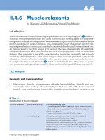
90 3. Grading Urothelial Neoplasms
Figure 3.24. High Grade Urothelial Carcinoma—bladder washing: A
cluster of malignant high grade urothelial cells is seen. Some malignant
cells have engulfed other malignant cells and show marked nuclear hyper-
chromasia. Cytoplasmic vacuolization, although not prominent, may be
seen in high grade urothelial carcinomas. In this case, there is not marked
nuclear overlap. (600x)
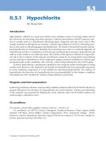
High Grade Urothelial Carcinoma 91
Figure 3.25. HighGrade Urothelial Carcinoma—bladder washing: In this
photomicrograph, numerous malignant cells are seen. The cells exhibit
high nuclear to cytoplasmic ratios, nuclear hyperchromasia and nuclear
membrane irregularities. The nuclear membrane appears thick in many
instances. (600x)
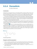
92 3. Grading Urothelial Neoplasms
Figure 3.26. High Grade Urothelial Carcinoma—voided urine: Degen-
erated malignant cells are seen admixed with benign squamous cells. Al-
though the nuclei are small and slightly degenerated, the nuclear mem-
branes are markedly thickened and irregular. Often, degeneration may limit
interpretation of high grade urothelial carcinoma. (600x)
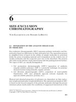
High Grade Urothelial Carcinoma 93
Figure 3.27. High Grade Urothelial Carcinoma—bladder washing: A
large malignant cell is seen in the center field. Admixed are numerous
neutrophils, benign urothelial cells and atypical squamous cells. In this
high grade urothelial carcinoma, squamous differentiation is seen. The
cytoplasm has a keratinized quality. Some types of high grade urothelial
carcinomas may exhibit more pronounced squamous differentiation and
may be difficult to separate from squamous carcinomas. (600x)
94 3. Grading Urothelial Neoplasms
Figure 3.28. High Grade Urothelial Carcinoma—bladder washing: Single
malignant cells are seen. These cells have high nuclear to cytoplasmic
ratios and thickened nuclear membranes. Several of the cells show stripped
nuclei. Numerous degenerated cells, crystals, squamous cells and debris
are present in the background. (600x)
High Grade Urothelial Carcinoma 95
Figure 3.29. High Grade Urothelial Carcinoma—catheterized urine: A
single neoplastic cell is seen in the center field. Some types of high grade
urothelial carcinomas show prominent nucleoli and less hyperchromatic
nuclei. In this case, abundant acute inflammation is admixed with debris.
The neoplastic cell has an enlarged nucleus and a high nuclear to cytoplas-
mic ratio. (600x)
96 3. Grading Urothelial Neoplasms
Figure 3.30. High Grade Urothelial Carcinoma—bladder washing: A true
tissue fragment in the center is composed of primitive epithelial cells with
high NC ratios and irregular nuclear outlines. Nuclear chromatin is granular
and nuclear shapes are variable. Compare these cells with surrounding
benign squamous and urothelial cells. (400x)
High Grade Urothelial Carcinoma 97
Figure 3.31. High Grade Urothelial Carcinoma—bladder washing: A true
tissue fragment consists of enlarged cells with high NC ratios. The nuclear
outlines are irregular as are the shapes. Chromatin is granular. Compare
with surrounding normal squamous and urothelial cells at the periphery of
the photograph. (600x)
98 3. Grading Urothelial Neoplasms
Figure 3.32. High Grade Urothelial Carcinoma—bladder washing: Cells
display characteristic variation in cellular size, NC ratio, cytoplasmic
shapes and nuclear irregularity. Contrast the obvious malignant cells with
the smaller cells with low NC ratios. (400x)
High Grade Urothelial Carcinoma 99
Figure 3.33. High Grade Urothelial Carcinoma—voided urine: High
grade lesions present with a variety of cellular features, including cyto-
plasmic vacuolization. Unless the cellular changes are consistently those
of an adenocarcinoma, such vacuoles should not persuade against the di-
agnosis of high grade urothelial carcinoma. Other cells in this photograph
do not have vacuolated cytoplasm and are characteristic of high grade
urothelial malignancy. (600x)
100 3. Grading Urothelial Neoplasms
Figure 3.34. High Grade Urothelial Carcinoma—bladder washing: Indi-
vidual cells have high NC ratios, irregular nuclear shapes and overall cellu-
lar enlargement. The nuclear chromatin is unevenly distributed. Compare
with surrounding benign squamous and urothelial cells. (600x)
High Grade Urothelial Carcinoma 101
Figure 3.35. High Grade Urothelial Carcinoma—bladder washing: Sev-
eral very large malignant cells display varying sizes, nuclear shapes, and
chromatin distribution. The cell in the center right could be infected with
polyoma virus, but its irregular nuclear shape is diagnostic of a cancer cell
rather than a benign decoy cell. (600x)
102 3. Grading Urothelial Neoplasms
Figure 3.36. High Grade Urothelial Carcinoma—voided urine: Voided
urine specimens may show few malignant cells that exhibit extensive de-
generation. In this case, a large malignant cell is seen in the center of the
field. The cell has an eccentric nucleus, and the nuclear chromatin is hy-
perchromatic and does not have the appearance of the nuclei seen in cells
infected with human polyoma virus. (600x)
High Grade Urothelial Carcinoma 103
Figure 3.37. High Grade Urothelial Carcinoma—voided urine: Several
malignant cells are seen in this photomicrograph, although the largest ma-
lignant cell is seen in the center. The nuclear to cytoplasmic ratio is not
markedly increased, although the nucleus is hyperchromatic and has an
irregular nuclear membrane. The cytoplasm is variegated. The nuclear size
is huge compared to red blood cells and acute inflammatory cells are seen
in the background. (600x)
104 3. Grading Urothelial Neoplasms
Figure 3.38. High Grade Urothelial Carcinoma—voided urine: A malig-
nant urothelial cell is seen in the center field. The nucleus is hyperchro-
matic and has a slightly irregular nuclear membrane. Compared to the
benign urothelial cell nuclei, the nucleus is huge. The cytoplasm is frothy
and slightly degenerated and an acute inflammatory background is seen.
In voided specimens, only a few malignant cells may be observed. (600x)
High Grade Urothelial Carcinoma 105
Figure 3.39. High Grade Urothelial Carcinoma—bladder washing: In this
bladder washing specimen, numerous degenerated malignantcells areseen.
In some cases of urothelial carcinoma, only few intact non-degenerated ma-
lignant cells are seen. These cells exhibit marked nuclear hyperchromasia
and slightly increased nuclear to cytoplasmic ratios. A background of acute
inflammation and several benign squamous cells are observed. (600x)
106 3. Grading Urothelial Neoplasms
Figure 3.40. Invasive High Grade Urothelial Carcinoma—bladder wash-
ing: Against a background of blood and inflammation, groups of tumor
cells suggest an invasive lesion. Such blood and debris are not univer-
sally an indicater of invasion in bladder lesions in contrast to the “tumor
diathesis” of cervical cancer. (200x)
High Grade Urothelial Carcinoma 107
Figure 3.41. Invasive Urothelial Carcinoma—bladder washing: Without
diagnostic tumor cells, a diagnosis of malignancy should not be made
despite the debris and blood in the background. (400x)
108 3. Grading Urothelial Neoplasms
Figure 3.42. Invasive High Grade Urothelial Carcinoma—bladder wash-
ing: Malignant spindle or fiber cells are one of the hallmarks of invasive
squamous carcinoma of the uterine cervix. Rarely seen in bladder cancer,
when present these cells are also indicative of invasion. Unfortunately, the
infrequency of this finding makes it of low utility. (200x)
High Grade Urothelial Carcinoma 109
Figure 3.43. Invasive High Grade Urothelial Carcinoma—bladder wash-
ing: Higher magnification of 3.42 verifies the malignant characteristics of
the fiber cells and the accompanying larger tumor cells. Note the relatively
clean background. (400x)