Emergency Vascular Surgery A Practical Guide - part 3 pdf
Bạn đang xem bản rút gọn của tài liệu. Xem và tải ngay bản đầy đủ của tài liệu tại đây (558.28 KB, 20 trang )
Chapter 3 Vascular Injuries in the Arm
36
3.5.2 Operation
3.5.2.1 Preoperative Preparation
Hemodynamically stable patients are placed on
their back with the arm abducted 90º on an arm
surgery table. The forearm and hand should be in
supination. Peripheral or central IV lines should
not be inserted on the injured side. Any continu-
ing bleeding is controlled manually directly over
the wound. If the site of injury is the brachial ar-
tery or distal to it, a tourniquet can be used to
achieve proximal control. It is then placed before
draping and should be padded to avoid direct skin
contact with the cuff. This minimizes the risk for
skin problems during inflation. The arm is washed
so the skin over the appropriate artery can be in-
cised without difficulty. The draping should allow
palpation of the radial pulse and inspection of fin-
ger pulp perfusion. One leg is also prepared in case
vein harvest is needed.
The position of the arm is the same for more
proximal injuries. Proximal control of high bra-
chial and axillary artery trauma may involve ex-
posure and skin incisions in the vicinity of the
clavicle and the neck, so for proximal injuries the
draping must also allow incisions at this level.
3.5.2.2 Proximal Control
For distal vessel injury, proximal control can be
achieved by inflating the previously placed tourni-
quet to a pressure around 50 mmHg above systolic
pressure. The cuff should be inflated with the arm
elevated to minimize bleeding by venous conges-
tion. After inflation, the wound is explored direct-
ly at the site of injury.
For more proximal injuries, control is achieved
by exposing a normal vessel segment above the
wounded area. The most common sites for proxi-
mal control in the arm are the axillary artery be-
low the clavicle, and the brachial artery (which is
what the artery is called distal to the teres major
muscle) somewhere in the upper arm. Some com-
mon exposures are described in the Technical Tips
box.
3.5.2.3 Exploration and Repair
Distal control is achieved by exploring the wound.
Sometimes this requires additional skin incisions.
The most common site for vascular damage in the
arm is the brachial artery at the elbow level. These
injuries occurs, for example, because of supracon-
dylar fractures in children and adults. In such
cases, exposure and repair of the brachial artery
through an incision in the elbow crease is appro-
priate. The anatomy is shown in Fig. 3.1, and a
brief description of the technique is given in the
Technical Tips box. Hematomas should be evacu-
ated to allow inspection of nerves and tendons.
Table 3.4. MESS: Mangled Extremity Severity Score (BP blood pressure)
Types Injury characteristics Points
Low energy
Medium energy
High energy
Massive crush
Stab wounds, simple closed fractures, small-caliber gunshot wounds
Open fractures, multiple fractures, dislocations, small crush injuries
Shotgun blasts, high-velocity gunshot wounds
Logging, railroad accidents
1
2
3
4
No shock (BP normal)
Transient hypotension
Prolonged hypotension
BP stable at the site and at the hospital
BP unstable at the site but normalizes after fluid substitution
BP <90 mmHg
1
2
3
No distal ischemia
Mild ischemia
Moderate ischemia
Severe ischemia
Distal pulses, no signs of ischemia
Absent or diminished pulses, no signs of ischemia
No signals by continuous-wave Doppler, signs of distal ischemia
No pulse; cool, paralyzed limb; no capillary refill
1
2
a
3
a
4
a
<30 years old patient
>30 years old patient
>50 years old patient
1
2
3
a
Points are doubled if ischemia lasts longer than 6 h.
37
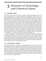
Fig. 3.2. The most proximal part of the axillary ar-
tery can be exposed through an incision parallel to
and just below the clavicle. Exposure of the brachial
artery is through an incision in the medial aspect of
the upper arm. This incision can be elongated and con-
nected with the clavicular incision to allow exposure
and repair of the entire axillary and brachial artery seg-
ments
TECHNICAL TIPS
Exposure for Proximal Control of Arteries in the Arm
Axillary Artery Below the Clavicle
An 8-cm horizontal incision is made 3 cm below
the clavicle (Fig. 3.2). The pectoralis major muscle
fibers are split parallel to the skin incision. The
pectoralis minor muscle is divided close to its
insertion. The nerve crossing the pectoralis minor
muscle can also be divided without subsequent
morbidity. The axillary artery lies immediately
below the fascia together with the vein inferiorly,
and the lateral cord of the brachial plexus is
located above the artery.
Brachial Artery in the Upper Arm
The incision is made along the posterior border of
the biceps muscle; a length of 6–8 cm is usually
enough (Fig. 3.3). The muscles are retracted medi-
ally and laterally, and the artery lies in the neuro-
vascular bundle immediately below the muscles.
The sheath is incised and the artery freed from the
median nerve and the medial cutaneous nerve
that surrounds it.
Brachial Artery at the Elbow
The incision is placed 2 cm below the elbow crease
and should continue up on the medial side along
the artery. If possible, veins transversing the
wound should be preserved, but they can be di-
vided if necessary for exposure. The medial inser-
tion of the biceps tendon is divided entirely, and
the artery lies immediately beneath it. By follow-
ing the wound proximally, more of the artery can
be exposed (Fig. 3.3). If the origins of the radial
and ulnar artery need to be assessed, the wound
can be elongated distally on the ulnar side of the
volar aspect of the arm. The median nerve lies
close to the brachial artery, and it is important to
avoid injuring it.
For supracondylar fractures, the brachial artery,
the median nerve, and the musculocutaneous
nerves must sometimes be pulled out of the frac-
ture site. Before the artery is clamped, the patient
is given 50 units of heparin/kg body weight IV. Re-
pair should also be preceeded by testing inflow
and backflow from the distal vascular bed by tem-
porary tourniquet or clamp release. It is often also
wise to pass a #2 Fogarty catheter distally to ensure
that no clots have formed. Occasionally, inflow is
questionable, and proximal obstruction must be
ruled out. This can be done intraoperatively by re-
trograde arteriography as described in Chapter 4
(p. 44) or by duplex scanning.
As a general principle, all vascular injuries in
the arms should be repaired, except when revascu-
larization may jeopardize the patient’s life. Arte-
rial ligation should be performed only when am-
putation is planned. Postoperative arm amputa-
tion rates are reported to be 43% if the axillary
artery is ligated and 30% at the brachial artery
level. Another exception is forearm injuries. When
perfusion to the hand is rendered adequate – as
assessed by pulse palpation and the Allen test –
one of these two arteries can be ligated without
3.5 Management and Treatment
Chapter 3 Vascular Injuries in the Arm
38
morbidity. In a substantial number of patients
with differing vessel anatomy, however, ligation of
either the ulnar or radial artery may lead to hand
amputation. If both arteries are damaged, the ul-
nar artery should be prioritized because it is usu-
ally responsible for the main part of the perfusion
to the hand.
For most arterial injuries, vein interposition is
necessary for repair. Veins are harvested from the
same arm, from parts of the cephalic or basilic
vein if the trauma is limited, or from the leg. The
saphenous vein in the thigh is suitable for axillary
and brachial artery repair, while distal ankle vein
pieces can be used for interposition grafts to the
radial and ulnar arteries. Before suturing the anas-
tomoses, all damaged parts of the artery must be
excised to reduce the risk of postoperative throm-
bosis. Rarely, primary suture with and without
patching can be used to repair minor lacerations.
Shunting of an arterial injury to permit osteo-
synthesis is rarely needed in the arm. Vascular in-
terposition grafting can usually be done with an
appropriate graft length before final orthopedic
repair. Also, extremity shortening due to fractures
is less of a problem in the arms (in contrast to
the legs), and orthopedic treatment without osteo-
synthesis is common especially in older patients.
Nevertheless, for some arm injuries shunting is a
practical technique that allows time for fracture
fixation, thus avoiding the risks of redisplacement
and repeated vessel injury. One example is injuries
to the axillary or brachial artery caused by a proxi-
mal humeral fracture, where the fragment needs to
be fixed in order to prevent such injuries. Another
example is humeral shaft fracture, which needs to
be rigidly fixed to abolish the instability that may
otherwise endanger the vascular graft. For more
details about shunting, see Chapter 9 (p. 111).
Veins should also be repaired if reasonably sim-
ple. If the vein injury is caused by a single wound
with limited tissue damage, concomitant veins to
the distal brachial artery can be ligated. For more
extensive injuries where the superficial large veins
are likely to be ruined, it is wise to try to repair the
deep veins. For very proximal injuries in the shoul-
der region, vein repair is important to avoid long-
term problems with arm swelling. It is also impor-
tant to cover the mended vessel segment with soft
tissue to minimize the risk for infection that may
involve the arteries.
3.5.2.4 Finishing the Operation
When the repaired artery or graft’s function is
doubtful and when the surgeon suspects distal
clotting, intraoperative arteriography should be
performed. The technique is described in Chap-
ter 10 (p. 128). After completion, all devitalized
tissue should be excised and the wound cleaned.
For penetrating wounds, damaged tendons and
transected nerves should also be sutured. This is
not worthwhile for most blunt injuries. Fascioto-
my should also be considered before finishing the
operation. As in the leg, long ischemia times and
successful repair increase the risk of reperfusion
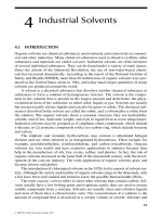
Fig. 3.3. Transverse incision in the elbow for exposing
the brachial artery and with possible elongations (dot-
ted lines
) when access to the ulnar and radial branches
as well as to more proximal parts of the brachial artery
is needed
39
and compartment syndrome, but the overall risk
for compartment syndrome is reported to be
less in the arm than in the leg. For a description of
arm fasciotomy techniques, we recommend con-
sulting orthopedic textbooks. After the wounds
are dressed, a fractured arm is put into a plaster
splint for stabilization.
3.5.2.5 Endovascular Treatment
In contrast to proximal arm vessel trauma, there
are few instances in distal injuries when endovas-
cular treatment is a feasible treatment option. Be-
cause the brachial artery and the forearm vessels
are easy to expose with little morbidity, open re-
pair during exploration of the wound is usually
the best option. Possible exceptions to this are
treatment of the late consequences of vascular
trauma, such as arteriovenous fistulas and pseu-
doaneurysms.
Especially in the shoulder region, including the
axilla, primary endovascular treatment is often
the best treatment option. Another circumstance
when endovascular treatment is favorable is bleed-
ing from axillary artery branches – such as the
circumflex humeral artery – due to penetrating
trauma. Active bleeding from branches, but not
from the main trunk, observed during arteriogra-
phy is preferably treated by coiling. The bleeding
branches are then selectively cannulated with a
guidewire and coiled, using spring coils or injec-
tions of thrombin to occlude the bleeding artery.
3.5.3 Management After Treatment
Postoperative monitoring of hand perfusion and
radial pulse is recommended at least every 30 min
for the first 6 h. When deteriorated function of the
repaired artery is suspected, duplex scanning can
verify or exclude postoperative problems. Appar-
ent occlusions should be treated by reoperation as
soon as possible. Compartment syndrome in the
lower arm may also evolve over time, and swelling,
muscle tenderness, and rigidity must also be mon-
itored during the initial days. For most patients,
treatment with low molecular weight heparin is
continued postoperatively. A common dose is
5,000 units subcutaneously twice daily.
Keeping the hand elevated as much as possible
may reduce swelling of the hand and arm as well as
problems with hematoma formation around the
wound. Early mobilization of the fingers facilitate
blood flow to the arm and should be encouraged.
3.6 Results and Outcome
The patency of arterial repair in the arm is often
excellent, but unfortunately, this appears to have
little impact on the eventual arm function. For
most patients in whom vessel trauma is associated
with nerve and soft tissue injury, it is the nerve
function that determines the outcome. Outcome
data after arterial repair in upper extremity inju-
ries have been reported in observational studies
and case series. One example is a review from the
United States of 101 patients with penetrating
trauma, including 13 axillary or subclavian cases.
Half of the patients had nerve injuries as well. At
follow-up the limb salvage rate was 99%, and all
patients who needed only vascular repair had ex-
cellent functional outcomes. Among arms that re-
quired nerve repair, 64% had severe impairment of
arm function. The corresponding figure for mus-
culoskeletal repair only was 25%.
A report from the United Kingdom included 28
cases of brachial artery injuries, of which six were
blunt. In this study, half of the patients had con-
comitant nerve injury and underwent immediate
nerve repair. All vascular repairs were successful,
but the majority of patients undergoing nerve re-
pair appear to have had some functional deficit at
follow-up.
Fortunately, it seems that function improves
over time in many patients. The risk factors for
poor outcome are similar to the ones used for the
MESS score – severity of the fracture and soft tis-
sue damage, length of the ischemic period, severity
of neurological involvement, and presence of
associated injuries.
3.7 Iatrogenic Vascular Injuries
The brachial artery is increasingly being used for
cannulation, both for vascular access and for en-
dovascular procedures. The latter requires large
introducer sheaths, and it is likely that we will ex-
perience an increase in the number of problems
related to this. Associated injuries are bleeding
3.7 Iatrogenic Vascular Injuries
Chapter 3 Vascular Injuries in the Arm
40
and thrombosis. (Both of these issues are dis-
cussed in Chapter 12.) Management of bleeding is
fairly straightforward. Bleeding is usually easy to
control by manual compression; exposure is sim-
ple; and repair is often accomplished by a few
simple sutures. Thrombosis is much less common
but is more complicated to handle. Management
should follow the guidelines given in Chapter 4.
Another problem that may be encountered is
related to arterial blood sampling from the radial
artery. Occasionally, thrombosis of this artery will
cause severe arm ischemia. This should then be
resolved by embolectomy and patch closure of the
injured vessel segment. Sporadically, vein graft in-
terposition is needed. Bleeding or an expanding
hematoma due to arterial puncture rarely occurs,
but pseudoaneurysm formation is not so infre-
quent. Such problems should be handled by sur-
gery, including proximal control and patch closure
of the injured vessel.
The radial artery is sometimes used as a graft
for coronary bypass procedures. This appears to
work extremely well, with little late morbidity in
the arm where the artery was harvested. We have
encountered occasional patients with mild hand
ischemia immediately after surgery, but only a few
cases who eventually needed revascularization.
For these rare patients, a vein bypass from the bra-
chial artery to the site where the ligature was
placed at harvest is the recommended treatment.
Further Reading
Fields CE, Lati R, Ivatury RR. Brachial and fore-
arm vessel injuries. Surg Clin North Am 2002;
82(1):105–114
McCready RA. Upper-extremity vascular injuries. Surg
Clin North Am 1988; 68(4):725–740
Myers SI, Harward TR, Maher DP, et al. Complex upper
extremity vascular trauma in an urban population.
J Vasc Surg 1990; 12(3):305–309
Nichols JS, Lillehei KO. Nerve injury associated with
acute vascular trauma. Surg Clin North Am 1988;
68(4):837–852
Ohki T, Veith FJ, Kraas C, et al. Endovascular ther-
apy for upper extremity injury. Semin Vasc Surg
1998;11(2):106–115
Pillai L, Luchette FA, Romano KS, et al. Upper-extrem-
ity arterial injury. Am Surg 1997; 63(3):224–227
Shaw AD, Milne AA, Christie J, et al. Vascular trauma
of the upper limb and associated nerve injuries. In-
jury 1995; 26(8):515–518
Stein JS, Strauss E. Gunshot wounds to the upper ex-
tremity. Evaluation and management of vascular
injuries. Orthop Clin North Am 1995; 26(1):29–35
ompson PN, Chang BB, Shah DM, et al. Outcome fol-
lowing blunt vascular trauma of the upper extrem-
ity. Cardiovasc Surg 1993; 1(3):248–250
Acute Upper Extremity Ischemia
4
CONTENTS
4.1 Summary 41
4.2 Background and Pathogenesis
41
4.3 Clinical Presentation
41
4.4 Diagnostics
42
4.5 Management and Treatment
42
4.5.1 Management Before Treatment 42
4.5.2 Operation 42
4.5.2.1 Embolectomy 42
4.5.2.2 Endovascular Treatment 43
4.5.3 Management After Treatment 43
4.6 Results and Outcome
43
Further Reading 44
4.1 Summary
History and physical examination are suf-
ficient for the diagnosis.
Few patients need angiography.
Embolectomy should be performed in most
patients.
It is important to search for the embolic
source.
4.2 Background and Pathogenesis
Acute ischemia in the upper extremity constitutes
10–15% of all acute extremity ischemia. The etiol-
ogy is emboli in 90% of the patients. The reason
for this higher rate compared with the leg is that
atherosclerosis is less common in arm arteries.
Emboli have the same origins as in the lower
extremity (see Chapter 10, p. 120) and usually end
up obstructing the brachial artery. Sometimes
plaques or an aneurysm in the subclavian or axil-
lary arteries is the primary source of emboli.
Embolization to the right arm is more common
than to the left due to the vascular anatomy.
For the 10% of patients with atherosclerosis and
acute thrombosis as the main cause for their arm
ischemia, the primary lesions are located in the
brachiocephalic trunk or in the subclavian artery.
Such pathologies are usually asymptomatic due to
well-developed collaterals around the shoulder
joint until thrombosis occurs, and they cause
either micro- or macroembolization.
Other less frequent causes of acute upper ex-
tremity ischemia are listed in Table 4.1.
4.3 Clinical Presentation
Acute arm ischemia is usually apparent on the
basis of the physical examination. The symptoms
are often relatively discreet, especially early after
onset. The explanation for this is the well devel-
oped collateral system circumventing the brachial
artery around the elbow, which is the most com-
mon site for embolic obstruction. The “six Ps” –
pain, pallor, paresthesia, paralysis, pulselessness,
Table 4.1. Less common causes of acute upper ex-
tremity ischemia
Cause Characteristics
Arteritis Lesions in distal
and proximal arteries
Buerger’s disease Digital ischemia in young
heavy smokers
Coagulation disorders Generalized
or distal thrombosis
Raynaud’s disease Digital ischemia
Chapter 4 Acute Upper Extremity Ischemia
42
poikilothermia – are applicable also for acute
arm ischemia, but coldness and color changes are
more prominent than for the legs. Accordingly,
the most common findings in the physical exami-
nation are a cold arm with diminished strength
and disturbed hand and finger motor functions.
Tingling and numbness are also frequent. The ra-
dial pulse is usually absent but is pounding in the
upper arm proximal to the obstruction.
Gangrene and rest pain appear only when the
obstruction is distal to the elbow and affects both
of the paired arteries in a finger or in the lower
arm. Ischemic signs or symptoms suggesting acute
digital artery occlusion in only one or two fingers,
imply microembolization.
4.4 Diagnostics
Only the few patients with uncertain diagnosis,
and those with a history and physical findings that
indicates thrombosis, need additional work-up.
Examples include patients with a history of chron-
ic arm ischemia (arm fatigue, muscle atrophy, and
microembolization) and bruits over proximal ar-
teries. Angiography should then be performed to
reveal the site of the causing lesion. Duplex ultra-
sound is rarely needed to diagnose acute arm isch-
emia but may occasionally be helpful.
4.5 Management and Treatment
4.5.1 Management Before Treatment
Even though symptoms and examination findings
may be so subtle that conservative treatment is
tempting, surgical removal of the obstruction is
almost always preferable. It has been suggested
that in patients with a lower-arm blood pressure
>60 mmHg embolectomy can be omitted, but such
a strategy has not to our knowledge been evaluated
systematically. In a patient series of nearly symp-
tomless acute arm ischemia, which was left to re-
solve spontaneously or with anticoagulation as the
only treatment, late symptoms developed in up to
45% of the cases. Surgical treatment is also fairly
straightforward. It can be performed using local
anesthesia and is associated with few complica-
tions.
Very often an embolus is a manifestation of
severe cardiac disease, so the patient’s cardiopul
-
monary function should be assessed and opti-
mized as soon as possible. Preoperative prepara-
tions include an electrocardiogram (ECG) and
laboratory tests to guide anticoagulation treat-
ment (see also Chapter 10, p. 25). Heparin treat
-
ment is started perioperatively and continued
postoperatively in most patients.
NOTE
Embolectomy is the treatment of choice
for almost all patients with diagnosis of
acute arm ischemia, regardless of the
severity of ischemia.
4.5.2 Operation
4.5.2.1 Embolectomy
As mentioned previously, the most common site
for embolic obstruction is the brachial artery. Em-
bolectomy of these clots is performed by expos-
ing the brachial artery as described in Chapter 3
(p. 37). The arm is placed on an arm table. We pre
-
fer to perform embolectomy using local anesthe-
sia. Often a transverse incision placed over the
palpable brachial pulse can be used. If proximal
extension of the incision is required, this should
be done in parallel with and dorsal to the dorsal
aspect of the biceps muscle. It has to be kept in
mind that 10–20% of patients may have a different
brachial artery anatomy. The most common varia-
tion is a high bifurcation of the radial and ulnar
arteries, and next in frequency is a doubled bra-
chial artery. The procedure is described in the
Technical Tips box.
Table 4.2. Frequency of signs and symptoms in pa-
tients with acute arm ischemia
Presentation Percentage
Pulselessness 96
Coldness 94
Pain 85
Paresthesia 45
Dysfunction 45
43
An alternative location for embolectomy in the
arm is to expose the brachial artery in the bicipi-
tal groove. A longitudinal incision starting 10 cm
above the elbow that is extended proximally is
then used.
TECHNICAL TIPS
Embolectomy via the Brachial Artery
Exposure of this vessel is described in Chapter 3.
A transverse arteriotomy in the brachial artery
is made as close as possible to the bifurcation
of the ulnar and radial arteries. The embolecto-
my is performed in proximal and distal direc-
tions with #2 and #3 Fogarty catheters. Separate
embolectomy in each branch should be done
if technically simple. The Fogarty catheter other-
wise slips down into the larger and straighter
ulnar artery. The route of the catheter can be
checked by palpation at the wrist level when
the inflated balloon passes. On the other hand,
restored flow in one of the arteries is usually
enough for a result that is sufficient for adequate
hand perfusion. The arteriotomy is closed
with interrupted 6-0 sutures, and distal pulses
and the perfusion in the hand are evaluated.
If the result is inadequate – poor backflow after
embolectomy, absence of pulse, a weak continu-
ous-wave Doppler signal, and questionable
hand perfusion – the arteriotomy should be
reopened and intraoperative angiography per-
formed (Table 4.3 and Chapter 10, p. 128).
If it is hard to achieve a good inflow, a proximal
lesion may cause the embolization or thrombosis.
More complicated vascular procedures are then
required to reestablish flow. The embolectomy
attempt is then discontinued and the patient taken
to the angiography suite for a complete examina-
tion. If practically feasible, an alternative is to
obtain the angiogram in the operating room. Fre-
quently, however, the preferred treatment is endo-
vascular, and this is better done in the angiography
suite. Occasionally the films will reveal a proximal
obstruction that needs open repair. Examples of
such are carotid-subclavian, subclavian-axillary,
and axillary-brachial bypasses.
4.5.2.2 Endovascular Treatment
Thrombolysis is as feasible for acute upper extrem-
ity ischemia as it is in the leg. The limited ischemia
that often occurs after most embolic events be-
cause of the collateral network around the elbow
also allows the time needed for planning and mov-
ing the patient to the angiosuite. The technique
involves cannulation in the groin with a 7-French
sheath. Long guide wires and catheters are re-
quired to reach the occluded site and makes iden-
tification of proximal lesions possible. A new arte-
rial puncture in the brachial artery may be neces-
sary for thrombolysis of distal occlusions.
It can be argued that thrombolysis in spite of
acceptable results, rarely is needed for treating this
disease because open embolectomy can be per-
formed under local anesthesia with good results
and little surgical morbidity. The advantages with
endovascular treatment are indeed limited. For
patients in whom suspicion of thrombosis is strong
or when proximal lesions are likely, it should be
attempted first. However, case series indicates that
results of thrombolysis are inferior for forearm
occlusions. In summary, thrombolysis is an alter
-
native but has little to offer in reducing risk or
improving outcome compared with embolectomy
for most patients.
4.5.3 Management After Treatment
Patients usually regain full function of their hands
immediately after the procedure, and postopera-
tive regimens consist of anticoagulation and a
search for the embolic source. Heparin or low
molecular weight heparin is administered as de
-
scribed in Chapter 10 (p. 129), usually followed by
coumadin. The search for cardiac sources may
advocate repeated ECGs, echocardiography, and
duplex ultrasound of proximal arteries.
4.6 Results and Outcome
The number of salvaged arms after surgical inter-
vention is very high, 90–95%, and arm function
is usually fully recovered. The remaining 5–10%
represents patients with extensive thrombosis
involving many vascular segments and most
branches of the distal arteries. The postoperative
4.6 Results and Outcome
Chapter 4 Acute Upper Extremity Ischemia
44
mortality is around 10–40% in most patient series,
reflecting that embolization often is a consequence
of severe cardiac disease. Postoperative mortality
is similar for thrombolysis to treat acute arm isch-
emia, while early technical success is slightly lower
or similar. Less favorable results with thromboly-
sis are achieved when the distal arteries also are
obstructed.
Further Reading
Baguneid M, Dodd D, Fulford P, et al. Management of
acute nontraumatic upper limb ischemia. Angiol-
ogy 1999; 50(9):715–720
Eyers P, Earnshaw JJ. Acute non-traumatic arm isch-
aemia. Br J Surg 1998; 85(10):1340–1346
Pentti J, Salenius JP, Kuukasjarvi P, et al. Outcome of
surgical treatment in acute upper limb ischaemia.
Ann Chir Gynaecol 1995; 84(1):25–28
Ricotta JJ, Scudder PA, McAndrew JA, et al. Manage-
ment of acute ischemia of the upper extremity. Am
J Surg 1983; 145(5):661–666
Whelan TJ Jr. Management of vascular disease of
the upper extremity. Surg Clin North Am 1982;
62(3):373–389
Table 4.3. Technique for retrograde intraoperative an-
giography
1. Control proximal to arteriotomy is achieved
by finger compression and/or vessel loop
2. Insert an angiography catheter or a small caliber
baby feeding tube through the arteriotomy in
retrograde direction
3. Place the tip of the catheter proximal to the
suspected obstructing lesion
4. Inject contrast under simultaneous fluoroscopy
in lateral projection with a C-arm
Abdominal Vascular Injuries
5
CONTENTS
5.1 Summary 45
5.2 Background 46
5.2.1 Background
46
5.2.2 Magnitude of the Problem
46
5.2.3 Etiology and Pathophysiology
46
5.2.3.1 Penetrating Injury
46
5.2.3.2 Blunt Injury
46
5.2.3.3 Pathophysiology
46
5.2.3.4 Associated Injuries
47
5.3 Clinical Presentation
47
5.3.1 Medical History
47
5.3.2 Clinical Signs and Symptoms
48
5.4 Diagnostics 48
5.5 Management and Treatment 50
5.5.1 Management Before Treatment
50
5.5.1.1 Treatment and Management
in the Emergency Department 50
5.5.1.2 Unstable Patients
50
5.5.1.3 Stable Patients
51
5.5.1.4 Laparotomy or Not?
. . . . . . . . . . . . . . . . . . 51
5.5.1.5 Renal Artery Injuries
51
5.5.2 Operation
52
5.5.2.1 Preoperative Preparation
52
5.5.2.2 Exploration
52
5.5.2.4 Vessel Repair
57
5.5.2.5 Finishing the Operation
60
5.5.3 Endovascular Treatment
60
5.5.4 Management After Treatment
60
5.6 Results and Outcome 61
5.7 Iatrogenic Vascular Injuries
in the Abdomen 61
5.7.1 Laparoscopic Injuries
61
5.7.2 Iliac Arteries and Veins
During Surgery for Malignancies
in the Pelvis 62
5.7.3 Iliac Artery Injuries
During Endovascular Procedures 62
5.7.4 Iatrogenic Injuries
During Orthopedic Procedures 62
Further Reading 63
5.1 Summary
Up to 25% of patients with abdominal
trauma may have major vascular injury.
Shock out of proportion to the extent of ex-
ternal injury suggests abdominal vascular
injury.
Isolated abdominal injury in patients with
shock suggests major vascular injury that
requires emergency laparotomy for con-
trol.
After the abdomen is entered, immediate
control of the supraceliac aorta should be
considered before continuing the opera-
tion.
Retroperitoneal hematomas should not be
explored right away unless they are actively
bleeding.
Stopping the procedure after the initial ex-
ploration for damage control to allow time
for resuscitation in the intensive care unit
is often a reasonable initial treatment.
If the patient’s condition allows and if en-
dovascular methods are available, consider
placing an aortic balloon from the left bra-
chial artery for temporary occlusion.
Chapter 5 Abdominal Vascular Injuries
46
5.2 Background
5.2.1 Background
Abdominal vascular trauma is fairly common in
modern civilian life and is a highly lethal injury,
with overall mortality around 40% in some
reported series. The main cause for this high
mortality relates to problems transporting injured
patients to the hospital fast enough to prevent ex-
sanguination. Furthermore, abdominal vascular
injuries are rarely isolated, and other organs are
often severely damaged as well. These factors
make it essential to resuscitate promptly and es-
tablish a rapid diagnosis.
The surgeon managing patients with major ab-
dominal injuries must be experienced with vascu-
lar surgical techniques and be able to expose the
aorta and its main branches, as well as the vena
cava. Dissection and the extensive organ mobili-
zation required for control and repair are often
difficult. It is therefore important to develop a
routine that can be employed during exploration
and control.
5.2.2 Magnitude of the Problem
Major abdominal vascular injury is seen in up to
25% of patients admitted with vascular trauma.
Blunt trauma is more common than penetrating
trauma in most European countries, while the op-
posite is reported in areas where gunshot wounds
are more frequent. Abdominal injury represents
10–20% of all traumas to the body caused by road
traffic accidents. Major vascular injury is estimat-
ed to occur in about 10% of cases of penetrating
stab wounds in the abdomen and in about 25% of
gunshot wounds. Blunt abdominal trauma affects
major vessels less frequently, estimates of below
5% is common in the literature.
NOTE
Major vascular injury is rather common
after abdominal trauma.
5.2.3 Etiology and Pathophysiology
5.2.3.1 Penetrating Injury
Penetrating injury creates the types of damage
that are common for most arteries – transection,
laceration, intimal dissection, and thrombosis, as
well as false aneurysms and arteriovenous fistula
formation. The first two are more common after
stab wounds. Gunshot wounds inflict more wide-
spread damage to the vessel wall, depending on
the bullet’s velocity. For example, high-velocity
missiles at speeds >700 m/sec cause up to 20 times
more damage than low-velocity projectiles. An
artery located within 10–15 cm of the trajectory
regularly thromboses after a high-velocity gun-
shot injury.
5.2.3.2 Blunt Injury
Typically, blunt injury to abdominal vessels occurs
after road traffic accidents or falls from heights.
Most commonly damaged are upper abdominal
arteries and veins such as the infrarenal aorta. The
mechanism is compression of the aorta against the
lumbar spine by the steering wheel, especially
when seat belts are not used. This causes intimal
tears and thrombosis of the aorta. Full-blown rup-
ture has also been reported. Vessel injuries are
much less frequent when seat belts are used. Avul-
sion of branches is also common and there is a
high incidence of associated injury to the small
arteries. Veins are usually not affected by blunt
trauma, except for the left renal vein. Major ab-
dominal injuries may cause avulsion of arteries; in
descending order of occurrence the vessels injured
are the left renal vein, the renal arteries, the supe-
rior mesenteric artery (SMA), and the abdominal
aorta just distal to the renal arteries.
5.2.3.3 Pathophysiology
When an artery is perforated, blood extravasates
into surrounding tissues, causing a hematoma that
counteracts the blood pressure and facilitates
spontaneous closure of the hole in the vessel.
When a vein is damaged, tamponade of the bleed-
ing often occurs, especially if retroperitoneal, un-
less the peritoneum is torn or is entered during
laparotomy. If vein damage is caused by a pelvic
fracture a cavity is created around the fragments,
preventing effective tamponade, and the bleeding
continues. Venous and arterial bleeding within
47
the mesentery is also enhanced by the same mech-
anism. The high blood flow through major arter-
ies in the abdomen makes spontaneous cessation
of bleeding less likely. Even the aorta, however, has
been reported to seal spontaneously after pene-
trating trauma when it is completely transected. If
an artery is partially lacerated, the severed ends
cannot contract; the hole is held open, and blood
flows more easily into the abdominal cavity. Pa-
tients rarely survive for long in this circumstance.
There are two principle mechanisms of vascu-
lar injury in blunt abdominal trauma: compres-
sion and deceleration forces. The former may cause
crush injuries and intramural hematoma or lacer-
ations. The latter cause stretching that creates ten-
sion between fixed and movable organs, leading to
avulsion or intimal disruption and thrombosis.
5.2.3.4 Associated Injuries
Any and all organs within the abdomen may be
injured in association with a major vessel injury. A
general rule is that for every major vascular injury,
three to four other organs are damaged as well.
The rate depends on the etiology of the trauma,
the location on the abdominal wall where the im-
pact or wound is located, and the direction of the
traumatic force. Table 5.1 gives an estimation of
the likelihood of injury to individual organs in
association with major vascular injury.
In general, blunt injury is more commonly as-
sociated with injury to many other organs, while
this is slightly less likely for penetrating trauma.
The small bowel is often injured by blunt trauma,
and the kidneys and spleen are frequently dam-
aged in both trauma types.
5.3 Clinical Presentation
5.3.1 Medical History
In patients who arrive to the emergency depart-
ment in shock with signs of penetrating or blunt
abdominal injury, the medical history does not
add much to the management, although informa-
tion about the mechanism of trauma is useful
when estimating the risk of associated injuries
(Table 5.1). Knowing exactly when the injury oc-
curred and when the patient became unconscious
may assist in predicting outcome.
Stable patients allows more time to gather in-
formation, and it is possible to ask direct questions
about the injury. This may provide important
clues about the possibility for major vascular
injury. For example, patients with contained he-
matomas are either stable or have a history of a
transient hypotensive period. This information is
easy to get from Emergency medical personnel.
Patients complaining of increasing abdominal
pain after either penetrating or blunt trauma
should be suspected of bleeding intraabdominally,
especially if the blood pressure is decreasing.
Shoulder pain and pain when breathing indicate
referred pain from blood irritating the diaphragm.
Patients should be asked about leg pain as an indi-
cation of arterial occlusion or embolization; this
is particularly important after blunt trauma. A
history of hematuria indicates renal or bladder
trauma.
Table 5.1. Probability of organ injury together with major arterial injury in the abdomen (compiled from seven
case series)
Stabbing Gunshot Blunt trauma
Liver + ++ +++
Pancreas ++ + ++
Stomach + ++ ++
Kidney +++ ++ ++
Spleen ++ + +++
Duodenum + + –
Small bowel + ++ ++
Colon+++++
5.3 Clinical Presentation
Chapter 5 Abdominal Vascular Injuries
48
5.3.2 Clinical Signs and Symptoms
The patient who presents with shock a short time
after injury to the abdomen should be presumed
to have a major vascular injury, with bleeding
directly into the peritoneal cavity. Increasing ab-
dominal distension or persistent hypotension de-
spite aggressive resuscitation are other signs sug-
gestive of continuing bleeding from an injured
vessel, liver, or spleen. Shock out of proportion to
the extent of external injuries, including fractures,
suggests abdominal vascular injury as the cause of
the bleeding. The finding of a mass during palpa-
tion, which is sometimes enlarging and pulsating,
strongly suggests major vessel damage. The ana-
tomical location gives some hint about the specific
vessel injured.
NOTE
Abdominal distension and shock out of
proportion to the extent of external
injuries indicate major vascular trauma.
In stable patients, assessment should include the
location of the wounds to assess the likelihood for
intraabdominal injury. As a general rule, all pen-
etrating wounds between the nipple line and the
groin should be presumed to have penetrated the
abdominal wall. Penetrating wounds in the mid-
line carry a substantial risk for aortic and vena ca-
val injury, but lateral wounds can also cause injury
to these structures. Wounds around the umbilicus
indicates that the bifurcation of these vessels is
likely to be affected. Entrance wounds located be-
low the umbilicus suggest iliac vessel injury. A tra-
jectory of a gunshot wound that passes the midline
also indicated major vascular trauma. It has to be
remembered, however, that it is notoriously diffi-
cult to assess trajectories, and bone and even the
muscle fascia may deflect bullets. The victim’s
body position at the time of injury can also influ-
ence which structures are damaged. Intraabdomi-
nal injuries may also be a result of wounds to the
back and buttocks.
Large hematomas tend to cause abdominal dis-
tension and tenderness in conscious patients. Ten-
derness may also be a result of peritonitis due to
contamination by perforated bowel or bowel isch-
emia. Blood in the urine, rectum, vagina, or a na-
sogastric tube also indicates intraperitoneal pene-
tration. Signs of a pelvic fracture should lead to a
high suspicion for iliac vessel damage.
Distal ischemia should also be excluded, and
palpation of pulses in the groins and distally is
obligatory after any major trauma. Particularly
after blunt trauma, distal ischemia may be the
only sign suggesting vessel damage. Unfortunate-
ly, 25% of patients who experience blunt trauma
causing some degree of arterial obstruction have
normal femoral pulses. Physical examination
should also include an assessment of the “six Ps”
(see Chapter 10, p. 121). In hemodynamically sta
-
ble patients with abnormal pulse examination, the
ankle–brachial index (ABI) should be measured
to aid in assessing limb ischemia. An ABI <0.9 –
especially unilaterally – implies some degree of
vessel obstruction. In general, trauma patients
tend to be young and therefore do not have sig-
nificant atherosclerosis, so an asymmetrical ABI
could be the only clue to an occult vascular injury.
Penetrating injury together with absent pulses
strongly indicates trauma to a major axial artery.
5.4 Diagnostics
In some circumstances, patients are so unstable
that they must be taken to the operating room for
laparotomy without diagnostic procedures. In
stable multitrauma patients in whom laparotomy
is not indicated for other reasons, additional
diagnostic measures may identify major vascular
injury, determine the extent of damage to other
organs, and facilitate treatment planning.
Ultrasonography can and should be performed
in most patients with abdominal trauma, regard-
less of their condition. The abdomen can be
scanned in the emergency department without
moving the patient and often takes less than 10 min
to perform. Its main objective is to detect hemo-
peritoneum as a possible source of hypotension. It
may also detect large hematomas and pseudoaneu-
rysms, but often misses retroperitoneal hemor-
rhage. Ultrasound also has low sensitivity for de-
tecting and excluding injuries to other organs such
as the intestines, liver, spleen, and kidneys.
Computed tomography (CT) has become a
valuable and widely used tool for evaluating most
stable patients with abdominal trauma. The CT
scan provides detailed information about the ret-
49
roperitoneal space, presence of hemoperitoneum,
active bleeding, false aneurysms, and damage to
other organs. The main limitation is its inability to
identify intestinal perforation, diaphragmatic in-
juries, and mesenteric tears. For blunt trauma it
gives information about the extent of damage to
the liver and spleen and thereby often identifies
patients who do not need laparotomy and those
who should undergo arteriography. Because CT is
unreliable in diagnosing intestinal perforation, it
has not been as valuable after penetrating trauma.
The use of contemporary spiral CT with intrave-
nous contrast has made it possible to detect active
bleeding, missile paths, and visceral perforation in
both blunt and penetrating trauma. For most ab-
dominal vascular injuries in stable patients, it is an
excellent screening tool, and when enhanced by
contrast, bleeding and vessel thrombosis can also
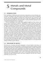
be diagnosed. Examples include detection of renal
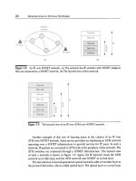
and visceral artery injuries, as seen in Fig. 5.1.
Some authors have recently suggested that CT
should be performed even in unstable patients in
order to reduce the number of unnecessary lapa-
rotomies. This concept depends in part on the
availability of CT, its location in the hospital and
on a strict management protocol.
Angiography is rarely used today to diagnose
arterial injury after abdominal trauma. Excep-
tions are stable patients with no signs of peritonitis
for whom a CT scan has given some indirect evi-
dence of arterial damage. The arteriogram is then
the initial step in an endovascular procedure
for definite treatment. Examples are arteriovenous
fistulas, pseudoaneurysm, active bleeding from
branch vessels, liver and spleen injuries, and pelvic
fractures. Other indications for angiography are
to diagnose suspected minor arterial lesions after
blunt trauma and to assess patients with signs of
organ or distal ischemia. Examples include aortic
and renal artery intimal tears and thrombosis.
A plain x-ray may be indicated in patients with
gunshot wounds in order to locate the bullet, to
facilitate estimation of the trajectory after apply-
ing markers at the entry and exit sites. If the bullet
is suspected to have passed through regions where
major vessels are located, angiography may be
indicated. Plain x-ray can also identify gas in the
abdominal cavity. Most of this information can
also be gained from CT.
Diagnostic peritoneal lavage (DPL) was the
standard way to diagnose intraabdominal bleed-
ing before the CT era. Because of its invasiveness
and the very high sensitivity in detecting even
minute intraabdominal bleedings that often does
not need surgical repair, it is much less often per-
formed today. Furthermore, it may not detect even
significant retroperitoneal bleeding. DPL is indi-
cated in unstable patients when it is vital to deter-
mine the source of bleeding and when ultrasound
is inconclusive and CT not possible to perform. It
may also be considered in stable patients when CT
and ultrasound are not available or in multitrau-
ma patients who require neurological or orthope-
dic operations and therefore will be inaccessible
for evaluation for long time periods. Technical
details about DPL are beyond the scope of this
text, so for descriptions on how to carry out DPL,
we recommend textbooks on abdominal trauma.
Intravenous pyelography (IVP) is a tool for di-
agnosing renal vascular injury that largely has
been replaced by CT. It may still have a place in the
operating room because it can be used during sur-
gery. The sign of renal vascular injury is lack of the
appearance of contrast in one of the kidneys. Its
main limitation is low sensitivity, and up to a third
of patients with vascular injury have normal IVPs
Laparoscopy has yet to find its place for evalu-
ating patients with suspected abdominal vascular
injury. It requires an operating suite and general
anesthesia, it cannot easily evaluate the retroperi-
Fig. 5.1. An example of computed tomography show-
ing retroperitoneal bleeding caused by blunt abdomi-
nal trauma
5.4 Diagnostics
Chapter 5 Abdominal Vascular Injuries
50
toneal space, and even small amounts of intraab-
dominal bleeding may disturb visualization. Its
main advantage is reported to be diagnosis of dia-
phragmatic injuries in stable patients.
5.5 Management and Treatment
5.5.1 Management Before Treatment
5.5.1.1 Treatment and Management
in the Emergency Department
The early management should follow the ABCs of
trauma resuscitation. Patients in shock should be
intubated and ventilated with 100% oxygen, and at
least two large intravenous (IV) lines should be
inserted, preferably in the upper extremity. Fluid
replacement through vascular access in the lower
extremities may extravasate and not reach the
heart if pelvic veins or the vena cava are injured.
The strategy and technique for obtaining rapid ve-
nous access in trauma are described in Chapter 11,
p. 137. When the lines are in place blood should be
drawn for routine analysis and blood typing and
cross-matching. Laboratory studies follow stan-
dard trauma management and should also include
acid-base balance, serum amylase, and urineanal-
ysis. Fluid resuscitation with warm lactated Ring-
er’s solution is continued or started. If the patient
has obvious severe blood loss, blood and plasma
are added as soon as possible. Platelet substitution
should also be considered. A Foley catheter and a
nasogastric tube should be inserted in all patients
with abdominal trauma. Hypothermia must be
prevented by all means.
Physical examination should be done during
the second survey, and the findings lead the man-
agement. For some patients, further diagnostic
procedures will determine whether they require
surgery or nonsurgical treatment. Ultrasound, for
instance, performed in the emergency department
can rule out or verify intraabdominal bleeding.
Patients with associated thoracic injury should
undergo chest x-ray to detect hemothorax and
other thoracic injuries as possible sources of
bleeding. However, as outlined below, CT is now
the most important diagnostic modality for stable
patients.
5.5.1.2 Unstable Patients
The management of unstable patients is summa-
rized in Table 5.2. Patients in shock with isolated
abdominal injury should undergo emergency lap-
arotomy. An abdominal ultrasound scan is needed
in multiply injured patients with injuries in the
thorax, head, or extremities. Unstable patients
with multiple injuries and a negative ultrasound
scan are a specific diagnostic problem. It may be
worthwhile to pursue the evaluation to rule out
abdominal bleeding as a possible cause of hypo-
tension in these patients. If they are in severe
shock, DPL may be indicated to rule out intra-
abdominal origin of the bleeding. CT may also
be an option in “less” unstable patients, especially
if there is improvement with resuscitation and CT
is readily available.
NOTE
Unstable patients with a negative ultra-
sound scan pose a particular diagnostic
problem when trying to exclude a major
vascular abdominal injury.
If DPL is negative, the cause of bleeding is likely to
be outside the abdomen, but false negatives can
occur. DPL may miss a serious retroperitoneal
bleeding. The ultimate management will then be a
matter of clinical judgment regarding whether the
patient will tolerate a CT scan or must be moved to
the operating room for emergency laparotomy.
The boxes in Table 5.2 indicating “maybe” repre-
sent circumstances in which clinical judgment is
especially important for the management.
If the patient is severely unstable and probably
not tolerates examination with CT, DPL could
possibly rule out intraperitoneal hemorrhage. A
positive DPL is an indication for surgery, while a
negative DPL points to the need for continued
evaluation as discussed above. More resuscitation,
for example, may be attempted followed by a CT
scan under close surveillance.
Emergency Thoracotomy
Patients with penetrating abdominal injury who
are unconscious and have prolonged severe hypo-
tension (<70 mmHg) but no other apparent inju-
ries causing the shock may occasionally be saved
by immediate proximal control of the aorta in
the emergency/operating room. Cross-clamping
51
of the descending thoracic aorta through a thora-
cotomy in the 4th or 5th interspace may then be
attempted if the patient is believed to have a realis-
tic chance of survival (e.g., became moribund in
the emergency department or lost measurable
blood pressure during the last part of the trans-
port to the hospital). The technique is briefly sum-
marized in Chapter 2 (p. 22). Aortic clamping be
-
fore laparotomy can facilitate perfusion through
the coronary and carotid arteries and prevent fur-
ther bleeding during laparotomy. Deterioration is
common in these severely ill patients when the ab-
dominal wall is incised and the tamponade it
maintains is released. It is disappointing, however,
how seldom this maneuver leads to the patient’s
survival.
5.5.1.3 Stable Patients
Stable patients with clinical signs of peritonitis af-
ter penetrating trauma should undergo laparoto-
my without delay for diagnostic procedures. All
others – with either blunt or penetrating trauma
– should be evaluated with CT to reveal the extent
of injury (Table 5.2). If ultrasonography in the
emergency department is performed routinely in
all trauma patients, it can be added to the diagnos-
tic process, but most stable patients admitted after
blunt trauma will need CT scanning regardless of
ultrasound findings. For example, surgery is indi-
cated for patients with ongoing active bleeding,
aortic thrombosis, or large hematomas caused by
organ injury. For other injuries, such as branch
vessel bleeding, renal or SMA thrombosis, and
pelvic arterial injuries, angiography followed by
endovascular treatment is often the best option.
Stable patients undergoing CT must be super-
vised at all times because they may become un-
stable quickly. Personnel must therefore be skilled
in assessing vital signs and the abdomen through-
out the examination.
5.5.1.4 Laparotomy or Not?
Unnecessary laparotomy is performed in up to
25% of patients with abdominal trauma and is as-
sociated with considerable morbidity and cost.
Nonoperative treatment has therefore grown in
popularity but has to be balanced against the price
of missed injuries. This approach has increased
the need for additional diagnostic procedures to
aid the decision process. Table 5.2 summarizes
these diagnostic modalities and how they can be
used for managing the patients.
Nonoperative treatment is particularly appeal-
ing in stable and multitrauma patients. Examples
of injuries that may be treated nonoperatively are
some liver, spleen, and renal injuries. For detailed
discussion on this subject, we recommend text-
books on trauma. Vascular injuries may be treated
without open surgery using endovascular meth-
ods. One example is embolization of bleeding pel-
vic vessels caused by pelvic fractures; another is
renal artery injuries.
5.5.1.5 Renal Artery Injuries
The most common type of renal vascular injury
after blunt trauma is thrombosis. This is usually
diagnosed by CT. For most blunt injuries, non-
operative treatment is appropriate if there are no
other indications for operative intervention, such
as when the diagnosis is made more than 12 h after
Table 5.2. Management of abdominal injuries when vascular damage is suspected
Patient’s
condition
Other
injuries
Ultrasonography Computed
tomography
Diagnostic
peritoneal lavage
Surgery
Finding Finding Finding
Unstable No No
(Yes
a
)
No No Yes
Yes Yes Positive No No Yes
Negative Maybe Maybe Positive Yes
Negative Maybe
Stable Yes/no No Yes Positive No Maybe
Negative No Observation
a
After blunt trauma, all patients should undergo ultrasonography.
5.5 Management and Treatment
Chapter 5 Abdominal Vascular Injuries
52
the trauma occurred. Reconstruction attempts
after renal ischemia of over 10–12 h are usually
futile, and the kidney will not regain its function
if this time limit is passed; however, successful
revascularization has been performed after 24 h of
ischemia. Exceptions when salvage may be tried
after longer ischemia times, are bilateral renal
ischemia, and patients with retrograde blood flow
as observed on an arteriogram indicating some
collateral supply.
Renal artery thrombosis following trauma can
usually be treated by angioplasty and stenting,
provided that rapid access to the angiosuite is pos-
sible. Also, minor lesions such as intimal flaps in
the renal arteries do not always need surgical
treatment. Such minor lesions should be treated by
observation. This includes patients with segmen-
tal parenchymal ischemia. Accordingly, surgical
reconstruction is saved for patients with active
bleeding and for situations when the diagnosis is
made during laparotomy.
5.5.2 Operation
5.5.2.1 Preoperative Preparation
The following section describes the recommended
procedure for a patient with active intraabdomi-
nal bleeding. It is also applicable for stable patients
in whom more time is initially available. Regard-
less, the patient should be prepped from the chin
to the knees so thoracic and groin vessel access
is possible if required. The saphenous vein must
also be accessible for harvest. After the patient is
prepped and draped and the surgeon is dressed
and ready, the patient is quickly anesthetized fol-
lowed by the start of the operation.
5.5.2.2 Exploration
Exploration
A midline incision from the xiphoid process to the
pubic bone is best for most situations. It is impor-
tant to divide fat and fascia for the entire length of
the wound before the peritoneum is incised. The
peritoneum is then opened rapidly – particularly
if the blood pressure drops after the abdomen is
entered – and the lesser omentum is opened and
widened using fingers. The aorta is palpated with
the index finger and can be occluded manually or
by compressing it against the spine with an aortic
occluder. To perform this maneuver it is some-
times necessary to mobilize the left lobe of the
liver to the right, as described in Chapter 7 (pp. 83,
84). If the hematoma is located above the trans-
verse mesocolon and aortic compression does not
rise the blood pressure, supraceliac or juxtaceliac
bleeding should be suspected. Extension of the in-
cision into the thoracic area to obtain occlusion of
the descending thoracic aorta is then recommend-
ed. This can be accomplished by dividing the
diaphragmatic crura and rarely requires median
sternotomy.
The aortic compression or occlusion is main-
tained while evacuating blood and blood clots. Re-
member that blood clots tend to accumulate close
to the bleeding site. Next, all sites where active
bleeding is noticed or suspected are packed with
laparotomy pads. Such pads usually stop even
quite substantial bleeding from the liver and
spleen as well as all venous bleedings, including
bleeding from the vena cava. Bleeding from the
aorta, iliac, celiac axis, SMA, and renal arteries, on
the other hand, will usually continue despite pack-
ing if the aortic occlusion is released and the pa-
tient not is hypotensive. Visual large arterial hem-
orrage may be handled without further dissection,
by ligature but one must be careful not to interrupt
the proximal SMA, aorta, or the renal arteries.
Temporary shunting may be a solution for these
vessels.
So far the whole procedure should take less
than 10–20 min.
NOTE
For patients in shock, the peritoneum is
left intact until the fascia is opened in its
entire length to preserve the peritoneal
tamponade as long as possible.
At this point, bleeding sources and their serious-
ness are assessed. If the patient is hypothermic
and has coagulopathy, the best decision could be
to stop the procedure, to temporarily close the ab-
domen and continue resuscitation in the intensive
care unit. This option, or “damage control” break,
may be considered even with some continuing ac-
tive bleeding. Damaged bowel segments are ligated
and injured ureters externalized before temporary
closure of the skin. The other option is to continue
53
the operation by focusing on definite control of
the most severe bleeding sites.
NOTE
Aortic compression at the supraceliac
level is often a good way to achieve
temporary proximal control while
assessing the damage.
Exposure and Control
Before the operation for definitive control contin-
ues, packing is reinforced by adding more pads.
Those packs should reapproximate disrupted tis-
sue planes if possible. Minor bleeding sites should
be left for later, unless they disturb the surgical
field. If the main bleeding appears to come from
the aorta at the suprarenal or juxtarenal level, the
manual supraceliac aortic occlusion is changed to
a clamp. During the time needed for the initial ex-
ploration described before, resuscitation can often
improve the patient’s condition enough to allow
temporary release of the aortic occlusion. If not, it
is often wise to wait a while before trying to clamp
the supraceliac aorta more permanently. The aorta
is freed by finger dissection, sporadically aided by
cutting the muscle fibers from the diaphragmatic
crus with a long-bladed pair of scissors. This
exposure is necessary for clamp placement. The
technique is further described in Chapter 7 (p. 84).
While the exposure also gives satisfactory proxi-
mal control to repair infrarenal aortic injuries,
the clamp should be moved to an infrarenal site as
soon as possible. If the patient’s condition allows
and endovascular methods are available, the place-
ment of an aortic balloon through a left brachial
or femoral artery access, is of great potential value.
This is best performed in the operating room. It
makes temporary occlusion of the aorta at differ-
ent levels possible in case of uncontrollable bleed-
ing during dissection. The technique is also de-
scribed in Chapter 7 (p. 83).
The technique for vascular exposure and final
control of other bleeding sites is described in the
Technical Tips box. Active bleeding from arteries
and veins around the liver hilus can be controlled
by using the “Pringle maneuver” – digital occlu-
sion of the hepatoduodenal ligament – followed by
careful dissection of the separate vessels.
NOTE
Stopping the procedure after the initial
exploration of damage control to allow
time for resuscitation in the intensive
care unit is often a reasonable initial
treatment.
TECHNICAL TIPS
Exposure of Different Intraabdominal Vascular Segments
Suprarenal Aorta and its Branches
The best way to expose the suprarenal aorta, the
origin of the SMA, the celiac axis, and the left renal
artery is to perform a “left medial visceral rota-
tion.” Divide the peritoneal reflection of the de-
scending colon, release the splenic flexure, and
cut the attachments between the spleen and the
diaphragm. Rotate the table slightly to the right
and move all viscera, including the colon, small
bowel, spleen, and the gastric fundus, to the right
side of the abdomen and cover all organs in large,
moist lap pads. This maneuver can be employed
either in a plane dorsal to the left kidney – which
will include the kidney with the viscera rotated to
the right – or ventral to the kidney. It is slightly
more difficult to find the appropriate dissection
plane for the latter approach, but this is more
practical for repairing most injuries. On the other
hand, including the left kidney with the rotated
viscera gives access to the posterior wall of the
aorta. When performed, little additional dissec-
tion enables proximal control using a Satinsky
clamp. The clamp is placed as distal as possible on
the aorta but sufficiently above the wounded area
to permit repair. Distal control is achieved by
clamps, balloons, or a Foley catheter.
Injuries to the portal vein are exposed and con
-
trolled by dividing the head of the pancreas be-
tween clamps or staplers to control the superior
mesenteric and splenic veins. Sometimes the gas-
troduodenal artery must be divided to facilitate
exposure. (See Fig. 5.2.)
5.5 Management and Treatment
Chapter 5 Abdominal Vascular Injuries
54
TECHNICAL TIPS
Exposure of Different Intraabdominal Vascular Segments (continued)
Vena Cava and Right Renal Vein
and Artery
Exposure of the infrahepatic vena cava and the
right side of aorta, including the portal vein and
the distal part of the SMA, is initiated by dividing
the attachments of the ascending colon, including
the hepatic flexure. Mobilize the colon, duode-
num, and the head of pancreas medially – perform
a full “Kocher maneuver” by dividing the lateral,
superior, and inferior attachments of the duode-
num – and cover the organs in lap pads and place
them under retractors. When the dissection is con-
tinued through the hematoma, the renal vein is
encountered first. To enable mobilization upward
and downward, it is banded and freed from sur-
rounding tissue. The renal artery is usually located
below or somewhat cranially to the vein. Proximal
control of the renal artery is accomplished either
on the left side of the vena cava or below the renal
vein. A DeBakey clamp is used unless the injury is
located close to the arterial origin; then partial
aortic occlusion with a larger clamp is necessary.
As shown in Fig. 5.2, venous control is accom-
plished by digital or sponge-stick compression of
the vena cava distal and proximal to the wounded
area. Another option for control during vena cava
repair is to insert a Foley catheter into the injured
vessel and inflate the balloon in the hole. Dorsal
cava injuries at the level of the renal vein some-
times necessitate mobilization and medial rota-
tion of the right kidney to expose the wounded
area. This approach is also the best way to expose
the portal vein within the head of the pancreas.
The reason why it works is that it is the most dorsal
structure in the portal triad (Fig. 5.3).
Retrohepatic Vena Cava
First, the inflow to the liver – the hepatic artery
and portal vein – is clamped together with the
bile duct. Use a small angled vascular clamp. Sec-
ond, the infrahepatic vena cava is freed as de-
scribed above and carefully cross-clamped proxi-
mal to the renal veins. Third, the proximal aorta
is exposed to be ready for cross-clamping if the
patient becomes hypotensive due to the cava
disruption. This exploration is performed through
the omentum minus as described in the main text.
The recommendation is to clamp the aorta if the
blood pressure falls to 60 mmHg or less. If possi-
ble, infrarenal cross-clamping is employed, espe-
cially if clamping is required for a long time to
achieve vascular repair. The fourth step is to mobi-
lize the liver by dividing all hepatic ligaments – the
falciform, teres, and right and left triangular liga-
ments – and clamp the suprarenal cava. This must
be done with care so the bleeding does not in-
crease. Control of the proximal vena cava can be
achieved below the diaphragm by continuing the
blunt and sharp dissection through the falciform
ligament. At this level the vena cava is freed cir-
cumferentially to permit clamping well above the
hepatic veins. Sometimes supradiaphragmatic ex-
posure is necessary. A right anterolateral incision
is then made in the diaphragm, and the dissection
is continued by opening the dorsal pericardial
fold until the suprahepatic vena cava is reached.
Finally, the liver is mobilized upward from the
right and left to expose as much as possible of the
retrohepatic vena cava.
Infrarenal Aorta
The aorta is exposed as for elective aortic proce-
dures. Wrap the small bowel in moist lap pads and
move it to the right side of the abdomen. Incise
the peritoneal reflection over the distal portions
of the duodenum and mobilize it to the right and
cephalad. Open the peritoneum directly over the
infrarenal aorta and free it from surrounding tis-
sue so that a clamp can be placed proximal to the
injury. Another clamp distally or a Foley catheter
inserted in the hole is used for distal control. If the
injury is in one of the common iliac arteries or is
close to the bifurcation, the iliac arteries must be
controlled distally. Dissection and clamping of
particularly the right common iliac artery must be
done with care so the iliac vein located under-
neath not is damaged. If the iliac veins are injured,
exposure sometimes necessitates temporary divi-
sion of the iliac artery to reach the injured vein.
Control is obtained by manual compression.
55
Fig. 5.2. Medial rightward rotation of the left viscera,
exposing the aorta from the diaphragm and all the
way down to the iliac arteries (“left medial visceral rota-
tion”)
Fig. 5.3. Peritoneal incision for a “Kocher maneuver”
to mobilize the duodenum, small intestine, and right
colon for a “right medial visceral rotation.” This allows
exposure of the entire inferior vena cava, right renal,
and iliac vessels
Iliac Arteries and Veins
The iliac arteries on the right side are found after
mobilizing the small bowel to the left and the ce-
cum proximally. The left-sided arteries are found
after mobilizing the sigmoid colon to the right
and incising the peritoneum. The arteries and
veins are usually quite easy to separate and con-
trol at this level.
5.5 Management and Treatment