Emergency Vascular Surgery A Practical Guide - part 5 pot
Bạn đang xem bản rút gọn của tài liệu. Xem và tải ngay bản đầy đủ của tài liệu tại đây (1.78 MB, 20 trang )
77
7.3.3 Dierential Diagnosis
Patients with a ruptured AAA who are not in
shock present with signs that are similar to a vari-
ety of other acute diseases in the abdomen or back.
To avoid misdiagnosis with conditions that do not
require emergency laparotomy, careful examina-
tion of the abdominal aorta is important.
Ruptured AAA, or symptomatic aneurysms
with incipient rupture, should be included in the
discussion about differential diagnosis in all ab-
dominal emergencies, particular in elderly men.
Kidney stones located in the ureter, diverticulitis,
constipation, intestinal obstruction, pancreatitis,
gastric or intestinal perforation, intestinal isch-
emia, vertebral body compression, and even acute
myocardial infarction are all primary diagnoses
that can be mixed up with a ruptured AAA. Of
course, there is a potential risk of sending a patient
home believing that, for example, a ureteral stone
has caused the trouble when AAA rupture is the
true diagnosis. A significant risk is also related to
performing a major operation because of a sus-
pected ruptured AAA in a patient who actually is
suffering from an acute myocardial infarction.
The only way to avoid this is to keep the AAA di-
agnosis in mind and to carefully examine the
patient.
Another important differential diagnosis is
aortic dissection. It is common that a patient will
initially have been treated at a smaller healthcare
unit or in the emergency department where an
ultrasound was performed and misinterpreted as
“dissection in an aortic aneurysm.” This misun-
derstanding is caused by the thrombus within the
AAA, which can be interpreted as a doubled aortic
lumen. There is, however, a clear distinction be-
tween rupture and dissection. Rupture is a true
burst of the aortic wall with bleeding out from the
vessel. Dissection starts with a tear in the inner
layer of the vascular wall through which the blood
passes and cause a longitudinal separation of the
layers, causing a double lumen. Rupture is com-
mon in AAA, but dissection is rare (see the infor-
mation on aortic dissection in Chapter 8).
7.3.4 Clinical Diagnosis
A summary of different clinical presentations of
AAA is presented in Table 7.2. These different sce-
narios can be used in determining the risk for the
presence of a ruptured AAA.
NOTE
The presentation of a patient with a
ruptured AAA varies, but in most cases
a classic triad is found:
– Abdominal pain
– Circulatory instability
– Tender pulsating mass
This combination of symptoms and
clinical findings should always be regard-
ed as a ruptured AAA until the opposite
is proven.
The purpose of Table 7.2 is to facilitate patient
management, and the remaining part of this chap-
ter is largely based on this table. It should be re-
membered, however, that patients might present
with a clinical picture that lies in between the cat-
egories.
7.4 Diagnostics
When an aid in detecting AAA is needed, a com-
puted tomography (CT) scan is the first choice for
all categories used in Table 7.2. When the suspi-
cion is strong and the risk for sudden deterioration
is considered high, the scan should be performed
quickly. The responsible surgeon should supervise
the procedure so that it can be stopped if neces-
sary and the patient transferred to the operating
room immediately. The CT scan should be per-
formed with contrast. The primary questions the
scan should answer are as follows: Is there an
AAA? Are there signs of rupture? What size is the
AAA, and how far proximally and distally does it
extend?
NOTE
In the classic case of a ruptured AAA,
no diagnostic tools except the physical
examination are needed.
7.4 Diagnostics
Chapter 7 Abdominal Aortic Aneurysms
78
To look for anything other than what is mentioned
above is unnecessary in an emergency work-up of
a patient with a suspected ruptured AAA. The di-
agnosis made by CT is easy, and typical findings
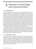
are demonstrated in Fig. 7.1.
Signs of rupture on the scan include a hemato-
ma and contrast that is visible outside the aortic
wall retroperitoneally. An early sign of rupture is
the presence of contrast in the thrombus and a
very thin aortic wall overlying it. The location of
the aneurysm in relation to the renal arteries is
important for planning an operation but rarely
influences the indication for surgery. It is impor-
tant to remember that a patient with a diagnosed
AAA and pain but with a CT scan showing no
signs of rupture needs to be managed as if the pa-
tient has impending rupture. Pain may precede
rupture, and the scan only answers the question of
whether a rupture is already present at the exami-
nation. Unfortunately, no signs can predict wheth-
er an AAA is going to rupture soon.
There is rarely a place for ultrasound when try-
ing to diagnose a ruptured AAA. Performed in the
operating room, it might occasionally be helpful
to exclude or verify the presence of an AAA.
When the patient is hemodynamically stable or
when the suspicion of rupture is low, the use of ad-
ditional diagnostic tests to exclude other illnesses
is encouraged. Examples of such diseases are pan-
creatitis and myocardial infarction. These can be
verified by electrocardiogram (ECG), a plain ab-
dominal x-ray, a CT scan, ultrasound, or urogra-
phy as well as by blood tests.
7.5 Management and Treatment
7.5.1 Management Before Treatment
7.5.1.1 Ruptured AAA
If the triad is present the patient needs to be oper-
ated without delay caused by preoperative exami-
nations or tests. The time available for making the
Table 7.2. Clinical ndings and management of ruptured aortic aneurysms (AAA abdominal aortic aneurysm,
OR operating room, CT computed tomography)
Pain Hemodynamic
instability
Pulsating
mass
Clinical diagnosis Measures
Yes Yes Yes Ruptured AAA
(classic triad)
Immediate transfer to OR
Yes Yes No Rupture suspected
(lack of mass may be due to
obesity or low blood pressure)
If history of AAA or signs peritonitis,
transfer to OR;
Perform ultrasound scan in the OR
or CT scan with the surgeon present
Yes No Yes Rupture possible
(may have an incipient rupture
or an inflammatory aneurysm)
Perform CT scan and consider urgent
surgery if diagnosis of AAA is made
Yes No No Rupture unlikely
(may have a contained rupture
if the patient obese or difficult
to palpate)
Perform CT or ultrasound scan
Fig. 7.1. Typical appearance on computed tomog-
raphy of a ruptured abdominal aortic aneurysm with
contrast in lumen, thrombus, calcications in the wall,
and a large retroperitoneal hematoma
79
correct decision regarding patient management is
usually limited. The following measures should
rapidly be done in the emergency department:
1. Obtain vital signs, medical history, and physi
-
cal examination.
2. Administer oxygen.
3. Monitor vital signs (heart rate, blood pressure,
respiration, SPO
2
).
4. Obtain informed consent.
5. Place two large-bore intravenous (IV) lines.
Insertion of central lines is time-consuming,
and to avoid delays it is better done in the
operating room after surgery has started.
6. Start infusion of fluids.
7. Obtain blood for hemoglobin, hematocrit,
prothrombin time, partial thromboplastin
time, complete blood count, creatinine, blood
urea nitrogen, sodium, and potassium, as well
as a sample for blood type and cross-match.
8. Catheterize the urinary bladder (this often has
to be done in the operating room to gain time)
and start recording urine output.
9. Administer analgesics, such as 2–3 mg mor
-
phine sulphate IV up to 15 mg, depending on
the patient’s vital signs, severity of pain, and
body weight.
10. Order eight units of packed red blood cells and
four of plasma.
The list suggested above may vary among different
hospitals. Remember to include pulses, including
femoral, popliteal, and pedal, in the physical ex-
amination. This is important as a baseline test in
case of thromboembolic complications to the legs
during surgery. It is also important to be cautious
about rehydration and administration of inotropic
drugs. The latter should be used only when the pa-
tient is in shock and when the low blood pressure
threatens to affect cardiac or renal function. The
aim should not be to restore the patient’s normal
blood pressure; a pressure of around 100 mmHg is
satisfactory if the patient’s vital functions are in-
tact. Hypotension may be an important factor
minimizing the bleeding and keeping it contained
within the retroperitoneal space. Too intense vol-
ume replacement and increased blood pressure
may initiate rebleeding.
As soon as possible, the patient should be taken
to the operating room and a vascular surgeon con-
tacted. If no surgeon with experience performing
AAA procedures is available, consider contacting
another hospital and presenting the case to the
vascular surgeon there. The patient may then be
referred to that hospital or the vascular surgeon
could come and perform the procedure if the pa-
tient’s condition does not allow transport. Even
stable patients might start to rebleed at any mo-
ment and should therefore not be transported too
liberally. If the patient is hemodynamically stable,
the start of operation should be delayed until an
experienced surgeon is available. However, if there
are signs of hemodynamic instability or manifest
shock despite treatment, the operation should be
initiated. The aim then is to achieve control of the
bleeding.
7.5.1.2 Suspected Rupture
The checklist described before is, by and large,
also valid when rupture is only suspected.
This category of patients is the most challeng-
ing, and generally applicable advice is difficult to
give. This category includes patients with a rup-
tured aneurysm but without a palpable pulsating
mass due to obesity and severe hypotension. There
are also many other life-threatening conditions
that should not be treated with surgery in this
group. One such condition is acute myocardial in-
farction, which also may start with thoracic and
abdominal pain and hypotension. Therefore, the
surgeon must rapidly decide whether to perform
an emergency operation or order diagnostic ex-
aminations to verify the diagnosis. In the case of
an actual rupture, it is evident that examinations
that delay the start of the operation are associated
with severe risk. Therefore, every such step should
be performed simultaneously with other preoper-
ative measures if possible. For example, ECG is
helpful in the diagnosis of myocardial infarction,
and ultrasound can verify or exclude the presence
of an AAA.
7.5.1.3 Possible Rupture
A tender pulsating mass supports the suspicion of
rupture. In a circulatory-stable patient with pos-
sible rupture, the following is done in the emer-
gency department:
1. Place an IV line and start a slow infusion of
Ringer’s acetate.
2. Order an emergency CT scan, with the patient
monitored by a nurse.
7.5 Management and Treatment
Chapter 7 Abdominal Aortic Aneurysms
80
If the CT scan shows an AAA >5 cm in diameter
without signs of rupture and the patient has not
displayed hemodynamic instability, the diagnosis
impending rupture should be considered. The
patient then needs surgery within 24 h. The
timing of the operation is based on the patient’s
condition and the hospital’s available resources.
While awaiting surgery, patients who need medi-
cal treatment to improve cardiac or pulmonary
function should receive it. In this category they are
also possible candidates for transfer to other hos-
pitals if necessary.
If the patient already has a known aneurysm at
admission, the management is also as described
above. However, if this known aneurysm has a di-
ameter <4 cm, rupture is unlikely. In such patients
the sign of a pulsating mass is also probably lack-
ing. A patient with a known small aneurysm who
is in shock should be resuscitated followed by a CT
scan. The possibility of cardiogenic shock due to
an acute myocardial infarction is a possibility that
has to be considered. If cardiac causes have been
excluded and the shock is refractory to treatment,
laparotomy is advised.
7.5.1.4 Rupture Unlikely
This category of patients should be evaluated with
regard to all possible differential diagnoses and
managed as any case of “acute abdomen.” To rule
out or verify AAA a CT scan or ultrasound is per-
formed. The risk for rupture is substantially less
for an AAA <5 cm in diameter than for larger
aneurysms. The patient should be admitted for
observation and worked up considering any other
causes of pain, such as kidney stone, pancreatitis,
gallstone, perforated duodenal ulcer, perforated
intestine, acute myocardial infarction, or vertebral
body compression. If the patient does not improve
and no other reasonable cause for the pain can be
identified, operation of the aneurysm should be
considered if it is large.
7.5.2 Operation
7.5.2.1 Starting the Operation
Elevated blood pressure in association with anes-
thesia induction can accentuate the retroperito-
neal bleeding. The patient should therefore be
scrubbed and draped and the surgeon ready to
start the operation before the patient is anesthe-
tized and intubated. The procedure starts with a
long midline incision from the xiphoid process to
the pubis. This allows fast and good access to the
abdomen. Proximal control of the aorta above the
aneurysm is of highest priority. The rest of the op-
eration includes reconstructing the aorta with a
straight aortic tube graft or an aortoiliac or aorto-
femoral bypass graft. The use of autotransfusion
of blood, a “cell saver,” is recommended. Resusci-
tation and anesthesia must be monitored closely.
The goal is to achieve optimal hemodynamics,
with a balance between infused volume and actu-
al, as well as expected, bleeding. The surgeon must
realize that it is sometimes necessary to stop the
procedure and maintain temporary bleeding con-
trol by tamponade or manual compression in or-
der to allow time for the anesthesiologist to com-
pensate for blood and fluid losses. Close contact
with the anesthesiologist is important during the
entire operation.
7.5.2.2 Exposure and Proximal Control
The conventional technique for exposure and
proximal control with a long midline incision and
incision of the dorsal peritoneum is recommend-
ed. The exposure must sometimes be modified
because of bleeding or presence of a hematoma.
Infiltration of blood in the tissue surrounding the
aneurysm makes it difficult to identify structures
such as the mesenteric, renal, and lumbar veins.
On the other hand, it often facilitates dissection of
the proximal neck by loosening the fibrous tissue
adjacent to the aorta.
In a hemodynamically stable patient it is rec-
ommended to apply a self-retaining retractor after
entering the abdomen. Preferably, a type that is
fixed to the table (such as the OmniTrac
tm
) is used.
This facilitates dissection by reducing protruding
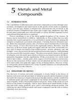
organs. After incision of the dorsal peritoneum
and mobilization of the duodenum to the right,
sharp and blunt dissection is used to carefully ap-
proach the anterior aspect of the aneurysmal neck
(Fig. 7.2).
The correct plane of dissection is reached when
the white and smooth surface of the aorta is visu-
alized. An important guide during the dissection
through the hematoma is the aortic pulse. Accord-
ingly, a weak pulse due to hypotension makes the
dissection more difficult. Exposure of the aneu-
81
rysmal neck is usually facilitated by the dissection
of tissue around the anterior aorta caused by the
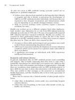
hematoma. Blunt dissection with a finger behind
the aorta in the “friendly triangle” can therefore
often be the easiest way to achieve control of the
aorta (Fig. 7.3).
When a finger can be pushed behind the aorta,
application of the aortic clamp is possible. In this
situation an angled Satinsky clamp is suitable.
When it is difficult to circumferentially free the
aorta, a straight clamp can be applied in an an-
teroposterior position just inferior to the renal ar-
teries, leaving the aorta adherent dorsally. This
often works well, but suturing the anastomosis
can be more difficult. The dissection behind the
aorta should be performed with great care to avoid
damage to the left renal vein, its gonadal branches,
and the lumbar veins. Bleeding during this part of
the dissection usually emanates from any of these
veins and is controlled by ligature, suture, or a
local tamponade. Another common source for
venous bleeding is the inferior mesenteric vein. It
can also be ligated. If profuse bleeding from the
ruptured aorta occurs during dissection control
can be obtained by several different strategies.
7.5.2.3 Other Options
for Proximal Control
There are ways to achieve proximal control of the
aorta that fit most situations. The recommenda-
tions listed below are ordered according to the
probability that they might be needed.
Fig. 7.2. Incision in the posterior
peritoneum for exposure of the
infrarenal aorta and the neck of
an abdominal aortic aneurysm.
The incision is placed in the angle
between the duodenum and the
inferior mesenteric vein, which
occasionally has to be divided for
good access. A 1–2-cm edge of the
peritoneum is left on the duode-
num to facilitate restoration of the
anatomy at closure
7.5 Management and Treatment
Chapter 7 Abdominal Aortic Aneurysms
82
1. Manual local compression or “a thumb in the
hole”
Apply local compression over the rupture with
one or several swabs, or try to seal it by putting
a finger or thumb into the hole in the aneu-
rysm. This method is convenient when the an-
eurysm ruptures suddenly during dissection of
the neck. It can often be followed by option
number two below.
2. Occlusion with balloon catheter
A Foley catheter, size 24-French or larger, is
inserted through the hole and the tip is placed
proximal to the aneurysmal neck. The balloon
is filled with saline until the bleeding dimin-
ishes; usually 15–20 ml is sufficient. The re-
maining bleeding is caused by backbleeding
from the distal vascular bed. If it is significant,
it has to be controlled before proceeding with
dissection of the aneurysmal neck. With this
technique the aorta is usually occluded at a su-
prarenal level and occasionally even higher.
When this method is used, the operation should
be continued as quickly as possible with expo-
sure of the neck of the aneurysm to allow an
aortic clamp to be applied in an infrarenal posi-
tion. The balloon should then be removed im-
mediately before the clamp is applied. Specially
designed balloon catheters for aortic occlusion
are also available to facilitate this method of
control.
3. Straight aortic clamp on the neck of the anu
-
erysm – anterior approach
If the patient is in severe shock and rapid aortic
control is necessary, there is little time for
circumferential dissection and exposure. A
straight clamp can then be applied as soon as
the dorsal peritoneum is divided and the duo-
denum retracted to the right. It is placed from
the ventral portion at the level of the neck. The
clamp is positioned by blunt dissection and
guided in place by the fingers. The surgeon
must be aware of the risk of damaging the vena
cava and should also check that the clamp bite
includes the entire aortic wall.
4. Manual compression of the subdiaphragmat
-
ic aorta
If the rupture is located on the anterior aspect
of the aneurysm and there is ongoing signifi-
cant bleeding within the peritoneal sac, an as-
sistant can achieve temporary proximal control
by manual compression of the subdiaphrag-
matic aorta. This is performed by simply plac-
ing the fist against the lesser omentum high up
under the xiphoid process and pushing down-
ward and cranially, thereby compressing the
aorta against the vertebral column. This gives
the surgeon an opportunity to visualize and
find the hole, followed by insertion of an oc-
clusive balloon as previously described.
Fig. 7.3. When an abdominal aortic aneurysm is pres-
ent the anatomy is often changed. The rst centimeters
of the infrarenal aorta (the neck of the aneurysm) are
usually angulated ventrally. The triangular space be-
tween the spine, the aneurysm, and its neck is called
the “friendly triangle” because its tissue usually allows
blunt dissection easily
83
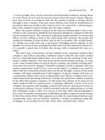
5. Straight clamp on subdiaphragmatic aorta
through the lesser omentum
Better control can be achieved by placing an
aortic clamp in the subdiaphragmatic position
(Fig. 7.4a–d). The technique is not so easy but is
useful when there is a very large hematoma sur-
rounding the neck of the aneurysm, indicating
that the rupture is located in that area. In such
a case there is considerable risk for uncontrol-
lable bleeding through the rupture when the
dorsal peritoneum is opened to expose the
aneurysmal neck. To achieve subdiaphragmatic
control, the lesser omentum is incised, the aor-
tic hiatus at the diaphragmatic crus is exposed,
and the aorta is clamped. The triangular liga-
ment must be divided to allow retraction of the
left liver lobe to the right. To avoid damage to
the ventricle and esophagus, these organs need
to be retracted to the left. Thereafter the muscle
fibers in the diaphragmatic crus are divided to
allow the straight clamp to be applied in an an-
teroposterior position. A straight clamp, how-
ever, has a tendency to slip off the aorta and
cause rebleeding, and repositioning of it is of-
ten necessary. This risk is increased if the mus-
cle fibers in the diaphragmatic crus are not cut
sufficiently. Great care must be taken to avoid
damaging the esophagus and vena cava. As
soon as possible, any supraceliac aortic occlu-
sion is replaced by one in an infrarenal posi-
tion.
6. Clamping of the thoracic aorta
Transthoracic control of the aorta can be used
in extreme situations. It is performed through a
low left-sided thoracotomy in the 5th–6th in-
tercostal space. The incision starts in the mid-
clavicular line and is extended dorsally as far as
possible. After the pleura is incised, the lung is
retracted anteriorly and caudally, after which
exposure of the thoracic aorta is relatively easy.
There are few disturbing surrounding struc-
tures. This technique, however, is associated
with increased postoperative morbidity and is
rarely necessary in the management of rup-
tured abdominal aortic aneurysms.
7. Proximal endovascular aortic control
In potentially technically challenging and se
-
vere cases of ruptured aortic or iliac aneurysms
in obese patients or in those with a “hostile” ab-
domen or traumatic injuries to large intraab-
dominal, retroperitoneal, or pelvic vessels, it
can be advantageous to start the procedure by
percutaneously inserting an intraluminal bal-
loon for proximal aortic control (Fig. 7.5). De-
pending on the location of the injury, this can
be done from the groin through the femoral ar-
tery or from the arm through the brachial ar-
tery. In the former situation, a supporting long
introducer left in place is often needed to pre-
vent dislocation by the bloodstream. This pro-
cedure requires the surgeon to have experience
in endovascular methods or an interventional
radiologist to be available for assistance. Briefly
the technique is as follows. The brachial artery
is punctured with a 12-French introducer. A
guide wire is inserted under fluoroscopy with
its tip then in the proximal aorta. A 100-cm
long catheter with a 46-mm compliant balloon
is inserted over the guide wire and connected to
a syringe with saline for insufflation. If the pa-
tient is in shock the balloon is immediately
insufflated by the surgeon for resuscitation.
Once positioned such an intraaortic balloon
can be temporarily insufflated when needed.
This might be a salvaging procedure in many
cases of extensive vascular injuries because it
controls hemorrhage while allowing dissection
of the injured segment. Subsequent application
of ordinary vascular clamps can then provide
better control. Aortic balloon occlusion can
also be valuable in extensive venous injuries in
the abdomen or pelvic area because the stopped
aortic inflow secondarily leads to diminished
venous bleeding.
7.5.2.4 Continuing the Operation
Proximal aortic control usually stabilizes the pa-
tient and the operation can proceed as in elective
operations for AAA. The iliac arteries are exposed.
The aorta and the iliac arteries are clamped, the
aneurysm incised, and the thrombus extracted.
If there are firm adhesions between the iliac artery
and the vein, dissection may be dangerous, poten-
tially causing severe bleeding by injuries to the
iliac vein. This can be avoided by using balloon
occlusion of the iliac arteries from inside the an-
eurysm once it has been opened. If there is back-
bleeding from lumbar arteries, the inferior mesen-
teric artery, or the median sacral artery, their ori-
gins are controlled with 2-0 suture from the inside
7.5 Management and Treatment
Chapter 7 Abdominal Aortic Aneurysms
84
Fig. 7.4. a The left triangular ligament is divided to
facilitate exposure of aorta at its diaphragmatic hilus.
b
The gastrohepatic omentum is divided longitudi-
nally, the lesser omental sac entered, and the aorta
digitally mobilized at the diaphragmatic crus.
c After
proximal subdiaphragmatic control is achieved by a
straight clamp, the posterior peritoneum is divided
and the neck of the aneurysm is palpated and digitally
dissected, as previously described, through the hema-
toma.
d A second clamp is then placed on the neck of
the aneurysm and the subdiaphragmatic clamp slowly
released
85
of the aneurysm. Ligature of the inferior mesen-
teric artery outside the aneurysm should be avoid-
ed because this is associated with a certain risk for
occlusion of arcade arteries that sometimes are
important collaterals in the intestinal circulation.
A straight tube graft or an aortobiiliac bypass
graft is used for the aortic reconstruction. A col-
lagen-coated woven Dacron graft is recommend-
ed; these types of grafts are presealed with albu-
min and do not need preclotting. A tube graft is
used if aorta is soft and not dilated at its bifur-
cation. If the dilation continues down into any of
the common iliac arteries or if there are extensible
calcifications in the bifurcation, a tube graft
should not be used. If the iliac arteries are calcified
or dilated extension of the graft limbs to the com-
mon femoral arteries may be necessary. This is
combined with ligation of the common iliac arter-
ies. The proximal anastomosis is usually sewn
with nonresorbable monofilic 3-0 or 4-0 suture.
When the graft is anastomosed to the iliac or fem-
oral arteries a 5-0 suture is used.
After the reconstruction is complete, the anas-
tomoses are checked for leakage and possible ob-
struction. Finally, the aneurysmal sac is wrapped
around the graft and the dorsal peritoneum closed
over it. Abdominal drains are never used because
even significant postoperative bleeding cannot be
drained. More about bleeding complications after
aortic surgery can be found in Chapter 12 (page
149). The most common causes for postoperative
bleeding are lumbar arteries not being secured
during the procedure, anastomotic leakage, or
veins that were not ligated but being temporarily
contracted during the operation and later dilated.
Because of the increased risk of bleeding, sys-
temic heparin should not be given to all patients
with ruptured aneurysms. Those hemodynami-
cally stable and with little operative bleeding
should be given heparin IV. A recommendation is
to use half the dose used for elective procedures.
Local heparinization should be administered by
infusing heparinized saline into the iliac arteries.
Liberal use of Fogarty catheters to remove clots
and emboli dislodged to the leg arteries from the
thrombus during dissection is also advocated. If
there is no backbleeding from either one of the
common iliac arteries, thrombectomy is manda-
tory.
Antibiotic prophylaxis should be administered
according to local protocols for operations involv-
ing synthetic vascular grafts. One suggestion is 2 g
Fig. 7.5. A balloon catheter occlud-
ing the aorta at a desired level is in-
serted through the brachial artery.
An alternative is to use a femoral
approach with a 16 French 55 cm
introducer, supporting the balloon
from below
7.5 Management and Treatment
Chapter 7 Abdominal Aortic Aneurysms
86
cloxacillin given at the start of the operation, with
the dose repeated after 4 h in prolonged proce
-
dures. Besides general perioperative IV fluids,
mannitol is recommended to maintain urinary
output.
7.5.2.5 What to do While Waiting
for Help
For surgeons without experience in AAA surgery
it is generally a good idea to wait for a more expe-
rienced colleague if the patient is reasonably
stable. While the surgeon is waiting for help the
patient should be prepared up to the point of
anesthesia induction. The surgeon scrubs and the
patient is also scrubbed and draped while the an-
esthesiologist closely monitors the patient’s vital
functions and hemodynamics. If the patient’s
blood pressure drops and cannot be maintained at
an acceptable level, the patient is anesthetized and
laparotomy is initiated without experienced help.
The goal is then to achieve control of the bleeding.
Besides the previously described techniques to
gain proximal control of the aorta, tamponade
with lots of swabs and compression with the fist
over the bleeding area is usually enough in this
situation. These simple measures combined with
IV fluids and inotropic drugs is often sufficient to
stabilize the patient until help arrives.
7.5.2.6 Endovascular Treatment
In recent years more than 300 patients with rup-
tured AAA or incipient rupture have been treated
with endovascular techniques. The results pre-
sented are observational studies and show that
endovascular repair of rupture is feasible. A large
percentage of the patients in these early series were
not in severe shock and the mortality rate aver-
aged around 10%. Furthermore, reduced post-
operative morbidity rates compared with conven-
tional open repair have been suggested.
One major benefit of endovascular treatment is
the possibility of obtaining rapid proximal control
by inserting an inflatable balloon from the groin
or through the brachial artery that occludes aorta.
This technique makes it possible to delay final
treatment until the patient is stabilized. Another
potential advantage may be that high-risk patients
can also be treated. Particularly favorable is the
possibility of using only local anesthesia and seda-
tion for repair.
The problems related to endovascular repair
include the availability and storage of suitable
grafts as well as logistical problems getting the pa-
tients worked up rapidly. Pretreatment evaluation
with CT angiography or digital subtraction arteri-
ography is necessary to evaluate the possibility
for endovascular repair and to plan the procedure.
The number of different grafts needed to meet in-
dividual requirements is minimized if a unilateral
aortoiliac tube graft is used in combination with
an occluder of the contralateral iliac system and a
femorofemoral crossover, as shown in Fig. 7.6.
Fig. 7.6. One alternative way to treat a ruptured AAA
with endovascular technique. A unilateral aortoiliac en-
dovascular graft decompresses the aortic aneurysm. A
coil in the right internal iliac artery and an occluder in
the left common iliac artery eliminate pressure caused
by backow, the latter deployed to allow retrograde
ow to the internal iliac artery from the groin. A femo-
rofemoral bypass restores perfusion of the left leg
87
The technique involves the following steps: The
patient is prepped and draped as for an elective
AAA procedure. The common femoral arteries
are surgically exposed if a bifurcated graft is
inserted or if unilateral aortoiliac tube grafts in
combination with a femorofemoral crossover
bypass are used. For tube grafts access of only one
common femoral artery is enough. One of the
femoral arteries is punctured and an introducer is
put in place, often a size 7 to 9-French. A guide
wire is inserted, an aortogram obtained, and land-
marks, either radiolucent (placed preoperatively)
or external (such as clamps), are used to assess the
length of the AAA. After systemic heparinization,
the sheath with the graft is introduced over the
guide wire to a level just below the renal arteries.
The sheath is then withdrawn somewhat to allow
proximal release of the graft. After final adjust-
ment of the proximal fixation level the system is
secured by angioplasty. The distal end of the endo-
luminal graft is deployed in the common, external
iliac, or common femoral artery with angioplasty
of stents. Depending on the conditions a hand-
sewn anastomosis is another option. An occluder
of the common iliac is inserted from the contralat-
eral femoral artery. If a bifurcated endoluminal
graft is used, the contralateral graft limb is insert-
ed through the same route. Finally, a completion
angiogram is performed after withdrawal of the
entire sheath.
A bifurcated aortobiiliac endoluminal prosthe-
sis as a primary alternative in rupture is also grow-
ing in popularity. The procedure requires a com-
pliant large-diameter balloon for aortic occlusion,
5 and 12-French introducers, Amplatz guide wires,
high-resolution fluoroscopy, and an assortment of
endoluminal aortic stent grafts with a body diam-
eters ranging from 22 to 34 mm and limb diame
-
ters of 12 to 24 mm.
7.5.3 Management After Treatment
The patient is treated in the intensive care unit un-
til circulatory, respiratory, and renal functions are
stable. This usually takes at least a couple of days.
The most common early postoperative complica-
tions are congestive heart failure, renal failure,
and ischemic colitis. The patient, often with con-
comitant coronary heart disease, is exposed to se-
vere stress during preoperative shock and aortic
clamping and declamping. Deterioration of cardi-
ac function with secondary hypotension that re-
quires inotropic treatment is common. Renal
function is also often impaired and occasionally
the patient requires dialysis. Almost all patients
have increased creatinine and blood urea nitrogen
elevations after operation for ruptured AAA.
These increases are also due to preoperative hypo-
tension and the stress of the operation. If the pa-
tient develops renal insufficiency with low urinary
output, dialysis should be considered at an early
stage.
The greatest risk for developing ischemic colitis
is in patients with a ruptured aneurysm and shock.
The severity of ischemic colitis varies from only
discharge of the mucosa to transmural necrosis.
Registration of pH at the wall of the sigmoid with
a tonometer can be used to determine the risk for
developing ischemic colitis. This condition is fur-
ther discussed in Chapter 12 on complications in
vascular surgery (page 145).
7.6 Results and Outcome
The 30-day mortality after surgery for ruptured
AAA averages from 30% to 50%, the variability
depending on whether the patient developed shock
and whether concomitant diseases were present.
For patients without shock, it is 20–25%, which
can be compared to 60–70% for those without.
The long-term results and prognosis for patients
who survive the initial postoperative period is
good. Outcome is even better than for patients
who have undergone elective aneurysm repair.
The reason for this is probably selection – the sick-
est patients die of rupture, and the survivors who
reach the hospital have fewer risk factors.
7.7 Unusual Types
of Aortic Aneurysms
7.7.1 Inammatory Aneurysm
An AAA can be symptomatic and cause pain
without actual or imminent rupture. The most
common cause for this pain is an inflammatory
reaction in and around the wall – an inflammato-
7.7 Unusual Types of Aortic Aneurysms
Chapter 7 Abdominal Aortic Aneurysms
88
ry AAA. CT, which then shows a thickened aneu-
rysm wall, verifies the presence of such a condi-
tion (Fig. 7.7). It could be presumed that the thick
wall prevents rupture, but rupture of inflamma-
tory AAAs is not uncommon. Because inflamma-
tory AAAs often are painful, separating them
from ruptured AAAs is a real diagnostic problem.
Elevated erythrocyte sedimentation rate (ESR) or
C-reactive protein (CRP) supports the diagnosis,
but CT is the only way to exclude it.
7.7.2 Aortocaval Fistula
A special form of AAA rupture occurs when the
bloodstream penetrates into the vena cava causing
an aortocaval fistula (Fig. 7.8). The patient typi-
cally develops sudden cardiac failure and cyanosis
of the lower extremities. The cardiac failure is due
to the large shunt and the discoloration of the legs
occurs because of venous stasis in combination
with the heart failure. At physical examination the
patient is positive for a bruit and a palpable aneu-
rysm in the abdomen. Treatment for an aortocaval
fistula is an emergency operation, but in most cas-
es some time for preoperative preparation is avail-
able. The operation follows the strategy for other
aneurysms as outlined previously and the fistula is
usually closed by suture from inside the aneurysm
while vena cava is controlled by manual compres-
sion proximally and distally.
7.7.3 Thoracoabdominal Aneurysm
A small number of aortic aneurysms engage the
suprarenal or thoracoabdominal parts of the aorta
including the orifices of the renal arteries, the
superior mesenteric artery, and the celiac trunk.
They originate in the thoracic part of the aorta or
anywhere below the level of the diaphragm. Man-
agement of rupture in such aneurysms is challeng-
Fig. 7.7. Typical appearance on computed tomogra-
phy of an inammatory abdominal aortic aneurysm
with its thick wall
Fig. 7.8. Computed tomography of
a patient with an abdominal aortic
aneurysm and a stula into the
inferior vena cava
89
ing, and if its extension is known prior to the
operation someone with experience should be
contacted before surgery begins. If such an aneu-
rysm is achieved during surgery for rupture, prox-
imal control is sought by one of the techniques
described previously. Endovascular repair is also
an option that needs to be considered.
7.7.4 Mycotic Aneurysm
Another special type of AAA is caused by a local
infection in an atherosclerotic and degenerated
aortic wall, known as mycotic or septic aneurysm.
Different from ordinary AAAs that are fusiform,
mycotic aneurysms are usually saccular (Fig. 7.9).
Patients with this type of AAA frequently have a
medical history that includes fever and malaise.
Elevated ESR, CRP, and other inflammatory pa-
rameters are also common. The most common
bacteria found in mycotic aneurysms and in the
patient’s blood are of the Salmonella species. It is
the infectious process in the wall that causes ero-
sion and subsequently rupture. Treatment is the
same as for other AAAs with the addition of long-
term antibiotics.
7.8 Ethical Considerations
Difficult and delicate ethical considerations often
arise when managing patients with ruptured AAA.
Accordingly, it has to be emphasized that the ad-
vice given above often needs to be modified in
very old patients, patients with dementia, and
patients with other serious medical conditions
implying only a short expected survival time. On
the other hand, patients who previously were de-
termined not suitable for elective repair because
of high risk should sometimes be considered for
repair of a ruptured AAA. When rupture has
already occurred the risk/benefit situation is com-
pletely different. The patient has little to lose by
undergoing an emergency operation.
Rupture of an AAA often occurs in elderly
patients and a complete medical history and infor-
mation about their present quality of life is fre-
quently missing when they are admitted. Because
nonsurgical management is associated with 100%
mortality, a policy of accepting every patient for
surgical treatment is advocated in many hospitals.
A certain selectivity, however, is often wise. It is
obvious that a patient who had cardiac arrest in
the ambulance and remains unconscious at ad-
mission, is anuric, and has ECG signs of myocar-
dial ischemia is extremely unlikely to survive sur-
gery. If the patient is 80 years old and also is known
to have dementia, difficulties ambulating, and
need for geriatric care, it is reasonable to avoid
surgery and instead give the patient terminal care
of high quality. Unfortunately, there are no reli-
able prognostic factors for treatment outcome for
the individual patient, but many studies report
relationships between presence of different risk
factors and survival.
A common conclusion in the literature, how-
ever, is that age should never be considered as a
contraindication to surgery. It is always the sur-
geon, the patient and relatives and their individual
judgment that finally decide whether to operate or
not. If one is in doubt, a good general rule is to be
liberal with repair attempts. It is always possible,
but difficult, to change such a strategy later during
Fig. 7.9. Angiographic appearance of a typical mycotic
aneurysm, with its saccular shape caused by local ero-
sion of the aortic wall and subsequent leakage of blood
into an aneurysmal sac consisting of a brous capsule
(not a true vascular wall)
7.8 Ethical Considerations
Chapter 7 Abdominal Aortic Aneurysms
90
the course when more information is available.
Accordingly, stopping the support of vital func-
tions and taking the patient to the floor for pallia-
tion is a viable option. Some factors in the postop-
erative course – large bleedings and cardiac, renal,
respiratory, and infectious complications – are
considered to be associated with a worse prognosis
and thus might indicate a suitable point at which
to make such a decision.
Further Reading
Bengtsson H, Bergquist D. Ruptured abdominal aortic
aneurysm: a population-based study. J Vasc Surg
1993; 18:74–80
Harris LM, Faggioli GL, Fiedler R et al. Ruptured ab-
dominal aortic aneurysms: factors aecting mortal-
ity rates. J Vasc Surg 1991; 14:812–820
Johansson G, Swedenborg J. Ruptured abdominal aortic
aneurysms: a study of incidence and mortality. Br J
Surg 1986; 73:101–103
Johnston KW. Ruptured abdominal aortic aneurysms:
six-year follow-up of a multicenter prospective
study. J Vasc Surg 1994; 19:888–900
Ouriel K, Geary K, Green RM, et al. Factors determin-
ing survival aer ruptured aortic aneurysm: the
hospital, the surgeon, and the patient. J Vasc Surg
1990; 12:28–33
Ohki T, Veith FJ. Endovascular gras and other im-
age-guided catheter-based adjuncts to improve the
treatment of ruptured aortoiliac aneurysms. Ann
Surg 2000; 232(4):466–479
Aortic Dissection
8
CONTENTS
8.1 Summary 91
8.2 Background
91
8.2.1 Magnitude of the Problem 91
8.2.2 Classication and Denition 92
8.2.3 Etiology 92
8.2.4 Pathophysiology 93
8.3 Clinical Presentation
94
8.3.1 Signs and Symptoms 94
8.3.2 Medical History . . . . . . . . . . . . . . . . . . . . . . 94
8.3.3 Physical Examination 96
8.4 Diagnostics
96
8.5 Management
97
8.5.1 Treatment in the Emergency
Department. . . . . . . . . . . . . . . . . . . . . . . . . . 97
8.5.2 Emergency Surgery 98
8.5.3 Type B dissection 98
8.5.4 Endovascular Treatment 98
8.6 Results and Outcome
99
Further Reading 100
8.1 Summary
Aortic dissection is one of the “great mas-
queraders,” so always suspect this diagno-
sis in any acute painful illness with a pulse
deficit.
A practical classification in the emergency
situation is type A, involving the ascend-
ing thoracic aorta and the arch and type B
involving the aorta distal to the left subcla-
vian artery.
Treatment of type A dissection is always
surgical.
Treatment of type B dissection is medical
in most cases and surgical if there is com-
plicating organ ischemia or bleeding.
Alert the attending thoracic or vascular
surgeon on call early during management,
especially in type A dissections.
8.2 Background
Dissection of the thoracic aorta represents a major
clinical problem that is extremely demanding to
manage even for experienced surgeons. Once it is
diagnosed this condition is usually managed by
an experienced specialist in thoracic or vascular
surgery. The responsibility for the diagnosis and
its primary management, however, mostly belongs
to the surgical or medical emergency physicians.
8.2.1 Magnitude of the Problem
The true prevalence and incidence of aortic dis-
section are unknown, but it has been reported
to have an annual occurrence of 5–10 cases per
Chapter 8 Aortic Dissection
92
million and to affect between 10,000 and 25,000
patients annually in the United States. Autopsy
studies in the United States and Denmark report
dissections in 0.2–0.8% and 0.2% of cases, respec-
tively. An age-adjusted mortality rate from aortic
dissections of 0.5–2.7% per 100,000 inhabitants
was calculated from 1950 to 1981. The overall inci-
dence of aortic dissections consequently is in the
same range or possibly up to two to three times
greater than that for ruptured abdominal aortic
aneurysm. It is two to five times more common in
men than in women, and maximum occurrence is
in the 5th decade of life. Still, in our era of modern
diagnostic methods, a majority of patients proba-
bly die with this disease undetected. Aortic dissec-
tion is a dramatic and dangerous condition with a
very high mortality: 20–50% of patients die within
the first 24–48 h, and up to 75% within the first
2 weeks. It is considered as one of the “great mas-
queraders,” with a wide range of presenting symp-
toms. Because the diagnosis is difficult, awareness
of aortic dissection in the differential diagnosis is
essential, as is rapid and correct management.
8.2.2 Classication and Denition
Aortic dissection is characterized by two or more
communicating flow channels originating from a
proximal intimal tear, with propagation of the
bloodstream within the medial layer. It should
be distinguished from an intramural hematoma,
which is a hemorrhage into the medial layer of the
aortic wall without an intimal tear. Intramural he-
matomas have a natural history similar to aortic
dissection and are treated similarly.
The most useful classification of aortic dis-
sections in the acute situation is the one proposed
by Daily (Stanford classification), as shown in
Fig. 8.1.
Other classic classifications are by DeBakey
and Crawford (thoracoabdominal aneurysms and
chronic dissections). The information in this chap-
ter is based on the Stanford classification because it
simplifies the acute management. A type-A dissec
-
tion always involves the ascending aorta, regard-
less of the distal extension. A type-B dissection
does not involve the ascending aorta. There is con-
sensus in the literature that an aortic dissection is
considered acute if the onset of symptoms occurred
within 14 days of presentation, and chronic if more
than 14 days have elapsed.
NOTE
The most practical classification in the
emergency situation is
– Type A: involving the ascending
thoracic aorta and the arch
– Type B: involving the aorta distal
to the left subclavian artery
8.2.3 Etiology
Aortic dissection is usually related to some kind of
degenerative changes in the aortic wall, in partic-
ular the media. Even if a dissection primarily
starts with a tear in the intima, its propagation
within the media varies considerably from almost
none to rapid progression along the entire length
of the aorta. The variation is related to the condi-
tion of the medial layer. Some congenital connec-
tive tissue defects are known to cause such degen-
eration, including Marfan’s syndrome, Turner
syndrome, and Ehlers–Danlos’ syndrome. Cystic
media necrosis is another predisposing condition.
The role of arteriosclerosis is often discussed. It is
present in older patients with hypertension pre-
Fig. 8.1. Classication of aortic dissection in types A
and B, according to Daily (Stanford)
93
senting with dissection and might constitute a
rare mechanism by penetration of atherosclerotic
ulcers extending through the intima into the me-
dia. This is, however, mostly considered coinci-
dental rather than causative, and some authors
even argue that atherosclerotic changes within the
aortic wall might be a barrier to the extension of a
dissection.
Arterial hypertension is the most important
predisposing factor. It is noteworthy that the sud-
den extreme hypertension associated with severe
physical exercise may cause aortic dissection in
younger persons. Also, pregnancy with its hyper-
circulation and hormonal changes affecting con-
nective tissue, is a certain risk factor particularly
during the last trimester and during labor. Fortu-
nately, aortic dissections in women are rare, but
50% of dissections occurring in women younger
than 40 years old do occur during pregnancy.
Iatrogenic injuries during coronary diagnostic
and therapeutic procedures with catheter manip-
ulations can also cause aortic dissection. Blunt
chest trauma in otherwise healthy persons may
cause aortic dissection, but such dissections are
usually very limited due to the minimal degenera-
tion in these structurally normal aortas.
NOTE
A degenerative process causing weaken-
ing of the aortic wall in combination with
hypertension is the most important
etiologic factor.
8.2.4 Pathophysiology
Type A dissection, constituting 60–70% of all aor-
tic dissections, is mostly seen in younger patients
with some elastic and connective tissue abnormal-
ity. It characteristically starts with a primary inti-
mal tear just distal to the sinotubular ridge in
the ascending aorta. This location is in the vicinity
of the cephalad extension of the aortic valve com-
missures. The tear is commonly transverse and
has a length corresponding to 50–60% of the
aortic circumference. The dissection process starts
in the intimal tear and its extension and direction
vary, as does the speed with which it propagates.
Typically, a type A dissection affects the right
lateral wall of the greater curvature of the ascend-
ing aorta. The dissection is usually directed ante-
gradely, but retrograde extension is also relatively
common. A primary entry in the ascending aorta
is associated with a great risk for bleeding into the
pericardium, causing cardiac tamponade.
Type B dissection usually starts with a primary
intimal tear in the descending thoracic aorta just
distal to the origin of left subclavian artery. This
type constitutes approximately 25% of all aortic
dissections. A patient with a type-B dissection is
typically older, in the 6th–7th decade of life, and
has thoracic aortic degeneration and hyperten-
sion.
Other possible but less common sites of the pri-
mary tear are the aortic arch, occurring in approx-
imately 10% of cases, and the abdominal aorta,
occurring in only 2%. As already mentioned, the
dissection in the aortic media can travel in a retro-
grade as well as an antegrade direction, causing
two flow channels with a false and a true lumen.
Secondary tears and reentries usually occur dis-
tally, allowing flow from the false into the true
lumen.
Rupture is the most common cause of death in
patients with aortic dissection and is mostly lo-
cated near the site of the primary intimal tear.
Consequently, a type-A dissection usually rup
-
tures into the pericardial sac, causing cardiac
tamponade, or an aortic arch rupture that bleeds
into the mediastinum. In addition, the close rela-
tion to the aortic valve commisures can result in
acute valve regurgitation due to prolapse of the
commissural attachments. Dissection into the
aortic root may also involve the coronary arteries,
leading to myocardial ischemia or infarction. A
type-B descending aortic dissection typically rup
-
tures into the left pleural cavity, and less frequent-
ly into the right.
As the dissection extends along the aorta it
will subsequently engage major important cere-
bral and visceral branches, possibly resulting in
threatening end-organ ischemia. The mechanisms
behind this are compression of the true lumen by
the false lumen or shearing of the branch by the
dissection process. A third possibility is disrup-
tion of an important dissection membrane, caus-
ing an intimal flap covering the orifice of a branch.
Such peripheral vascular complications occur in
25–30% of patients with aortic dissection and can
critically affect cerebral, renal, visceral, and lower
8.2 Background
Chapter 8 Aortic Dissection
94
extremity perfusion. Because the dissection in the
descending aorta mainly engages its left perimeter,
the left renal and left iliac arteries are at higher
risk than the right ones.
NOTE
Peripheral vascular complications occur
in 25–30% of cases of dissection of the
aorta.
8.3 Clinical Presentation
8.3.1 Signs and Symptoms
Acute aortic dissection patients can display a
variety of symptoms, and affected individuals
can develop symptoms mimicking those of almost
any other acute medical or surgical condition.
Aortic dissection must be considered in patients
presenting with symptoms indicating acute arte-
rial occlusion and an acute illness that seems to
involve unrelated organ systems.
8.3.2 Medical History
The most dominant symptom is severe pain, which
is migrating or nonmigrating and experienced by
more than 90% of patients. When analyzing the
pain its typical characteristics are evaluated; if it
is: sudden, severe, new, ripping or tearing, and
constant. The pain is typically related to the loca-
tion of the dissection and its propagation distally
into different aortic segments. In proximal dissec-
tion, the most common pain location is the ante-
rior chest. The pain frequently radiates into the
neck and jaws and can be associated with swallow-
ing difficulties. As the dissection propagates dis-
tally, the pain migrates to an interscapular loca-
tion followed by pain in the midback, lumbar, and
groin regions (Fig. 8.3).
Abdominal pain might be severe in patients
suffering from visceral or renal ischemia. As pre-
viously mentioned, the left renal artery is more
likely to be compromised, which may explain why
severe left flank pain mimicking ureteral colic is
often included in the reported history. One should
always include questions about hypertension, car-
diac disease, peripheral vascular disease, connec-
tive tissue abnormalities (such as Marfan’s, Turner,
and Ehlers–Danlos’ syndromes), cystic media ne-
Table 8.1. Dierential diagnoses in aortic dissection
Possible differential diagnoses
Coronary ischemia
Myocardial infarction
Aortic regurgitation without dissection
Aortic aneurysm with dissection
Musculoskeletal pain
Mediastinal tumors or cysts
Pericarditis
Gall bladder disease
Pulmonary embolism
Stroke
Visceral or lower extremity ischemia
without dissection
Fig. 8.2. Mechanisms of branch occlusion and organ
malperfusion in aortic dissection.
a False lumen expan-
sion causes compression of a side branch.
b The orice
of a side branch is disrupted by dissection, and its inner
layers are impacted distally
95
crosis, diagnostic or therapeutic catheter manipu-
lations, or intense exercise.
Secondary effects of aortic dissection with or-
gan malperfusion necessitate a thorough and com-
plete history to include previous, present, and un-
dulating symptoms of the following:
Cerebral ischemia: stroke, loss of conscious
-
ness, focal neurological symptoms
Spinal ischemia: paraplegia or parapareses
Renal ischemia: flank pain, hematuria, dimin
-
ished urinary output
Visceral ischemia: severe abdominal pain
(for further details of typical symptoms, see
Chapter 6, p. 67)
Lower-extremity ischemia: loss of pulse, loss of
sensory or/and motor function, severe pain and
coldness (see Chapter 10, p. 122)
Cardiac malperfusion: angina pectoris or
symptoms of acute congestive heart failure
Possible differential diagnoses are listed in Table
8.1.
Fig. 8.3. a Radiation of pain in type
A dissection. The pain is usually
referred to the neck, anterior chest,
and interscapular area.
b Radiation
of pain in type B dissection. Pain
is primarily interscapular, but with
distal progression of dissection,
pain is often referred to the lower
back and groin
8.3 Clinical Presentation
Chapter 8 Aortic Dissection
96
NOTE
Be aware of aortic dissection as an
important differential diagnosis in any
acute case presenting with sudden painful
illness, in particular if it is associated with
symptoms or signs of organ ischemia.
8.3.3 Physical Examination
Complete and repeated physical examinations are
of paramount importance in diagnosing and man-
aging patients with suspected or verified acute
aortic dissection since this condition can affect so
many different organ systems and has a dynamic
course.
The typical patient presents with paradoxical
physical findings. He or she is frequently pale,
restless, and in preshock or shock, and has an ap-
pearance indicating poor peripheral perfusion but
with a paradoxically high blood pressure. Eighty
percent of the patients have arterial hypertension
at admission. The high blood pressure is second-
ary to underlying essential hypertension, elevated
catecholamine levels due to severe pain, or occlu-
sion of the renal arteries or even the thoracic or
abdominal aorta.
Twenty percent of the patients have a low blood
pressure instead. This is usually secondary to car-
diac tamponade or rupture, or to acute congestive
heart failure secondary to acute aortic insuffi-
ciency. Another possible explanation is pseudo-
hypotension secondary to mechanical obstruction
from the dissection of one or both subclavian
arteries.
Auscultation of the chest is of vital importance.
A cardiac murmur indicates aortic regurgitation.
The first heart sound is diminished or absent due
to elevated end diastolic ventricular pressure.
There might be an S3 gallop rhythm. A continu-
ous murmur usually indicates rupture into the
right atrium. A pericardial friction rub indicates
leakage into the pericardial sac. Auscultation of
the lungs might reveal signs of pulmonary edema.
Loss of alveolar breath sounds can be found after
leakage or rupture into one or both of the pleural
cavities. Jugular venous distension is also a com-
mon finding.
A complete and repeated neurological exami-
nation is mandatory. Horner’s syndrome, loss of
consciousness, loss of sensory or motor function,
paraparesis, paralysis, or paraplegia might be pres-
ent. Acute cerebral vascular occlusion is for obvi-
ous anatomic reasons, more common in proximal
dissection, but fortunately neurological deficits
occur in only about 20% of those patients.
Lower extremity paralysis in the examina-
tion is caused by shearing off or compression of
major arteries feeding the spinal cord (intercos-
tal-T8–L1). Another possible explanation is occlu
-
sion of the thoracic or abdominal aorta, causing
ischemia of the lower body including peripheral
nerves. The clinical distinction is important be-
cause spinal cord ischemia has a poor prognosis,
while a peripheral nerve ischemia has a better
prognosis if treated. This distinction can be made
by examining peripheral pulses. The latter condi-
tion is usually combined with loss of pulses in the
groins and distally in the affected lower extremi-
ties.
Repeated examination of peripheral pulses as
well as blood pressures in the arms and ankles are
important indicators of the extension of a dissec-
tion and its consequences of organ malperfusion.
Repeated examinations are important in order to
follow the development. A peripheral pulse may
disappear, or a pulse deficit may be dynamic and
resolve spontaneously, which is reported to occur
in one-third of the patients. Such a dynamic course
is probably related to redirection of flow from the
false into the true lumen after spontaneous fenes-
tration of the aortic septum known as the reentry
phenomenon.
A new pulse deficit is found in approximately
60% of the patients.
8.4 Diagnostics
An electrocardiogram (ECG) should be obtained
in the emergency department. Low voltage might
indicate pericardial tamponade, and ST–T wave
changes could indicate myocardial ischemia.
The following blood tests should be ordered:
complete blood cell count, arterial blood gases,
protrombin and thromboplastin times, serum
electrolytes, creatinine, blood urea nitrogen, liver
enzymes and lactate.