Endoscopic Extraperitoneal Radical Prostatectomy - part 4 ppsx
Bạn đang xem bản rút gọn của tài liệu. Xem và tải ngay bản đầy đủ của tài liệu tại đây (810.46 KB, 20 trang )
Chapter 5
F. Koenig et al.
5
52
leads to the highest tidal volume is kept constant in
order to reduce the pressure difference. In general, we
aim for tidal volumes of 6–8 ml/kg, respiration rate of
12–16/min and an inspiratory:expiratory (I:E) ratio of
1:1.5. When using this approach the end-expiratory
CO
2
increase can be maintained within justifiable
limits. Only in a few patients with a significant end-
tidal CO
2
increase (~ 50 mmHg; seen only with long
operation times) is the ventilation frequency initially
increased to 24–28/min. Nevertheless, in some cases,
very high end-expiratory CO
2
values are achieved,
which can be more easily tolerated (permissive hyper-
capnia) (Fig. 5.1).
If problems (i.e. bleeding complications) occur
during the operation the pressure is increased by the
surgeon up to 20 mmHg. This should be applied only
in close coordination with the anaesthetist, since it is
always associated with difficulty regarding mechani-
cal ventilation and may last for only a short time (10–
15 min).
5.8 Postoperative Pain
In a recent study by Hoznek et al., the mean dose of
morphine and the mean duration of its administra-
tion was 53.1% and 44.4% lower, respectively, after
extraperitoneal than after transperitoneal radical
prostatectomy. Although the difference was not sta-
tistically significant, the authors considered it clini-
cally relevant. In addition, abdominal tenderness and
shoulder pain, commonly observed among LRPE pa-
tients, were not reported in their extraperitoneal se-
ries [11].
Our initial experience with “minimally invasive
treatment” of prostate cancer includes 70 cases per-
formed transperitoneally in 2000 and 2001. Due to
the lack of experience at that time we postoperatively
transferred all patients to the intensive care unit. Ad-
ministration of analgesia was started intraoperatively,
with a peripherally acting analgesic (metamizole 1–2
g) and continued postoperatively using a patient-con-
trolled analgesia (PCA) pump (with piritramide).
Later, the PCA pumps were omitted due to the fact
that self-administered bolus doses and the total quan-
tities of the opiates were small.
In December 2001 we completely changed the
technique to a completely extraperitoneal access. The
patients no longer had to be postoperatively trans-
ported to the ICU. They were postoperatively moni-
tored in a postanaesthetic care unit (PACU) or inter-
mediate care unit, although they could have been
observed in a regular recovery room. With increasing
experience, it became obvious that there is no need
for a PCA pump and regular administration of anal-
gesics.
5.9 Role of Carbon Dioxide
Retroperitoneal insufflation of CO
2
is used in several
urological procedures despite the potential risk of
CO
2
accumulation. The retroperitoneal space offers
less of a barrier to CO
2
diffusion than the peritoneum,
it is very vascular, it contains adipose tissue and, un-
like the peritoneal cavity, it is not a limited cavity.
These features result in greater absorption of CO
2
during retroperitoneal than during intraperitoneal
laparoscopy.
In certain studies, there was a difference in the ex-
tent of CO
2
absorption during retroperitoneoscopy,
compared with intraperitoneal laparoscopy. In an ex-
perimental pig model, the increase of CO
2
pressure
was independent of the intra- or retroperitoneal gas
insufflation [7]. On the contrary, in an experimental
dog model, the absorption of CO
2
was greater after
intra- than after extraperitoneal insufflation [12].
In humans, some authors did not observe greater
absorption of CO
2
in retroperitoneoscopic renal sur-
gery than in transperitoneal laparoscopy [13]. How-
ever, others found that CO
2
absorption was greater in
patients when the retroperitoneal approach instead of
the transperitoneal one was used in renal surgery [14].
In addition, Streich et al. reported that retroperitoneal
CO
2
insufflation results in more CO
2
absorption than
peritoneal insufflation. Persistence of absorption of
CO
2
after the end of surgery was observed [3].
Large variations in CO
2
production can be ob-
served during retroperitoneoscopic surgery. Proper
management of the ventilator could maintain end-
tidal CO
2
within normal values, risking a change in
lung volumes and dead space that could affect the al-
veolo-arterial CO
2
difference.
It is a fact that CO
2
output remained high after ret-
roperitoneoscopy while CO
2
output decreased imme-
diately on cessation of peritoneal insufflation. Persis-
tent accumulation of CO
2
during the early
postoperative period should be considered in the post-
operative care of patients after retroperitoneoscopy.
Chapter 5
53
Anaesthesia Considerations in Radical Prostatectomy
An additional CO
2
pressure effect on the cerebral
circulation is caused in patients in Trendelenburg po-
sition for prostate and bladder surgery. A rise in CO
2
blood tension increases cerebral blood flow, whereas
intra-abdominal pressure and central haemodynamic
effects reduce cerebral blood flow. In addition to an
osmotic diuresis towards the end of the procedure,
the routine for the prolonged head-down position in-
cluded restricted fluid loads and maintenance while
so positioned. Increases in ventilation rates adjust the
rises in end-tidal CO
2
.
5.10 Summarised Recommendation
Co-operation between the surgeon and the anaesthe-
siologist is of paramount importance for safe and ef-
fective performance of EERPE. Appropriate anaes-
thetic equipment and trained personnel must be
available. In Table 5.1 we present a brief but meticu-
lous description of the entire anaesthesiological man-
agement of the EERPE patient.
Table 5.1. Standardised anaesthesiological management of EERPE
1. Preoperative visit
Preoperative day
•
Patient history
•
Physical status
•
Information of patient about specific content of operation and anaesthesia details (only general anaesthesia is performed;
additional regional anaesthesia is not needed due to low postoperative pain)
•
Twelve-lead electrocardiogram
•
Laboratory investigations (basic blood test, electrolytes, coagulation status, glucose)
•
Chest X-ray, spirometry, echocardiography only in cases with severe cardiac or pulmonary comorbidities
•
In night before operation, administration of long-acting benzodiazepines (e.g. 5 mg nitrazepam or 1 mg flunitrazepam)
•
Prophylaxis of thromboembolic events with fractionated heparin (replacement of pre-existing long-acting medication
with short-acting anticoagulatory agents; interruption of acetylsalicylic acid intake for 3–5 days)
Operative day
•
Continuation of comedications
•
Premedication with midazolam (5–10 mg p.o. 1 h before anaesthesia initiation)
2. In the operating room
Patient positioning
•
Supine position
•
Right arm: noninvasive blood pressure measurement. Arm is attached to body and fixed under a sheath (no arm holder;
fixation of this arm should be performed when patient is conscious to prevent inadvertent nerve injuries)
•
Left arm: SaO
2
measurement. One intravenous cannula preoperatively, and one after induction of anesthesia. Elongation
of infusion line with injection port to upper end of table. Arm is positioned on arm holder fixed to operating table (use of
silicone protective gel)
•
ECG (standard leads; if needed, additional leads II and V5 for detection of ischemic events)
•
Extended monitoring (invasive arterial blood pressure measurement only in patients with high cardiac risk; temperature
measurement and rewarming blankets only if needed)
•
Belt with a silicone protective sheath under it, on upper part of thorax (prevent movement of patient during operation in
the case of extreme Trendelenburg positioning); never use mechanical restraints on shoulder area because of danger to
plexus brachialis
Anaesthesia
Induction
•
Opioid injection (i.e. 20–30 µg/kg alfentanil)
•
Induction with 1.5–2 mg/kg propofol (in the case of pre-existing cardiac comorbidities or hypotensive patients,
0.2–0.3 mg/kg etomidate is preferable)
•
Relaxation (i.e. 0.6 mg/kg rocuronium)
•
Intubation with RAE tube; connection with extension (patient’s head is not accessible during operation; safe positioning
protecting airway system on patient’s head; avoidance of pressure damage to patient’s face; eye protection by ointment)
Continuation as balanced anaesthesia
•
Isoflurane or desflurane with O
2
/air mixture (FiO
2
0.4)
Chapter 5
F. Koenig et al.
5
54
References
1. Drummond GB, Duncan MK (2002) Abdominal pres-
sure during laparoscopy: eects of fentanyl. Br J Anaesth
88:384–388
2. Seed RF, Shakespeare TF, Muldoon MJ (1970) Carbon
dioxide homeostasis during anaesthesia for laparoscopy.
Anaesthesia 25:223–231
3. Streich B, Decailliot B, Perney C, Duvaldestin P (2003) In-
creased carbon dioxide absorption during retroperitoneal
laparoscopy. Br J Anaesth 91:793–796
4. Hodgson C, McClelland RMA, Newton JR (1970) Some
eects of the peritoneal insuation of carbon dioxide at
laparoscopy. Anaesthesia 25:382–390
5. Baird JE, Granger R, Klein R, Warriner CB, Phang PT
(1999) e eects of peritoneal carbon dioxide insuation
on hemodynamics and arterial carbon dioxide. Am J Surg
177:164–166
6. Hirvonen EA, Poikolainen EO, Paakkonen ME, Nuutinen
LS (2000) e adverse hemodynamic eects of anesthesia,
head-up tilt, and carbon dioxide pneumoperitoneum dur-
ing laparoscopic cholecystectomy. Surg Endosc 14:272–277
7. Coskun F, Salman MA (2001) Anesthesia for operative en-
doscopy. Curr Opin Obstet Gynecol 13:371–376
8. Stolzenburg J-U, Rabenalt R, Do M, Ho K, Dorschner W,
Waldkirch E, Jonas U, Schütz A, Horn L, Truss MC (2005)
Endoscopic extraperitoneal radical prostatectomy (EE-
RPE) – oncological and functional results aer 700 proce-
dures. J Urol 174:1271–1275
9. Stolzenburg J-U, Truss MC (2003) Technique of laparo-
scopic (endoscopic) radical prostatectomy. BJU Int 91:749–
757
10. Stolzenburg JU, Aedtner B, Oltho D, Koenig F, Rabenalt
R, Filos K, McNeill A, Liatsikos EN (2006) Anaesthesia
considerations for endoscopic extraperitoneal and lapa-
roscopic transperitoneal radical prostatectomy. BJU Int
98:508–513
11. Hoznek A, Antiphon P, Borkowski T, Gettman MT, Katz
R, Salomon L, Zaki S, de la Taille A, Abbou CC (2003) As-
sessment of surgical technique and perioperative morbid-
ity associated with extraperitoneal versus transperitoneal
laparoscopic radical prostatectomy. Urology 61:617–622
12. Wolf JS, Carrier S, Stoller ML (1995) Intraperitoneal ver-
sus extraperitoneal insuation of carbon dioxide as for
laparoscopy. J Endourol 9:63–66
13. Ng CS, Gill IS, Sung GT, Whalley DG, Graham R, Sch-
weizer D (1999) Retroperitoneoscopic surgery is not as-
sociated with increased carbon dioxide absorption. J Urol
162:1268–1272
14. Wolf JS Jr, Monk TG, McDougall EM, McClennan BL,
Clayman RV (1995) e extraperitoneal approach and
subcutaneous emphysema are associated with greater ab-
sorption of carbon dioxide during laparoscopic renal sur-
gery. J Urol 154:959–963
Table 5.1. (Continued)
•
Optimised ventilation monitoring is performed and the supply of the narcotic agent is provided by the volatile anaesthe-
tic. When intravenous method is applied, supply of anaesthetic can be interrupted because of poor access to long infusion
lines.
•
Continuation of opiates (i.e. 30–60 µg/kg/h alfentanil) until beginning or end of anastomosis (depending on speed of
suturing by surgeon)
•
Intermittent relaxation if needed (TOF monitoring of muscle relaxation)
•
Ventilation
Pressure controlled
PEEP = 5 mbar
Ventilator frequency 10–14 per minute
Time inspiration/expiration (t
i
:t
E
) = 1:1.5
Pressure difference between P
exp
and P = 20 mbar
•
In the case of CO
2
increase
Elevation of minute volume with increase of ventilatory frequency
If ineffective, increase of PEEP with same pressure difference
The level of PEEP is titrated to have a tidal volume of 6–8 ml/kg body weight
The pressure difference between expiratory and inspiratory is increased to maximal 25 mbar
Reduction of CO
2
insufflation pressure (decrease in intra-abdominal pressure)
If CO
2
is continuously increasing, high CO
2
values are tolerated (risk/benefit balance)
Recovering from anaesthesia
•
Extubation in operating room
•
Transport with O
2
insufflation via face mask or nasal line and monitoring to intermediate care unit (at least SaO
2
)
3. Postoperatively
•
Pain therapy with non-opioid analgesics, e.g. paracetamol (acetaminophen) or metamizol, and rarely opioids,
e.g. piritramide on demand
Contents
6.1 Introduction . . . . . . . . . . . . . . . . . . . . . . . . . . . . . . . . . . . . 56
6.2 Can Pelvic Lymphadenectomy
Alter Prognosis? . . . . . . . . . . . . . . . . . . . . . . . . . . . . . . . . 56
6.3 Patient Selection
for Pelvic Lymphadenectomy . . . . . . . . . . . . . . . . . . . . 57
6.4 Anatomical Boundaries
in Pelvic Lymphadenectomy . . . . . . . . . . . . . . . . . . . . . 58
6.5 Histopathological Analysis
of Pelvic Lymphadenectomy Tissue . . . . . . . . . . . . . . 60
6.6 Surgical Techniques
in Pelvic Lymphadenectomy . . . . . . . . . . . . . . . . . . . . . 61
6.7 Sentinel Lymph-Node Mapping
in Prostate Cancer . . . . . . . . . . . . . . . . . . . . . . . . . . . . . . . 61
6.8 Imaging Pelvic Lymph Nodes
in Prostate Cancer . . . . . . . . . . . . . . . . . . . . . . . . . . . . . . . 62
6.9 Conclusion . . . . . . . . . . . . . . . . . . . . . . . . . . . . . . . . . . . . . 62
References . . . . . . . . . . . . . . . . . . . . . . . . . . . . . . . . . . . . . . 63
Pelvic Lymphadenectomy
in the Management of Prostate Cancer
Sivaprakasam Sivalingam ∙ H. Schwaibold
6
Chapter 6
S. Sivalingam ∙ H. Schwaibold
6
56
6.1 Introduction
In the era before prostate-specific antigen (PSA),
when patients presented in advanced stages of pros-
tate cancer, pelvic lymphadenectomy was the only
available tool to provide staging information prior to
commencement of surgical treatment for the prostate
cancer. Contemporary imaging modalities such as bi-
lateral pedal lymphangiography lacked sufficient sen-
sitivity to replace surgical lymph-node staging.
With the advent of PSA testing, the emphasis in
prostate cancer management shifted gradually from
treatment of disease to screening for disease. The in-
creasing recourse to PSA testing and transrectal ul-
trasound-guided prostate biopsy meant that prostate
cancers were diagnosed at earlier stages of their evo-
lution. Medical treatment of prostate cancer with hor-
mones was also beginning to gain momentum. These
factors led many clinicians to question the practice of
routine pelvic lymphadenectomy for prostate cancer
staging. Alternative strategies were devised for select-
ing patients who required lymph-node staging prior
to initiation of treatment. Prognostic tools and nomo-
grams were developed, integrating the patient‘s indi-
vidual clinical and histological findings. These tools
were then used to select patients with a high risk of
lymph-node metastasis for surgical exploration.
The follow-up of patients who developed biochem-
ical recurrence after radical prostatectomy in indi-
vidual centres suggests that despite low prediction of
metastasis by nomograms, these patients may have
harboured micrometastasis in their lymph nodes at
the time of their initial presentation. This reinvigo-
rated some experts to re-explore the role of „limited“
and „extended“ pelvic lymphadenectomy in the past
few years. Newer developments in surgery such as
laparoscopic surgery and robotic surgery have also
fuelled this interest in pelvic lymphadenectomy as
they offer a minimally invasive procedure which is
more acceptable to the patient and promise reduced
morbidity compared to the open procedure.
Despite major developments in prostate cancer
treatment over the past two decades, the role of pelvic
lymphadenectomy in prostate cancer treatment has
yet to be clearly defined. Unlike other pelvic malig-
nancies such as colorectal cancer [1] and gynaecologi-
cal cancer [2], where guidelines exist, prostate cancer
lacks any clear consensus on surgical management of
pelvic lymph-node disease.
The practice of performing pelvic lymphadenec-
tomy today varies significantly among urological sur-
geons, with some performing the procedure only in
high-risk patients, based on preoperative parameters,
while others offer the procedure to all patients under-
going localised treatment of their prostate cancer. The
key questions are as follows:
•
Does pelvic lymphadenectomy per se alter the
prognosis of the disease?
•
Which patients should undergo pelvic lymphade-
nectomy?
•
What are the relevant anatomical boundaries for
optimal pelvic lymph-node dissection?
•
How should the lymph-node tissue be evaluated
in the histopathology laboratory?
•
Which surgical technique should be used?
In the following sections we discuss these questions,
the current practice, its related issues and the scien-
tific evidence behind the rationale for performing
pelvic lymphadenectomy in patients with prostate
cancer.
6.2 Can Pelvic Lymphadenectomy
6.2 Alter Prognosis?
The question of whether pelvic lymphadenectomy is a
staging tool, or in addition has a therapeutic impact
in the era of prostate cancer stage migration and
lymph-node micrometastasis, is still unanswered.
Bhatta-Dar et al. retrospectively examined the role
of pelvic lymph-node dissection (PLND) in 336 men
who underwent radical prostatectomy [3]. In this se-
ries, the patients had a PSA level of <10 ng/ml, a Glea-
son score of <7 and were of clinical stage T1 or T2.
They were divided into 140 men who had PLND and
196 men who did not undergo PLND during radical
prostatectomy based on the discretion of the operat-
ing surgeon. The two groups were evenly matched in
terms of age, family history of prostate cancer, race,
clinical staging and PSA. The boundaries of the PLND
included the external iliac vein, pelvic sidewall, obtu-
rator nerve, bifurcation of the common iliac artery
and inguinal ligament. The PLND group had a 0.7%
metastasis rate in the lymph nodes.
The results showed no statistically significant dif-
ference in biochemical relapse rates after a mean fol-
low-up of 60 months. On the multivariate analysis,
PLND did not appear to be an independent predictor
Chapter 6
57
Pelvic Lymphadenectomy in the Management of PC
of the outcome. The estimated 6-year biochemical re-
lapse-free survival rate also showed no statistical sig-
nificance. This was a retrospective non-randomised
study and, in terms of prostate cancer natural history,
had a relatively short follow-up interval.
Salomon et al., in a prospective, non-randomised
study, compared 43 men with preoperative PSA of
<10 ng/ml and a Gleason score of <7 who did not un-
dergo PLND prior to perineal prostatectomy with 25
men with similar PSA and Gleason score who under-
went PLND during retropubic prostatectomy [4]. The
actuarial 5-year recurrence rates for both groups were
not statistically significantly different. Although this
was a prospective study, the patient numbers were
small and the follow-up interval short. The authors of
this study did not describe the surgical dissection
template used in the lymphadenectomy procedure,
which is crucial, as will be described later.
Both the above studies incorporated only patients
with low risk of lymph-node metastasis, based on
their preoperative parameters. Such a narrow selec-
tion of subjects makes it difficult to draw any conclu-
sion on the efficacy of PLND as a therapeutic modal-
ity, as most patients in these studies would have had
low risk of lymph-node metastasis anyway.
Bader et al. retrospectively examined the impact of
lymph-node metastasis on disease progression and
survival [5]. They performed a meticulous lymphad-
enectomy incorporating the internal, external and
obturator lymph-node packets during radical prosta-
tectomy in 367 patients with organ-confined prostate
cancer. The preoperative patient selection criteria in
terms of PSA level and Gleason grade were broader in
this study than in the two studies described above.
Ninety-two patients (25%) had histologically proven
lymph-node metastasis. Cox regression analysis
showed that the probability for PSA relapse, symp-
tomatic progression and tumour-related death in-
creased with each additional lymph node involved.
Forty per cent of patients with only one positive lymph
node remained without signs of clinical or chemical
progression after a follow-up of 45 months.
Frazier et al., in a retrospective analysis of 156 cas-
es from the pre-PSA screening era, showed a similar
survival advantage in patients with low-volume pelvic
lymph-node involvement [6].This suggests that not all
lymph-node-positive cases automatically have a poor
prognosis.
In low-volume lymph-node metastasis, pelvic
lymphadenectomy may have a therapeutic impact and
confer a survival advantage. In order to demonstrate
this point, a prospective multicentre randomised trial
recruiting patients with low, medium and high risk of
lymph-node metastasis is required. They should all be
subjected to a standardised protocol (identical surgi-
cal dissection template and technique of histological
analysis) during the lymphadenectomy procedure.
They should also all receive identical treatments for
their prostate cancer and be followed up for at least 10
years. At present, however, there are no such pub-
lished data to demonstrate the therapeutic impact of
pelvic lymphadenectomy.
6.3 Patient Selection for Pelvic
6.2 Lymphadenectomy
The published literature has recorded an apparent
"fall" in lymph-node metastasis rates from 20–40% in
the 1970s and 1980s to the present rate of about 6% [7,
8]. This downward pathological stage migration of
prostate cancer is thought by many to be secondary to
early detection by virtue of PSA screening programs.
Prostate cancer nomograms were developed with
the aim of predicting the prognosis in patients with
prostate cancer. Partin's Table and the Memorial
Sloan Kettering Cancer Center Prostate Nomogram
are examples of such tools that have been used to
identify preoperatively patients who are at increased
risk of lymph-node metastasis [9, 10].
Nomograms are useful in the clinical setting when
discussing treatment options with patients as they
provide means of quantifying the individual patient's
risk in terms of morbidity and mortality. However,
their usefulness as an aid for the clinician's decision
making has yet to be fully explored. Ross et al. cata-
logued 42 different types of prostate cancer nomo-
grams in their review in 2001 [11]. They concluded
that more comparative studies on the predictive ac-
curacy of the various published nomograms were
needed. The authors also felt that more studies com-
paring the predictive accuracy of nomograms with
that of an expert clinician's judgement were neces-
sary. Such comparisons will help determine the true
usefulness of prostate cancer nomograms in routine
clinical practice.
These nomograms have several drawbacks:
•
They rely on results of historical lymphadenecto-
my series without proper definition of the ana-
tomical boundaries of the lymph-node dissection.
Chapter 6
S. Sivalingam ∙ H. Schwaibold
6
58
In most cases they rely on a form of limited dissec-
tion [8–10].
•
Digital rectal examination is subjective and not
easily reproducible due to inter-observer variabil-
ity.
•
Discrepancy in Gleason grade exists between Tru-
Cut prostate biopsies and the final prostatectomy
specimen. Burkhard et al. showed a 24% rate of
undergrading and 12% overgrading between Tru-
Cut prostate biopsy samples and the final histo-
pathological examination of the radical prostatec-
tomy specimen [12].
•
Recent changes in histopathological reporting
with a trend towards higher grade assignment
means that the Gleason grade data used to estab-
lish these prediction nomograms are not compa-
rable to the modern patient [13].
6.4 Anatomical Boundaries
6.2 in Pelvic Lymphadenectomy
Anatomical dissections and lymphographic studies
have demonstrated that prostate lymphatics drain via
three pathways [14, 15]:
•
Ascending ducts – draining into the external iliac
lymph nodes
•
Lateral ducts – draining into the hypogastric
lymph nodes
•
Posterior ducts – draining into the sacral lymph
nodes
Schuessler et al. [16] described the various forms of
pelvic lymphadenectomies performed on prostate
cancer patients (Table 6.1).
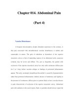
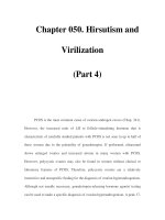
The distribution of the pelvic lymph nodes is il-
lustrated in Fig. 6.1.
Bader et al. demonstrated that the anatomical
boundaries in lymphadenectomy influence the accu-
racy of detection of lymph-node metastasis [17]. In
their retrospective study, they were able to demon-
strate that the sampling of the internal iliac lymph
nodes was important. Eighty-eight of the 365 patients
in their series had positive lymph nodes. Of these 88
patients, 17 (19%) had positive nodes confined exclu-
sively to the internal iliac area. In an earlier study,
McDowell et al. showed a high incidence (29%) of
lymph-node metastases also confined exclusively to
the internal iliac nodes [18].
Burkhard et al. demonstrated that when a meticu-
lous lymphadenectomy (internal, external iliac and
obturator lymph-node packets) was carried out in pa-
tients with localised prostate cancer, even “low risk”
patients with a PSA level of 10 ng/ml and preoperative
cytological grade <3 had a 10% lymph-node metasta-
sis rate [12].
On the other hand, Clark et al.’s prospective ran-
domised evaluation of 123 patients undergoing radi-
cal prostatectomy concluded that an extended dissec-
tion (common iliac, internal iliac, external iliac,
obturator fossa and presacral nodes) does not improve
the accuracy of lymph-node staging compared to a
limited dissection (external iliac and obturator fossa
nodes) [19]. This publication, however, exhibited sev-
eral shortcomings: its patient population was at low
risk of lymph-node metastasis based on preoperative
parameters and the patient numbers were too small
for an equivalence study. The histopathological eval-
uation protocol was also not fully described. The ran-
domisation procedure assigned the extended dissec-
Table 6.1. Description of lymphadenectomies by Schuessler in 1993
Terminology Anatomical boundaries of lymphadenectomy
Standard lymphadenectomy Common iliac artery, external iliac artery, genitofemoral nerve, hypogastric
vessels and obturator fossa nodes
Extended lymphadenectomy As standard plus presacral and lateral sacral nodes
Modified or limited lymphadenectomy Numerous variations:
As standard with omission of common iliac artery, +/– hypogastric vessels
and external iliac artery or vein as lateral margin instead of genitofemoral
nerve
Obturator fossa lymphadenectomy Obturator fossa nodes only
Chapter 6
59
Pelvic Lymphadenectomy in the Management of PC
tion only unilaterally, while on the contralateral side a
limited dissection was carried out. This meant that
the investigators could have potentially missed lymph-
node metastasis pick-up from the extended dissection
in cases where the prostate cancer focus was only uni-
lateral. These factors can affect the interpretation of
the efficacy of the surgical techniques that were com-
pared.
The total number of lymph nodes removed during
a lymphadenectomy procedure is of importance in
order to maintain the accuracy of the staging proce-
dure. Weingärtner et al. showed through their work
on cadaveric pelvic dissections that approximately 20
lymph nodes must be present in the histopathological
specimen to ensure an adequate and representative
pelvic lymphadenectomy [20].
Fig. 6.1. Pelvic lymph
nodes dissected during
pelvic lymphadenectomy
Table 6.2. Lymph-node metastasis pick-up rates from published series
Series
Limited
Standard
Modified
Extended
Unclassified
Heidenreich [23] (n=203) 12% 26%
Parkin [26] (n=50) 24%
Shackley [27] (n=27) 7%
Clark [19] (n=123) 2% 3%
Allaf [24] (n=4000) 1% 3%
Stone [25] (n=189) 7% 23%
Bader [17] (n=365) 24%
Schuessler [16] (n=86) 23%
Lezin [28] (n=44) 27%
Herrell [21] (n=68) NA
Golimbu [22] (n=30) 50%
McDowell [18] (n=217) 59%
NA, not available
Extent of lymphadenectomy
Chapter 6
S. Sivalingam ∙ H. Schwaibold
6
60
Examination of current papers on the subject of
pelvic lymphadenectomy quickly demonstrates the
lack of standardisation in nomenclature and surgical
practice. Terminologies such as standard, limited or
modified, extended and obturator fossa lymphade-
nectomy are loosely used to describe the surgical pro-
cedure. The anatomical boundaries denoted by these
terminologies are inconsistent, as shown by a review
of existing publications [16–19, 21–28].
The lymph-node metastasis rates obtained in dif-
ferent lymphadenectomy series are also variable,
partly due to the variation in the surgical practice
(Table 6.2).
The general lack of consistency creates an unclear
picture of the outcomes from the procedure. There-
fore, there is a need to reach an international consen-
sus on the standardisation of the anatomical bound-
aries and establish an agreed nomenclature to describe
the surgical boundaries. Surgical lymphadenectomy
performed in breast cancer [29] and gastric cancer
[30] patients has been stratified into clearly defined
levels of surgical dissection. Similar stratification
would be invaluable for pelvic lymphadenectomy in
prostate cancer and would ensure uniformity of sur-
gical practice. It would also aid in the interpretation
and comparison of future studies on pelvic lymphad-
enectomy in prostate cancer.
6.5 Histopathological Analysis
6.2 of Pelvic Lymphadenectomy Tissue
The role of frozen section diagnosis in the assessment
of pelvic lymph nodes at the time of radical prostatec-
tomy has shown varying use over the last 20 years. In
1986, Epstein et al. reviewed 310 patients, who had
had frozen sections and found that the frozen sections
detected 67% of positive lymph nodes, which were
grossly uninvolved and all the patients with grossly
involved lymph nodes [31]. These authors concluded
that this was a useful technique in grossly uninvolved
nodes. Since that time PSA testing and nomograms
predicting positive lymph nodes have been developed.
Young et al. in 1999 examined the cost and accuracy
of frozen sections before radical prostatectomy and
found a false-negative rate of 33% (similar to Epstein
et al.) [32]. These authors estimated the cost of meta-
static cancer detection to be £7,516 (ca. €11,000). In
view of this and the false-negative rate, they concluded
that frozen section was not routinely warranted.
Beissner et al. in 2002 found an even higher false-
negative rate of 70%, but by stratifying patients into
low, intermediate and high risk based on nomograms
they improved the sensitivity [33]. They concluded
that low-risk patients (stage T2 or less, PSA level of
10 ng/ml or less and Gleason score of 6 or less) gain no
benefit from frozen section, whilst the intermediate
group (stage T2 or less and Gleason score 7 and/or
PSA level of 10.1–20 ng/ml) gain minimal benefit.
The high-risk patients who have elected for surgery
do gain benefit as they have high risk of positive
lymph nodes and detecting such nodes could save
them the morbidity of radical prostatectomy.
A variety of histopathological techniques have
been used to examine lymph nodes in the laboratory.
Pelvic lymph nodes are usually not visible but are pal-
pable and are extensively replaced by adipose tissue,
leaving just a small rim of lymphoid tissue at the edge.
Some authors feel that this makes these nodes impos-
sible to count accurately [34], whilst others use fat-
dissolving techniques (e.g. acetone or xylene) to count
them [5, 20]. Fat clearance has been shown to increase
lymph-node harvest in colorectal carcinoma [35], but
the lymph nodes in the colonic mesentery are well de-
fined and lack the degree of fat infiltration seen in
pelvic nodes. This discrepancy makes it impossible to
assess surgical technique solely on the basis of the
number of lymph nodes harvested by the patholo-
gist.
Studies in colorectal cancers have shown that the
more lymph nodes recovered the more accurate the
staging [36]. Similar work looking at the pelvic lymph
nodes from prostate cancer patients came to the same
conclusion [37]. This study showed that the number
of lymph nodes recovered varied from 5 to 40, and in
those with 13 or more nodes the metastatic lymph-
node involvement was twice as high as in those with
lower lymph-node counts. Colectomy specimens dif-
fer considerably from pelvic lymphadenectomies, as
in the latter all the tissues submitted by the surgeon
can be blocked and examined, which is not feasible in
colectomies. As a result the number of lymph nodes
may not be known but the status of all of them is.
When the lymph nodes have been processed and
examined, further histopathological techniques can
affect the positive rate in the lymph nodes. The posi-
tive rate has been shown to increase in colorectal car-
cinomas with examination of the lymph nodes at sev-
eral levels [38], and this has also been shown in prostate
cancer [39]. Wawroschek et al. showed that by exam-
Chapter 6
61
Pelvic Lymphadenectomy in the Management of PC
ining lymph nodes at several levels combined with
immunohistochemistry the node positive rate in low-
risk patients increased from 5% to 11% but there was a
smaller increase in the intermediate-risk patients,
from 34% to 37%. The cautionary note about using
immunohistochemistry to detect micrometastasis is
that we are uncertain of the prognostic significance.
It is essential that the histopathological techniques
used to detect metastasis are considered when data
from various pelvic lymphadenectomy studies are
compared.
6.6 Surgical Techniques in Pelvic
6.2 Lymphadenectomy
Both open and laparoscopic pelvic lymphadenectomy
have been described in the literature. Some centres
perform laparoscopic pelvic lymphadenectomy before
a planned perineal prostatectomy, radical radiothera-
py, brachytherapy or cryotherapy.
Lymph-node harvest during laparoscopic lymph-
adenectomy is comparable to its open counterpart in
centres where the learning curve has been overcome
[21, 40]. Parkin et al.’s series demonstrated that the
laparoscopic procedure has low complication rates
[26]. Solberg et al.’s 132 cases (94 open cases and 38
laparoscopic cases) had a significantly lower lympho-
cele complication rate in the laparoscopic group [41].
Improved magnification and a meticulous need for
haemostasis during laparoscopy may be a contribut-
ing factor to this observation.
Two routes of laparoscopy are described in the lit-
erature: the transperitoneal and the preperitoneal ap-
proach. The preperitoneal approach results in less in-
terference from small bowel loops and it mimics the
standard open approach. The transperitoneal ap-
proach, however, gives better access to the internal
iliac and presacral lymph nodes for those who attempt
a more extended resection and has a lower lympho-
cele rate. The latter is due to the communication be-
tween the preperitoneal and the intraperitoneal
space.
Overhead costs are higher in laparoscopic lymph-
adenectomy than in its open counterpart [21, 28]. In
high-caseload laparoscopic units, however, the costs
can be reduced by substituting disposable kits with
reusable ones and tailoring laparoscopic kits specifi-
cally to the surgeon’s requirements so that instru-
ments are not unnecessarily desterilised.
6.7 Sentinel Lymph-Node Mapping
6.2 in Prostate Cancer
Research has been conducted to determine whether
pelvic lymphadenectomy in prostate cancer can be
targeted to metastatic lymph nodes, thereby sparing
patients the morbidity arising from extensive PLND.
This meant that researchers revisited the concept of
the ‘sentinel lymph node’ in the context of prostate
cancer.
Wawroschek was among the first to demonstrate
the feasibility of sentinel lymph-node (SLN) mapping
in prostate cancer patients [42]. Technetium-99m-la-
belled nanocolloid radio-isotope was injected into
each lobe of the prostate under transrectal ultrasound
guidance in 348 patients. The ‘hot nodes’ were then
identified with preoperative lymphoscintigraphy and
intraoperative gamma probe detection. After the sur-
gical removal of the SLNs (hot nodes), the patients
underwent either modified or extended lymphade-
nectomy based on preoperative risk stratification.
The SLN technique demonstrated a metastasis rate of
24%. Most of the histopathologically proven meta-
static SLNs were found in the external and internal
iliac node packets. The authors concluded that with a
pelvic lymphadenectomy confined to the obturator
fossa, they would have missed approximately 60% of
the metastatic cases in their series.
A recent pilot study on SLN in prostate cancer by
Brenot-Rossi et al. showed a 15% metastasis rate [43].
All cases of metastasis involved the SLNs. 50% of the
SLNs in this series were distributed along the internal
iliac lymph-node packets. The study comprised only
27 men, and the additional surgical lymphadenecto-
my performed after the removal of the SLN included
lymph nodes from the obturator and external iliac re-
gion only.
The authors of the first study made no comments
on the anatomical boundaries of the comparative pel-
vic lymphadenectomy, while the authors of the sec-
ond study did not include the non-SLN internal iliac
lymph nodes. This is an important point, as the ex-
tent of the dissection directly impacts on the lymph-
node metastasis rates.
The SLN mapping technique in prostate cancer has
limitations. Unlike breast cancer and melanoma of
the skin, it is not possible to “visualise” the tumour
within the prostate to deliver a peritumoral injection
of the radio-isotope contrast. Therefore, the SLNs
identified may not be histopathologically infiltrated
Chapter 6
S. Sivalingam ∙ H. Schwaibold
6
62
with tumour. In practise, this could give rise to false-
negative results if the pelvic lymphadenectomy were
purely limited to such SLNs. It may not always be pos-
sible to get the collimator close enough to SLNs in dif-
ficult-to-reach anatomical locations such as the pre-
sacral and pararectal regions. It is not known how
many of these nodes may have been missed in these
studies. In addition, the ‘background noise’ generated
by the prostate might impede the detection of ‘hot
nodes’ in the periprostatic area.
6.8 Imaging Pelvic Lymph Nodes
6.2 in Prostate Cancer
Radiologists have long sought to find means of deter-
mining lymph-node metastasis without subjecting pa-
tients to the surgeon’s scalpel. Bilateral pedal lym-
phangiography initially showed promise, but it later
became evident that this modality lacked sensitivity
[44]. Percutaneous lymph-node biopsy of suspicious
radio-opaque lymph nodes on lymphangiography was
used as a means of enhancing the sensitivity [45]. This
modality eventually gave way to CT in the late 1970s.
At present, the assessment of lymph-node metasta-
sis with imaging modalities such as CT and MRI of
the pelvis still lacks sufficient sensitivity to replace
the ‘gold standard’ of surgical lymphadenectomy.
Wolf et al. reviewed 25 studies on CT and MRI pelvic
imaging which used histopathological confirmation
of lymph-node metastasis. They revealed that CT and
MRI had a combined sensitivity of only 36% and
specificity of 97% in detecting lymph-node metastasis
in prostate cancer [46].
Borley et al.’s contemporary series compared the
sensitivity and specificity of both CT and MRI with
surgical lymphadenectomy in 55 high-risk prostate
cancer patients. They found a sensitivity of 27.3% for
MRI and 0% for CT [47].
ProstaScint is a technique which utilises capromab
pendetide, a monoclonal antibody that specifically
targets the prostate-specific membrane antigen. This
form of functional imaging is fused with images tak-
en from CT or MRI to provide a detail map of cancer
infiltration within the lymph nodes. However, a clini-
cal trial by Ponsky et al. showed a low sensitivity of
17% and specificity of 90% for this modality [48]. The
false-positive rate was unacceptably high at 89%. This
clinical trial had some limitations: it included only 22
subjects, and the histopathological analysis was surgi-
cally limited to obturator fossa and common iliac
nodes only.
The utilisation of PET scans in prostate cancer
lymph-node staging has been explored with a multi-
tude of tracers. Of these, only [
11
C]choline or acetate
tracer appears to have emerged as suitable for the as-
sessment of lymph nodes [49]. De Jong et al. exam-
ined 67 patients with histopathologically proven
lymph-node metastasis using [
11
C]choline PET and
demonstrated sensitivity of 80% and specificity of
96% [50]. Although these results are promising, more
trials with larger numbers of patients will be needed
to confirm and validate these findings.
Intravenous lymphotropic superparamagnetic
nanoparticles have been explored in prostate cancer
lymph-node staging during MRI scanning. This tech-
nique revealed far better sensitivity (90.5% vs 35.4%)
and specificity (97.8% vs 90.5%) than the convention-
al MRI technique in the 80 cases studied [51]. The
sensitivity of this technique was, however, low (41.1%)
for lymph nodes with diameter <5 mm on the short
axis. This is particularly important because patients
who have microscopic metastatic disease in the lym-
phatic sinusoids may be the ones who would benefit
the most from a pelvic lymphadenectomy.
In this study, the baseline lymphadenectomy per-
formed for the comparison was rather limited by in-
cluding only the obturator fossa lymph-node package,
except in nine cases where a more extensive explora-
tion was carried out. The sensitivity and specificity of
this novel MRI technique may therefore have been
overestimated. The findings of this single-centre
study also require confirmation before our daily clin-
ical practice is altered as a result.
6.9 Conclusion
The practice of pelvic lymphadenectomy in prostate
cancer needs standardisation of both the nomencla-
ture and methodology of the surgical procedure. Pa-
tient selection and the technique of histopathological
analysis also need to be harmonised. We need stan-
dardised protocols to improve our understanding of
lymph-node metastasis in prostate cancer. Only this
approach will enable us to obtain a better picture of
the impact of pelvic lymphadenectomy both in stag-
ing and potentially in treatment.
The key question of whether pelvic lymphadenec-
tomy has a therapeutic role in prostate cancer man-
Chapter 6
63
Pelvic Lymphadenectomy in the Management of PC
agement has not been answered comprehensively.
There is some evidence pointing in the same direc-
tion as in other cancers, i.e. the smaller the volume of
lymph-node metastasis, the better the prognosis. A
large retrospective multicentre examination of the
long-term biochemical recurrence rates and survival
of patients with documented nodal disease after pel-
vic lymphadenectomy and radical prostatectomy
(without adjuvant therapy) is now overdue. Only a de-
tailed analysis of those data can elucidate the role of
pelvic lymphadenectomy as a therapeutic modality in
prostate cancer.
However, in order to be able to prove or dismiss the
concept of therapeutic pelvic lymphadenectomy, a
prospective randomised trial would still be necessary.
It is unlikely that any future such trial can give us the
answer, due to the fact that many patients today un-
dergo localised treatment of their prostate cancer
with curative intent although many of them might
not succumb to the cancer if it were left untreated.
New developments in prostate cancer prognosticators
could help us to define the patient population who
“truly” need radical treatment and those who might
benefit from inclusion of pelvic lymphadenectomy in
their treatment protocol.
For the moment, however, each individual clini-
cian will have to decide if and when to perform pelvic
lymphadenectomy for patients with prostate cancer.
References
1. Nelson Heidi, Petrelli N, Carlin A, et al (2001) Guidelines
2000 for colon and rectal cancer surgery. J Natl Cancer
Inst 93:583–596
2. Benedet JL, Bender H, Jones H 3rd, Ngan HY, Pecorelli S
(2000) Staging classication and clinical practice guide-
lines in the management of gynaecological cancers. FIGO
Committee on Gynecologic Oncology. Int J Gynaecol Ob-
stet 70:209–262
3. Bhatta-Dar N, Reuther AM, Zippe C, Klein EA (2004)
No dierence in six-year biochemical failure rates with
or without pelvic lymph-node dissection during radical
prostatectomy in low risk patients with localised prostate
cancer. Urology 63:528–531
4. Salomon L, Hoznek A, Lefrère-Belda MA, Bellot J, Cho-
pin DK, Abbou CC (2000) Nondissection of pelvic lymph-
node does not inuence the results of perineal radical
prostatectomy in selected patients. Eur Urol 37:297–300
5. Bader P, Burkard FC, Markwalder R, Studer UE (2003)
Disease progression and survival of patients with positive
aer radical prostatectomy. Is there a chance for cure? J
Urol 169:849–854
6. Frazier HA 2nd, Robertson JE, Paulson DF (1994) Does
radical prostatectomy in the presence of positive pelvic
lymph-node enhance survical? World J Urol 12:308–312
7. Fowler JE, Whitmore WF Jr (1981) e incidence and
extent of pelvic lymph-node metastases in apparently lo-
calised prostate cancer. Cancer 47:2941–2945
8. Partin AW, Kattan MW, Subong EN, et al (1997) Com-
bination of prostate-specic antigen, clinical stage, and
Gleason score to predict pathological stage of localised
prostate cancer. A multi-institutional update. JAMA
147:1445–1451
9. Partin AW, Mongold LA, Lamm DM, Walsh PC Epstein JI,
Pearson JD (2001) Contemporary update of prostate can-
cer staging nomograms (Partin Tables) for the new mil-
lennium. Urology 58:843–848
10. Cagiannos I, Karakiewicz P, Eastham JA, et al (2003) A
preoperative nomogram identifying decreased risk of pos-
itive pelvic lymph-nodes in patients with prostate cancer. J
Urol 170:1798–1803
11. Ross PL, Scardino PT, Kattan MW (2001) A catalog of
prostate cancer nomograms. J Urol 165:1562–1568
12. Burkhard FC, Bader P, Schneider E, Markwalder R, Studer
UE (2002) Reliability of preoperative values to determine
the need for lymphadenectomy in patients with prostate
cancer and meticulous lymph-node dissection. Eur Urol
42:84–92
13. Kondylis FI, Moriarty RP, Bostwick D, Schellhammer
PF (2003) Prostate cancer grade assignment: the eect of
chronological, interpretive and translational bias. J Urol
170:1189–1193
14. Gil-Vernet JM (1996) Prostate cancer: anatomical and sur-
gical considerations. Br J Urol 78:161–168
15. Raghavaiah NV, Jordan WP Jr (1979) Prostatic lymphog-
raphy. J Urol 121:178–181
16. Schuessler WW, Pharand D, Vancaillie TG (1993) Lapa-
roscopic standard pelvic node dissection for carcinoma of
the prostate: is it accurate? J Urol 150:898–901
17. Bader P, Burkhard FC, Markwalder R, Studer UE (2002) Is
limited lymph-node dissection an adequate staging proce-
dure for prostate cancer? J Urol 168:514–518
18. McDowell GC 2nd, Johnson JW, Tenney DM, Johnson DE
(1990) Pelvic lymphadenectomy for staging localised pros-
tate cancer. Indication, complication and results in 217
cases. Urology 35:476–482
19. Clark T, Parekh DJ, Cookson MS, et al (2003) Randomised
prospective evaluation of extended versus limited lymph-
node dissection in patients with clinically localised pros-
tate cancer. J Urol 169:145–148
20. Weingärtner K, Ramaswamy A, Bittinger A, Gerharz EW,
Voge D, Riedmiller H (1996) Anatomical basis for pelvic
lymphadenectomy in prostate cancer: results of an autop-
sy study and implications for the clinic. J Urol 156:1969–
1971
21. Herrell DS, Trachtenberg J, eodorescu D (1997) Stag-
ing pelvic lymphadenectomy for localised carcinoma of
the prostate: a comparison of 3 surgical techniques. J Urol
157:1337–1339
22. Golimbu M, Morales P, Al-Askari S, Brown J (1979) Ex-
tended pelvic lymphadenectomy for prostatic cancer. J
Urol 121:617–620
Chapter 6
S. Sivalingam ∙ H. Schwaibold
6
64
23. Heidenreich A, Varga Z, Knobloch RV (2002) Extended
pelvic lymphadenectomy in patients undergoing radical
prostatectomy: high incidence of lymph-node metastasis.
J Urol 167:1681–1686
24. Allaf ME, Palapattu GS, Trock BJ, Carter HB, Walsh PC
(2004) Anatomical extent of lymph-node dissection: im-
pact on men with clinically localised prostate cancer. J
Urol 172:1840–1844
25. Stone NN, Stock RG, Unger P (1997) Laparoscopic pelvic
lymph-node dissection for prostate cancer: comparison of
the extended and modied techniques. J Urol 158:1891–
1894
26. Parkin J, Keeley FX Jr, Timoney AG (2002) Laparoscopic
lymph-node sampling in locally advanced prostate cancer.
BJU Int 89:14–18
27. Shackley DC, Irving SO, Brough WA, O’Reilly PH (1999)
Staging laparoscopic pelvic lymphadenectomy in prostate
cancer. BJU Int 83:260–264
28. Lezin SM, Cherrie R, Cattolica EV (1997) Comparison of
laparoscopic and minilaparotomy pelvic lymphadenec-
tomy for prostate cancer staging in a community practice.
Urology 49:60–64
29. [No authors listed] (1998) Axillary dissection. e steer-
ing committee on clinical practice guidelines for the care
and treatment of breast cancer. Canadian Association of
Radiation Oncologists. CMAJ 158 [Suppl 3]: S22–26
30. Bonencamp JJ, Hermans J, Sasako M, et al (1999) Extended
lymph-node dissection for gastric cancer. NEJM 340:908–
914
31. Epstein JI, Oesterling JE, Eggleston JC, Walsh PC (1986)
Frozen section detection of lymph-node metastases in
prostatic carcinoma: accuracy in grossly uninvolved pel-
vic lymphadenectomy specimens. J Urol 136:1234–1237
32. Young MPA, Kirby RS, O’Donoghue EPN, Parkinson MC
(1999) Accuracy and cost of intraoperative lymph-node
frozen sections at radical prostatectomy. J Clin Pathol
52:925–927
33. Beissner RS, Stricker JB, Speights VO, Coeld KS, Spieker-
man AM, Riggs M (2002) Frozen section diagnosis of met-
astatic prostate adenocarcinoma in pelvic lymphadenec-
tomy compared with nomogram prediction of metastasis.
Urology 59:721–725
34. Winstanley AM, Sandison A, Bott SRJ, Dogan A, Parkin-
son MC (2002) Incidental ndings in pelvic lymph-nodes
at radical prostatectomy. J Clin Pathol 55:623–626
35. Scott KW, Grace RH (1989) Detection of lymph-node me-
tastases in colorectal carcinoma before and aer fat clear-
ance. Br J Surg 76:1165–1167
36. Goldstein NS (2002) Lymph-node recoveries from 2427
pT3 colorectal resection specimens spanning 45 years:
recommendations for a minimum number of recovered
lymph-nodes based on predictive probabilities. Am J Surg
Pathol 26:179–189
37. Barth PJ, Gerharz EW, Ramaswamy A, Riedmiller H
(1999) e inuence of lymph-node counts on the detec-
tion of pelvic lymph-node metastasis in prostate cancer.
Pathol Res Pract 195:633–636
38. Verrill C, Carr NJ, Wilkinson-Smith E, Seel EH (2004)
Histopathological assessment of lymph-nodes in colorec-
tal carcinoma: does triple levelling detect signicantly
more metastases? J Clin Pathol 57:1165–1167
39. Wawroschek F, Wagner T, Hamm M, et al (2003) e in-
uence of serial sections, immunohistochemistry and
extension of pelvic lymph-node dissection on the lymph-
node status in clinically localised prostate cancer. Eur Urol
43:132–137
40. Rukstalis DB, Gerber GS, Vogelzang NJ, Haraf DJ, Straus
FH 2nd, Chodak GW (1994) Laparoscopic pelvic lymph-
node dissection: a review of 103 consecutive cases. J Urol
151:670–674
41. Solberg A, Angelsen A, Bergen U, Haugen OA, Viset T,
Klepp O (2003) Frequency of lymphocoele aer open and
laparoscopic pelvic lymph-node dissection in patients
with prostate cancer. Scand J Urol Nephrol 37:218–221
42. Wawroschek F, Hamm M, Weckermann D, Vogt H, Harz-
mann R (2003) Lymph-node staging in clinically localised
prostate cancer. Urol Int 71:129–135
43. Brenot-Rossi I, Bastide C, Garcia S, et al (2005) Limited
pelvic lymphadenectomy using the sentinel lymph-node
procedure in patients with localised prostate carcinoma: a
pilot study. Eur J Nucl Mol Imaging 32:635–640
44. Spellman MC, Castellino RA, Ray GR, Pistenma DA, Bag-
shaw MA (1997) An evaluation of lymphography in local-
ized carcinoma of the prostate. Radiology 125:637–644
45. Wallace S, Jing BS, Zornoza J (1977) Lymphangiography in
the determination of the extent of metastatic carcinoma:
the potential value of percutaneous lymph node biopsy.
Cancer 39 [Suppl 2]:706–718
46. Wolf JS Jr, Cher M, Dall’Era M, Presti JC Jr, Hricak H,
Carroll PR (1995) Prostate cancer: the use and accuracy of
cross sectional imaging and ne needle aspiration cytol-
ogy for detection of pelvic lymph-node metastases before
radical prostatectomy. J Urol 153 [Suppl 3S]: 993–999
47. Borley NC, Fabrin K, Sriprasad S, Mondani N et al. (2003)
Laparoscopic pelvic lymph-node dissection allows sig-
nicantly more accurate staging on “high risk” prostate
cancer compared to MRI or CT. Scand J Urol Nephrology
37:382–386
48. Ponsky LE, Cherullo EE, Starkey R, Nelson D, Neumann
D, Zippe CD (2002) Evaluation of pre-operative ProstaS-
cint scans in the prediction of nodal disease. Prostate Can-
cer Prostatic Dis 5:132–135
49. Schoder H, Larson SM (2004) Positron emission tomog-
raphy for prostate, bladder and renal cancer. Semin Nucl
Med 34:274–292
50. de Jong IJ, Pruim J, Elsinga PH, Vaalburg W, Mensink HJ
(2003) Preoperative staging of pelvic lymph-node in pros-
tate cancer by 11C-choline PET. J Nucl Med 44:331–335
51. Harisinghani MG, Barentsz J, Hahn PF, Deserno WM
et al. (2003) Nonivasive detection of clinically occult
lymph-node metastases in prostate cancer. N Engl Med J
348:2491–2499
Contents
7.1 Operative Set-up and Trocar Placement . . . . . . . . 66
7.2 Pelvic Lymph Node Dissection . . . . . . . . . . . . . . . . . 75
7.3 „Wide-Excision“ EERPE. . . . . . . . . . . . . . . . . . . . . . . . . 77
7.4 Nerve-sparing EERPE . . . . . . . . . . . . . . . . . . . . . . . . . . 91
7.5 Apical Dissection . . . . . . . . . . . . . . . . . . . . . . . . . . . . . 103
7.6 Anastomosis . . . . . . . . . . . . . . . . . . . . . . . . . . . . . . . . . 109
7.7 EERPE and Hernia Repair
with Mesh Placemen . . . . . . . . . . . . . . . . . . . . . . . . . . 114
Technique of Endoscopic Extraperitoneal
Radical Prostatectomy – Step by Step
Jens-Uwe Stolzenburg ∙ Robert Rabenalt ∙ Minh Do ∙ Evangelos Liatsikos
7
Chapter 7
J U. Stolzenburg ∙ R. Rabenalt ∙ M. Do ∙ E. Liatsikos
7
66
In the present chapter we will go through the technique of EERPE. For didactic reasons we will present
every single step of the operation with a drawing and a corresponding endoscopic image. Both conventional
(wide excision) and intrafascial nerve-sparing procedures will be presented step by step. The authors
gratefully acknowledge the assistance of Mr. Jens Mondry in preparing the computer imagings and
Mr. Gottfried Müller in preparing the black and white figures (hand drawings).
7.1 Operative Set-up and Trocar Placement
The patient is placed in a classical Trendelenburg position, or in a dorsal supine position with legs slightly
apart. With increasing experience we realised that there was no need for an extreme Trendelenburg position
and that 10° head-down tilt was sufficient for performing the procedure, since the bowel did not interfere with
this procedure. In addition, even long operating times in difficult cases can be managed without cardiopulmo-
nary limitations. We prefer a single video monitor at the foot end so that the surgeon and the assistant can
work without any problem of mirror imaging. The monitor is placed at the bottom of the operating table as
close as possible to the surgeon’s eye level. The surgeon stands to the left of the patient with an assistant op-
posite. The camera holder stands behind the head of the patient. The scrub nurse can also act as camera
holder because the procedure requires only few instruments.
Chapter 7
67
Technique of EERPE – Step by Step
The first step of the procedure is to create a preperitoneal space and the placement of the first trocar. A 12- to
15-mm incision is made 1 cm below and lateral to the umbilicus (right paraumbilical incision) (lower right).
We prefer not to make the incision in the midline, avoiding the linea alba adhesions. The left image shows the
layers of the abdominal wall. After the right infraumbilical incision, a blunt dissection is performed down to
the anterior rectus sheath. The anterior rectus sheath is slightly horizontally incised (upper right), and this
opening is enlarged by blunt dissection (to avoid any bleeding) with the help of scissors. Specially designed
hooks used by the assistant are very helpful (see Chap. 3.1). As soon as the horizontal fascial incision is per-
formed the longitudinal muscle fibres of the rectus muscle become visible.
The patient is draped on the table with a chest belt. Care should be taken not to exert too much tension. The
belt serves only to stabilise the patient. The right arm is draped adherent to the patient’s body, and the left arm
is accessible by the anaesthesiologist. Two intravenous peripheral lines are placed in the left arm. Extensions
should be placed by the anaesthesiologist so access can be ascertained after covering the head of the patient.
There is no need for central lines.
Chapter 7
J U. Stolzenburg ∙ R. Rabenalt ∙ M. Do ∙ E. Liatsikos
7
68
A balloon trocar is introduced superior to the posterior rectus sheath and insufflated under direct visual con-
trol. The posterior rectus sheath is absent inferior to the arcuate line. That is why the balloon dilation creates
the preperitoneal space. The epigastric vessels are first seen ventrally compressed through the balloon (upper
right). The epigastric vessels and the pubic arch are the main landmarks during balloon dilation. The pubic
arch becomes visible towards the end of the dissection.
Only blunt dissection is used to separate the muscle fibres of the rectus muscle. Again the specially designed
hooks are very helpful. The assistant has to change the direction of the hook retraction from horizontal to
vertical. The posterior rectus fascia is then exposed (upper right). Be aware that the thickness of the muscle
differs from patient to patient. In very obese patients longer hooks are needed.
The space between the rectus muscle and the posterior rectus sheath is bluntly developed by finger dissec-
tion in the direction of the preperitoneal space (lower right). Be careful with epigastric vessels on the right side.
Vigorous dissection could injure them. Extensive blunt finger dissection is not needed.
Chapter 7
69
Technique of EERPE – Step by Step
A 5-mm trocar (trocar number 3) is then positioned two fingers medial to the right anterior superior iliac spine
(in the line from the spine to the umbilicus). Another 5-mm trocar (trocar number 4) is placed in the right
pararectal line (on the same theoretical line from the spine to the umbilicus) taking care to avoid injury of the
epigastric vessels.
The balloon trocar is removed and stay sutures are then placed at either end of the anterior rectus sheath inci-
sion. An optical trocar (Hassan type) is introduced and fixed (trocar number 1). After insufflation with CO
2
(12 mmHg), a 5-mm trocar (trocar number 2) is positioned 2 fingertips left, lateral to the midline (one-third of
the way from the umbilicus to the pubic arch).
Chapter 7
J U. Stolzenburg ∙ R. Rabenalt ∙ M. Do ∙ E. Liatsikos
7
70
Operating bimanually through trocars number 3 and 4, dissection is continued bluntly to the left preperito-
neal space. Follow the pubic arch from right to left and then identify the iliac vessels and spermatic cord. Go
ventrally and dissect underneath the epigastric vessels to the left side. Most dissection can be performed blunt-
ly. If the assistant is not experienced, the surgeon should then change his position and move to the right side of
the patient for this step.
Injury to the epigastric vessels can be caused during insertion of the fourth trocar (pararectal line, right iliac
fossa). For this reason this is the most dangerous trocar for bleeding. Injury to the epigastric vessels can be
avoided by careful inspection of the abdominal wall via the laparoscope before trocar insertion. The fourth
trocar position has to be varied medially or laterally (see arrows) aiming to avoid epigastric vessel injury. An-
other way to reduce the risk of injury is to use a Versastep trocar.
Chapter 7
71
Technique of EERPE – Step by Step
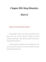
This figure summarises the position of all trocars. The imaginary lines drawn in the pictures are helpful for
orientation (from umbilicus to anterior superior iliac spine – continuous lines; pararectal line – interrupted
lines). The numbers give the sequence of trocar placement.
The final 12-mm trocar is placed approximately three finger breaths medial to the left anterior superior iliac
spine (on a hypothetical line from the spine to the umbilicus). Avoid placement too distally or too close to the
iliac spine, because this can cause problems during apical dissection and anastomosis. This is the trocar
through which all the lymph nodes are extracted. Furthermore, the needles are inserted and extracted through
this trocar.