Endoscopic Extraperitoneal Radical Prostatectomy - part 8 pps
Bạn đang xem bản rút gọn của tài liệu. Xem và tải ngay bản đầy đủ của tài liệu tại đây (1.13 MB, 20 trang )
Chapter 8
J U. Stolzenburg et al.
8
132
8.2.4 Obturator Nerve Injury
The obturator nerve is responsible for the innervation
of the medial thigh adductor muscles. Nerve injury is
rare and can occur during lymphadenectomy by elec-
trofulguration, complete transection, or entrapment
by clips. When electrofulguration is the cause of in-
jury, the symptoms usually subside after 6 weeks. In
the case of iatrogenic nerve transection, some authors
advocate a microsurgical epineural end-to-end ten-
sion-free anastomosis.
In our series we encountered a 0.2% rate of tempo-
rary obturator nerve apraxia, treated successfully
with neurotropic drugs and physiotherapy. We never
experienced complete nerve transection.
8.2.5 Lymphoceles
Lymphoceles occur due to leakage from transected
lymphatic vessels. Diagnosis and treatment depend
on size, site, and possible infections. Significant lym-
phoceles may cause pelvic pain as well as voiding
problems after catheter removal. Later symptoms can
be deep venous thrombosis followed by leg oedema
with concomitant pain. A very rare complication is
the development of hydronephrosis. Infected lym-
phoceles are often associated with febrile conditions.
Percutaneous drainage, sclerotherapy, or laparoscopic
transperitoneal fenestration may be performed.
When a small symptomatic lymphocele is diag-
nosed by ultrasonography, percutaneous drainage
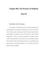
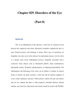
Fig. 8.12. Major leak aer dislocation of the catheter. In the
case of accidental catheter dislocation (c) due to extreme tension
of the catheter, a major leak (a, b) can be created. is requires
insertion of ureteral mono J catheters (d). e urethral catheter
should be advanced within the bladder and its balloon should be
inated with 20 ml. Both ureteral and urethral catheters should
remain in place for a minimum of 2 weeks. ese patients
will develop a secondary cavity at the site of initial dislocation
(e, f). e complete healing process of this “additional” cavity
can take 1 month or longer. e nal cystography (f) that is
always performed before catheter removal and shows the “ab-
normal” healing process without any extravasation
Chapter 8
133
Troubelshooting
and sclerotherapy can be performed as a first-line
treatment, but its success rate is under 50%. The per-
cutaneous drain should be closed for 1 day to evaluate
the effect of sclerotherapy treatment. If lymph pro-
duction continues, a laparoscopic fenestration should
be performed.
Patients with infected symptomatic lymphoceles
(fever, leucocytosis, increased C reactive protein) are
initially treated by percutaneous drainage and antibi-
otic coverage. Laparoscopic fenestration is performed
when the patient has recovered his normal condition.
Access for the fenestration is achieved through the
periumbilical trocar (minilaparotomy), the site of
previous placement of the laparoscope during the EE-
RPE. In contrast to EERPE, lymphocele fenestration
requires a transperitoneal approach. In most cases the
lymphocele is clearly visible and the fenestration is
performed starting ventrally and concluding dorsally,
taking care not to injure the ureter or the iliac vein. If
the site of lymphatic collection is not evident, methyl-
ene blue can be injected percutaneously into the lym-
phocele with the aid of ultrasonographic guidance, or
injected via the percutaneous drainage tube.
8.2.6 Miscellaneous
Rare untoward postoperative events include perineal
pain, pubic osteitis, urosepsis, penile haematoma and
perineal haematoma. If a perineal haematoma causes
voiding disorders, it should be drained under perineal
ultrasonographic guidance.
Contents
9.1 Introduction . . . . . . . . . . . . . . . . . . . . . . . . . . . . . . . . . 136
9.2 Mechanical Means . . . . . . . . . . . . . . . . . . . . . . . . . . . . 136
9.3 Electrosurgical Tools. . . . . . . . . . . . . . . . . . . . . . . . . . 136
9.3.1 Monopolar Electrocautery . . . . . . . . . . . . . . . . . . . . 137
9.3.2 Bipolar Electrocautery . . . . . . . . . . . . . . . . . . . . . . . . 137
9.3.3 The LigaSure Sealing System . . . . . . . . . . . . . . . . . . 137
9.4 Ultrasonic Energy Device . . . . . . . . . . . . . . . . . . . . . 137
9.5 Lasers for Haemostasis . . . . . . . . . . . . . . . . . . . . . . . 138
9.6 Tissue Sealants . . . . . . . . . . . . . . . . . . . . . . . . . . . . . . . 138
9.6.1 Fibrin Glues . . . . . . . . . . . . . . . . . . . . . . . . . . . . . . . . . . 138
9.6.2 Haemostatic Gelatine Matrix . . . . . . . . . . . . . . . . . . 139
9.6.3 Human Fibrinogen and Thrombin Fleece . . . . . .140
9.6.4 Experimental Tissue Sealants
in Radical Prostatectomy . . . . . . . . . . . . . . . . . . . . . 141
9.6.5 Possible Adverse Events of Tissue Sealants . . . . 141
References . . . . . . . . . . . . . . . . . . . . . . . . . . . . . . . . . . . 141
Haemostasis in Radical Prostatectomy
Evangelos N. Liatsikos ∙ Paraskevi Katsakiori ∙ Jens-Uwe Stolzenburg
9
Chapter 9
E. N. Liatsikos ∙ P. Katsakiori ∙ J U. Stolzenburg
9
136
9.1 Introduction
Adequate haemostasis is essential in every surgical
procedure. Uncontrolled bleeding hinders the sur-
geon‘s work and potentially threatens the patient‘s
life. Particularly, during laparoscopic radical prosta-
tectomy, even small amounts of blood may critically
impair the view at a site where vision is already re-
stricted a priori. For this reason, haemostasis in lapa-
roscopic procedures focuses mainly on primary pre-
vention of bleeding.
There are various methods of securing surgical
haemostasis, including mechanical means (sutures,
ligatures or staples), vessel coagulation (electrocau-
tery or ultrasonic energy) and tissue sealing. Fre-
quently, more than one type of procedure is needed to
achieve satisfactory haemostasis. The application of
mechanical devices is time consuming, requires good
access to the vessels and leaves a foreign material in-
side the patient, which may lead to complications.
Haemostatic clips are utilised for the mechanical liga-
tion of vessels with a diameter of 3–7 mm. Stapling
devices are costly for multiple single-vessel applica-
tions [1]. Electrocoagulation systems are quickly ap-
plied and do not introduce foreign materials. They
are capable of sealing vessels with a diameter up to
2–3 mm. However, possible lateral thermal damage
and potential tissue necrosis impede their applica-
tion. In addition, they are unreliable for vessels with a
diameter >2 mm [2]. Tissue sealants can be applied
with or without clips or staples and are capable of pro-
viding satisfactory haemostasis alone or in conjunc-
tion with other haemostatic methods.
This chapter provides an overview of the various
methods of haemostasis.
9.2 Mechanical Means
Mechanical means of haemostasis include mechani-
cal compression, sutures, clips and staples [3]. The
same principles are used in both open and laparo-
scopic radical prostatectomy. Proper tissue dissection
and early identification of the supplying blood ves-
sels, preferably before bleeding occurs, are necessary.
Dissection with a laparoscopic styptic stick helps to
control bleeding from the adjacent vessels.
Local compression with a sponge in the case of un-
controllable venous bleeding provides the surgeon
with time to elaborate further strategies for final hae-
mostasis. Local compression by itself may sometimes
be sufficient. If not, the application of tissue sealants
in combination with local mechanical compression
may adequately seal large vessels, even the vena cava.
Suturing techniques in laparoscopic radical pros-
tatectomy differ from those in open surgery and re-
quire advanced laparoscopic skills. Freehand intra-
corporeal suturing is preferable to external knotting
because it avoids excessive traction during suturing.
The use of endo-loops may be of great help, particu-
larly for surgeons inexperienced in endoscopic sutur-
ing. During the application of endo-loops, however, a
significant amount of healthy tissue is sacrificed.
Moreover, the loops may slip off due to tissue isch-
aemia, and loops that remain in place may loosen.
Laparoscopic vascular clips are the preferred tool
for sealing blood vessels. Small amounts of bleeding
may still occur, however, either due to malposition of
clips or because the enclosed bundles of tissue are too
small. Titanium clips tend to slip off during further
dissection. For this reason, at least two to five clips are
needed for safe control of vessels with a diameter of
3 mm.
Vascular endo-staplers with 2.0- to 2.5-mm jaw
width and various lengths have been used to achieve
safe occlusion of major vessels and vascular pedicles.
The modern endo-staplers are bulky instruments that
require 12-mm access ports, utilise three lines of sta-
ples for safe vascular control and provide the cutting
simultaneously. These devices are costly, single-use
instruments and require training before use. The lap-
aroscopic surgeon must always use the appropriate
vascular jaw width (not the tissue width) and must
ensure that the entire vessel is within the stapler line
before firing.
9.3 Electrosurgical Tools
Electrosurgery has been widely used in open surgery
for obtaining adequate haemostasis. Monopolar elec-
trocautery was the first tool to be adapted for laparo-
scopic procedures. However, owing to the high risk of
thermal injury in the surrounding tissues during the
application of electrocautery, new energy sources have
been employed. Ultrasonic coagulation systems have
been used in radical prostatectomy with better hae-
mostatic effect, less thermal damage and better func-
tional results.
Chapter 9
137
Haemostasis in Radical Prostatectomy
9.3.1 Monopolar Electrocautery
Although monopolar electrocautery provides ade-
quate haemostasis, its use is restricted by potential
complications. By limiting the time of application and
the maximum current force, the complications can be
minimised. Electrical bypass may occur at sites of low
impedance or damaged insulation. This is the reason
why we do not use any monopolar energy during EE-
RPE. The safety of monopolar electrocautery may be
secured by active electrode monitoring. In the case of
any break in the integrity of the insulation, the in-
strument is immediately shut off and the monitoring
device does not allow activation if the foot pedal is
depressed. Another potential drawback is that re-us-
able scissors may lose their sharpness after extensive
use of monopolar current during dissection. This
problem can be solved by using single-use scissors
blades for re-usable instruments. Modern re-usable
instruments are thought to be safer.
A haemostatic monopolar cautery device that has
been utilised in handling capillary bleeding is the ar-
gon beam coagulator [3]. This device is a monopolar
cautery instrument that uses an argon jet to propel
blood away from the surgical field. Although it has
proved efficacious in control of minor capillary bleed-
ing, argon beam coagulation alone cannot be success-
fully used for tissue dissection. Additionally, it is not
suitable for managing significant bleeding or haem-
orrhage from larger vessels.
9.3.2 Bipolar Electrocautery
Bipolar electrocautery has been proposed instead of
monopolar and bulk clipping in order to obtain ade-
quate haemostasis and safer dissection and to mini-
mise possible thermal injury of adjacent tissues [4, 5].
Bipolar coagulating forceps have already been used
during radical retropubic prostatectomy for coagula-
tion of the vascular plexus [6]. Radical prostatectomy
always involves a considerable risk of thermal and
electrical injury of the neurovascular bundles and the
branches of the pelvic plexus. Significant reductions
in intraoperative blood loss and in the need for trans-
fusion during or after the operation were described.
Furthermore, the visibility was improved, allowing
maximal preservation of the urethral length, com-
plete extirpation of all apical prostatic notches and
improved application of the nerve-sparing technique
compared to the standard approach. Urogenital func-
tion at 14 months after operation was comparable to
that with the standard method, assuming that the
parasympathetic nerves and the ventral urethral wall
did not suffer any negative thermal effect.
9.3.3 The LigaSure Sealing System
The LigaSure vessel-sealing system was developed in
1995. It works by coagulating the walls of the target
vessel by means of bipolar energy. The feedback-con-
trol mechanism ensures that the adjacent tissues are
not charred by overcoagulation. This instrument is
effective in sealing vessels with a diameter of 1–7 mm
and results in a high burst strength and permanent
seal while limiting the lateral thermal damage [7].
The LigaSure system has already been used in open
radical prostatectomy for sealing the pelvic lymphatic
tissues and for ligating the lateral pedicles (from the
base to the apex of the prostate), the puboprostatic
ligaments and the dorsal vein complex. Total opera-
tion time and the need for blood transfusion were sig-
nificantly reduced with the use of LigaSure, compared
to conventional ligation [7, 8].
The safety of blood vessel control with the LigaS-
ure system has also been demonstrated in a porcine
experimental study. The seals created by LigaSure,
were stronger than those accomplished with other en-
ergy-based ligation methods (ultrasonic coagulation
and standard bipolar coagulation). The seals obtained
by the application of LigaSure were able to withstand
a minimum of three times the normal systolic pres-
sure [9].
9.4 Ultrasonic Energy Device
The piezoelectric ultrasonic energy device (UED –
SonoSurg, Olympus; AutoSonix, Tyco; UltraCision,
Ethicon) simultaneously excises and coagulates tissue
with the application of high-frequency ultrasound.
Dissection and cavitation are achieved using frequen-
cies of 23.5 and 55.5 kHz. The UED minimises col-
lateral damage, avoids tissue carbonisation and re-
duces potential thermal injury compared to
monopolar energy sources. Use of the UED is limited
to vessels with diameter <4 mm. In larger vessels, ad-
equate haemostasis cannot be achieved with the sole
use of a UED. The same problem may occur at the
Chapter 9
E. N. Liatsikos ∙ P. Katsakiori ∙ J U. Stolzenburg
9
138
Santorini plexus [3]. Nevertheless, many groups have
used this instrument in laparoscopic radical prosta-
tectomy due to its excellent haemostatic properties.
Heat production is a source of concern, as uninten-
tional thermal injuries may occur whenever dissect-
ing close to neural structures in laparoscopic surgery.
In contrast to bipolar energy, a 23.5 and 55.5 kHz ul-
trasonically activated device minimises macroscopic
tissue charring. In addition the heat production is
much slower than monopolar electrosurgery.
Owaki et al. found that the blade of the ultrasonic
shears becomes hot after use, increasing to 63°C after
3 seconds and 150°C after 30 s. They suggested that
contact of the blade with neural structures immedi-
ately following use caused recurrent laryngeal nerve
injury in their series of patients undergoing endo-
scopic parathyroid surgery. This is important to note,
since the surgeon has no indication of the tempera-
ture of the instrument tips while performing laparo-
scopic surgery, and there is relatively little space for
the dissipation of heat [10, 11]. However, the UED is
certainly safe when performing a wide-excision EE-
RPE. For example, we have never had problems with
rectum or obturator nerve injuries caused by the use
of UED. In nerve-sparing procedures the UED should
be used more as a dissecting tool than a cutting tool
and should not be activated for a long time near the
neurovascular bundle. To date there are no clinical
human studies comparing the effect of UED and cold
scissors dissection during nerve-sparing radical pros-
tatectomy.
9.5 Lasers for Haemostasis
There are no clinical data on the use of laser devices
for achieving adequate haemostasis during open or
laparoscopic radical prostatectomy. The most com-
mon applications of laser in the field of urology are
the incision of urethral/ureteral strictures, ablation of
superficial transitional cell carcinoma, bladder neck
incision, prostate resection and lithotripsy of urinary
calculi [13–15]. Laser prostatectomy has emerged as
an alternative to the traditional transurethral resec-
tion for the treatment of benign prostatic hypertro-
phy, aiming to significantly reduce blood loss [16].
9.6 Tissue Sealants
Tissue sealants have successfully been used in the
management of adequate haemostasis in various op-
erations, with or without sutures and staples. A num-
ber of tissue sealants – commercial and noncommer-
cial – are available, including fibrin glues,
cyanoacrylates, polymethylmethacrylates and gela-
tine products [17–21]. Fibrin sealants seem to be the
optimal tissue adhesives, since both the adhesive and
the degradation products are biocompatible.
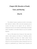
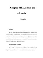
9.6.1 Fibrin Glues
The fibrin glues consist of thrombin and fibrinogen,
the plasma derivatives at the end of the clotting cas-
cade (Fig. 9.1). Initially, fibrin sealants contained hu-
man fibrinogen and bovine thrombin. The use of a
nonhuman protein could potentially cause an ana-
phylactic reaction or development of antibodies
against bovine factor V and subsequent cross-reac-
tion with human factor V. Therefore, recent commer-
cial sealants use human thrombin rather than its bo-
vine equivalent. Other key components that a fibrin
sealant may contain are fibronectin, factor XIII and
aprotinin.
Aprotinin is a natural protease inhibitor, derived
from bovine lung, that impedes clot lysis by inhibit-
ing trypsin, plasmin and kallikrein as well as convert-
ing plasminogen to plasmin. However, some research-
ers have suggested that the aprotinin is not only
unnecessary for achieving a stable clot but also entails
the rare risk of anaphylaxis. Clotting factor XIII is
used to cross-link fibrin monomers into polymers,
providing a mechanically stable clot resistant to fibri-
nolysis [22]. It is added to or co-purified with fibrino-
gen. Factor XIII is a pro-enzyme that is activated by
thrombin in the presence of calcium ions. After its ac-
tivation, the polymerisation of fibrin monomers oc-
curs within 3 min. Fibronectin enhances the migra-
tion of fibroblast and fibroblastic growth into areas of
fibrin seal application and therefore participates in
wound healing. In purified preparations of fibrino-
gen, however, fibronectin may be absent.
Careful and proper application of the fibrin sealant
is needed in order to achieve optimal adhesion. If fi-
brin sealant is applied to two surfaces for approxima-
tion, the surfaces should be brought into contact im-
mediately, before the polymerisation of the agent. If
Chapter 9
139
Haemostasis in Radical Prostatectomy
fibrin is applied only to one surface and allowed to po-
lymerise, it acts as an anti-adhesive agent, preventing
the adherence of the two surfaces [20]. The two com-
ponents of the fibrin sealant can be applied sequen-
tially or simultaneously to the surgical field by means
of a dual-syringe system – with or without using an
endoscopic delivery system –, spraying or sponge ap-
plication. Commonly, the dual-syringe system enables
simultaneous application of equal amounts of fibrino-
gen and thrombin through a blunt-tipped needle. A
long applicator needle with a dual-lumen adapter is
available for introducing the agent during laparoscop-
ic procedures. Alternatively, the material can be ap-
plied by mixing equal amounts of the two components
and spraying with forced sterile gas [20].
To date, fibrin glues have been used for haemostat-
ic or adhesive effect in various urosurgical applica-
tions such as kidney-sparing surgery, orchiopexy, py-
eloplasty and fistula repair. Their success varies with
the depth of the resection and the blood pressure. In
radical prostatectomy, fibrin glues have been utilised
for obtaining adequate haemostasis with satisfactory
results [23–25]. Tissue sealants behave differently in
contact with urine. Their adhesive capacity may be
reduced because of the fibrinolytic activity of uroki-
nase. Sealants with a lower concentration of aprotinin
or sealants containing an antifibrinolytic agent may
delay the degradation of the fibrin clot [19].
Another fibrin glue is Tisseel fibrin glue (Baxter,
Austria) which contains human fibrinogen, human
activated thrombin, calcium chloride solution, bovine
aprotinin, fibronectin and factor XIII. When Tisseel
initially comes in contact with urine, it tends to main-
tain a solid form which consequently, turns to a semi-
solid gelatinous state that is still present at 5 days. Tis-
seel has been tested in the formation of the
urethrovesical anastomosis after radical retropubic
prostatectomy [26]. This agent proved to have both
haemostatic and tissue adhesive properties.
9.6.2 Haemostatic Gelatine Matrix
FloSeal (Baxter, Germany) is a two-component seal-
ant consisting of a bovine gelatine-based matrix and a
bovine-derived thrombin component [27, 28]. The
gelatine matrix contains bovine collagen, cross-linked
with glutaraldehyde. The matrix can be prepared eas-
ily and can be applied in 2 hours. When in contact
with normal or sanguineous urine, FloSeal stays in a
fine particulate suspension.
The urological application of FloSeal has been de-
scribed with satisfactory haemostatic results [27, 28].
FloSeal and Gelfoam were used in clipless, cautery-
free, nerve-sparing, robotic radical prostatectomy by
Ahlering et al. [29]. Intraoperative handling of haem-
orrhage was satisfactory and only 4 of 17 cases re-
quired further management with sutures. No postop-
erative bleeding events were described.
Fig. 9.1. Physiological pathway
to brin
Chapter 9
E. N. Liatsikos ∙ P. Katsakiori ∙ J U. Stolzenburg
9
140
9.6.3 Human Fibrinogen
9.6.5 and Thrombin Fleece
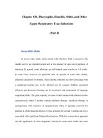
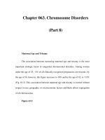
The main representative of this category is TachoSil
(Nycomed, Austria), a dry, equine fibrin adhesive-
coated collagen sponge. Its mechanism of action – like
other fibrin glues – is reproduction of the last step of
the clotting cascade (Fig. 9.2). It consists of a fixed,
solid layer that contains human thrombin and fibrin-
ogen. This layer is anchored on the surface of a colla-
gen carrier. A special fan-like Endo-doc carrier is
used to ensure controlled application of the dry
fleece.
TachoSil is a further development of Tachocomb
and Tachocomb H and differs from them by the ab-
sence of bovine aprotinin and by containing purely
human coagulation agents. Tachocomb contained
human fibrinogen, bovine thrombin and bovine
aprotinin, while Tachocomb H contained human fi-
brinogen and thrombin and bovine aprotinin.
When the coated collagen fleece comes in contact
with fluids (e.g. normal saline, body fluids, bleeding
surface), the components of the layer dissolve, diffuse
into the wound cavities and start to react. The colla-
gen fleece helps to tamponade the wound and there-
fore keeps the coagulation components in the bleed-
ing area. The required time for gluing is 3–5 min, and
during this time the TachoSil must be pressed gently
onto the surface of the wound. After its proper appli-
cation, the sealed surface can be used for further bi-
polar coagulation or sutures if needed, without jeop-
ardising the seal. Additionally, TachoSil separates
tissues, providing an anti-adhesive effect to the adja-
cent structures. TachoSil is degraded within weeks or
months after its application, either by fibrinolysis and
cellular phagocytosis of the fibrin clot or by layer-by-
layer degradation of the collagen patch by absorptive
granulation tissue, followed by conversion into a
pseudocapsule consisting of endogenous connective
tissue.
With the use of TachoSil, various vessel or paren-
chymatic defects can be sealed. A recently published
study reviews the application of TachoSil in 408 pa-
tients with haemorrhagic risk factors or operations
associated with an expected increase of bleeding [30].
The operations were performed on various organs,
such as liver, vascular system, heart, spleen, thorax
and kidney, and the results supported the efficacy and
safety of TachoSil as a haemostatic agent. In addition,
when compared to argon beam coagulation, TachoSil
proved superior in obtaining effective and fast intra-
operative haemostasis during liver resection [31].
During nerve-sparing EERPE in patients with
prostate cancer, TachoSil seems to provide adequate
haemostasis without jeopardising the clinical out-
come. We performed a pilot study evaluating the use
of TachoSil during cautery-free EERPE in 20 consecu-
tive patients (unpublished data). The total operative
time was 128 min (range 75–210 min). No patient
needed blood transfusion or conversion to open sur-
Fig. 9.2. Blood coagulation and
degradation of clot and collagen
patch. e active components of
the TachoSil® coating are shaded
Chapter 9
141
Haemostasis in Radical Prostatectomy
gery. Fourteen of 20 patients were fully continent at 3
months after operation and only one patient needed
more than two pads per day. At 6 months, 12 of these
14 men (85.7%) reported full continence and no pa-
tient reported needing more than two pads a day. Six
out of 20 patients (30%) and 9 out of 15 patients (60%)
were potent at 3 and 6 months, respectively. All the
patients who reported being potent at 3 months post-
operatively were 43–55 years of age. At 6 months, all
the patients aged 43–55 years were potent, but only 1
out of 7 (14.3%) aged 56–73 years reported potency.
Potency is defined here as a score of 21 points or more
on the IIEF-5 questionnaire. The use of TachoSil
seems to be safe, as no intra- or postoperative bleed-
ing was reported, and the potency results are very
promising.
9.6.4 Experimental Tissue Sealants
9.6.5 in Radical Prostatectomy
The use of cyanoacrylates has been restricted due to
their rapid degradation to cyanoacetate and formalde-
hyde, each of which can lead to significant tissue tox-
icity. This problem led to the development of cyanoac-
rylates with longer alkyl chains which show slower
formation of these toxic products. 2-Octyl-cyanoac-
rylate (2-OCA) is an agent of this type which is utilised
for skin closure. 2-OCA can be used only as a second-
ary haemostatic factor since it is not able to provide
adequate haemostasis by itself. In an experimental ca-
nine model, 2-OCA was used in order to form a water-
tight, vesicourethral anastomosis during open total
prostatectomy, with disappointing results [32].
9.6.5 Possible Adverse Events
9.6.5 of Tissue Sealants
Tissue sealants have been used in a wide variety of
applications over the last 30 years. However, their use
has been limited by some potential complications,
such as inflammatory or allergic reactions and viral
infections [18–21].
Anaphylactic reaction to bovine thrombin is an ex-
tremely rare reaction. However, sudden and severe
hypotension resulting in death has been reported af-
ter application of bovine thrombin to a deep hepatic
wound [33]. In most of the recently developed com-
mercial sealants, bovine thrombin has been replaced
by human thrombin, avoiding this potential compli-
cation. Additionally, allergic reactions have been re-
ported with the use of other nonhuman agents such
as aprotinin. The frequency of hypersensitivity to in-
travenous injection of aprotinin is reported to be ap-
proximately 10%.
Bovine thrombin may cause the so-called immu-
nologically induced coagulopathy [34]. In this case,
the patient may develop antibodies to plasma proteins
in bovine thrombin preparations. Many of these plas-
ma proteins are clotting factors or glycoproteins in-
volved in coagulation. The developed antibodies to
these bovine proteins may cross-react with human
homologues, leading to significant anticoagulation
results.
The possibility of transmission of infection by fi-
brin sealants has long been a matter of concern and
debate [18–20]. Like any other blood product, com-
mercial fibrin sealants bear the theoretical risk of vi-
ral transmission. However, no cases of serious viral
transmission have been reported since the develop-
ment of commercial fibrin sealants. Careful donor
selection strategies help to decrease viral transmis-
sion risk. Additionally, recent advances in viral inac-
tivation technology further reduce the risk of trans-
mission of hepatitis A, B and C and HIV. Various
techniques can be applied for viral inactivation,, in-
cluding vapour heating, steam treatment, pasteurisa-
tion, irradiation, solvent detergent extraction and
nanofiltration [34].
Finally, caution should be taken during the appli-
cation of fibrin sealants to avoid the direct injection
of the agent into large blood vessels, with the atten-
dant risk of thromboembolic complications. To date,
prolonged inflammation has not been reported for fi-
brin sealants.
References
1. Nelson MT, Nakashima M, Mulvihill SJ (1992) How secure
are laparoscopically placed clips? Arch Surg 127:718–720
2. Kennedy JS, Stranahan PL, Taylor KD, Chandler JG (1998)
High-burst-strength, feedback-controlled bipolar vessel
sealing. Surg Endosc 12:876–878
3. Klingler CH, Remzi M, Marberger M, Janetschek G (2006)
Haemostasis in laparoscopy. Eur Urol 50:948–57
4. Chien GW, Mikhail AA, Orvieto MA, Zagaja GP, Sokol-
o MH, Brendler CB, Shalhav AL (2005) Modied clipless
antegrade nerve preservation in robotic-assisted laparo-
scopic radical prostatectomy with validated sexual func-
tion evaluation. Urology 66:419–423
Chapter 9
E. N. Liatsikos ∙ P. Katsakiori ∙ J U. Stolzenburg
9
142
5. Harrell AG, Kercher KW, Heniford BT (2004) Energy
sources in laparoscopy. Semin Laparosc Surg 11;201–209
6. Stief CG (2003) Apical dissection during radical prostatec-
tomy without ligature. World J Urol 21:139–143
7. Daskalopoulos G, Karyotis I, Heretis I, Delakas D (2004)
Electrothermal bipolar coagulation for radical prostatec-
tomies and cystectomies: a preliminary case-controlled
study. Int Urol Nephrol 36:181–185
8. Sengupta S, Webb DR (2001) Use of a computer-controlled
bipolar diathermy system in radical prostatectomies and
other open urological surgery. ANZ J Surg 71:538–540
9. Landman J, Kerbl K, Rehman J, Andreoni C, Humphrey
PA, Collyer W, Olweny E, Sundaram C, Clayman RV
(2003) Evaluation of a vessel sealing system, bipolar elec-
trosurgery, harmonic scalpel, titanium clips, endoscopic
gastrointestinal anastomosis vascular staples and sutures
for arterial and venous ligation in a porcine model. J Urol
169:697–700
10. Owaki T, Nakano S, Arimura K, Aikou T (2002) e ultra-
sonic coagulating and cutting system injures nerve func-
tion. Endoscopy 34:575–579
11. Ong AM, Su LM, Varkarakis I, Inagaki T, Link RE, Bhay-
ani SB, Patriciu A, Crain B, Walsh PC (2004) Nerve spar-
ing radical prostatectomy: eects of hemostatic energy
sources on the recovery of cavernous nerve function in a
canine model. J Urol 172:1318–1322
12. Stolzenburg J-U, Truss MC (2003) Technique of laparo-
scopic (endoscopic) radical prostatectomy. BJU Int 91:749–
757
13. Fried NM (2006) erapeutic applications of lasers in
urology: an update. Expert Rev Med Devices 3:81–94
14. Wollin TA, Denstedt JD (1998) e holmium laser in urol-
ogy. J Clin Laser Med Surg 16:13–20
15. Kabalin JN (1996) Holmium:YAG laser prostatectomy: re-
sults of U.S. pilot study. J Endourol 10:453–457
16. Chun SS, Ravzi HA, Denstedt JD (1995) Laser prostatec-
tomy with the holmium: YAG laser. Tech Urol 1:217–221
17. Morikawa T (2001) Tissue sealing. Am J Surg 182:29S–
35S
18. MacGillivray TE (2003) Fibrin sealants and glues. J Card
Surg 18:480–485
19. Albaba DM (2003) Fibrin sealants in clinical practice. Car-
diov Surg 11:5–11
20. Shekarriz B, Stoller ML (2002) e use of brin sealant in
urology. J Urol 167:1218–1225
21. Schexneider KI (2004) Fibrin sealants in surgical or trau-
matic hemorrhage. Curr Opin Hematol 11:323–326
22. Buchta C, Hedrich HC, Macher M, Hocker P, Redl H (2005)
Biochemical characterization of autologous brin sealants
produced by Cryoseal and Vivostat in comparison to the
homologous brin sealant product Tissucol/Tisseel. Bio-
materials 26:6233–6241
23. Boeckman WR, Jakse G (1994) Reconstruction of the
urethra aer radical perineal prostatectomy using brin
sealing. In: Schlag G, Melchior H, Wallwiener D (ed) Gy-
necology and obstetrics: Urology, vol 7. Springer, Berlin
Heidelberg New York, p 103
24. Lobel B, Ordonez O, Olivo JF, Cipolla B, Milon D, Leveque
JM, Guille F (1991) Radical prostatectomy and biologic
glue. Prog Urol 1:440–444
25. Patel R, Caruso RP, Taneja S, Stifelman M (2003) Use of
brin glue and Gelfoam to repair collecting system inju-
ries in a porcine model: implications for the technique of
laparoscopic partial nephrectomy. J Endourol 17:799–804
26. Diner EK, Patel SV, Kwart AM (2005) Does brin sealant
decrease immediate urinary leakage following radical ret-
ropubic prostatectomy? J Urol 173:1147–1149
27. User HM, Nadler RB (2003) Applications of FloSeal in
nephron-sparing surgery. Urology 62:342–343
28. Richter F, Tullmann ME, Turk I, Deger S, Roigas J, Wille
A, Schnorr D (2003) [Improvement of hemostasis in lapa-
roscopic and open partial nephrectomy with gelatine
thrombin matrix (FloSeal)]. Urologe A 42:338–346
29. Ahlering TE, Eichel L, Chou D, Skarecky DW (2005) Feasi-
bility study for robotic radical prostatectomy cautery-free
neurovascular bundle preservation. Urology 65:994–997
30. Haas S (2006) e use of a surgical patch coated with hu-
man coagulation factors in surgical routine: a multicenter
postauthorization surveillance. Clin Appl romb He-
most 12:445–450
31. Frilling A, Stavrou GA, Mischinger H-J, De Hemptinne
B, Rokkjaer M, Klempnauer J, orne A, Gloor B, Becke-
baum S, Ghaar MFA, Broelsch CE (2005) Eectiveness
of a new carrier-bound brin sealant versus argon beamer
as haemostatic agent during liver resection: a randomized
prospective trial. Langenbecks Arch Surg 390:114–120
32. Grummet JP, Costella AJ, Swanson DA, Stephens C, Cro-
meens DM (2002) Vesicourethral anastomosis with 2-
octyl cyanoacrylate adhesive in an in vivo canine model.
Urology 60:935–938
33. Gibble JW, Ness PM (1990) Fibrin sealant: the perfect op-
erative sealant? Transfusion 30:741–747
34. Radosevich M, Goubran HI, Burnouf T (1997) Fibrin seal-
ant: scientic rationale, production methods, properties,
and current clinical use. Vox Sang 72:133–143
Contents
10.1 Installation and Robot Connection . . . . . . . . . . . .144
10.2 Robotic Radical Extraperitoneal
Prostatectomy . . . . . . . . . . . . . . . . . . . . . . . . . . . . . . . 147
References . . . . . . . . . . . . . . . . . . . . . . . . . . . . . . . . . . . 159
Extraperitoneal Robotic Radical
Prostatectomy: – Operative Technique –
Step by Step
Hubert John ∙ Matthew T. Gettman
10
Chapter 10
10
144
The extraperitoneal approach for conventional lapa-
roscopic prostatectomy was proposed by Raboy 1997
[1] and popularized by Bollens [2], Hoznek and Ab-
bou [3], Dubernard [4] and Stolzenburg [5]. The fea-
sability of an extraperitoneal access for robotic sur-
gery was reported in 2003 by Gettman and Abbou
[6].
The extraperitoneal approach avoids potential
small-bowel injuries, allows only a moderate Tren-
delenburg position and is more comparable to the
standard open retropubic radical prostatectomy. This
chapter demonstrates step by step the extraperitoneal
technique that has been used since 2002 by the first
author and performed now in over 400 cases [7]. The
transperitoneal access is chosen only after laparo-
scopic hernia repair with preperitoneal mesh implant,
after kidney transplantation or further extensive ret-
roperitoneal surgery.
The access is similar to the technique described in
Chap. 7. A short oblique subumbilical incision of 3–
4 cm is made. Two Langenbeck retractors expose the
anterior rectus fascia, which is incised vertically over
a 1 cm length. The retractors are used to split the left-
sided muscle fibers of the rectus sheath and expose
the posterior rectus fascia and peritoneum. Blunt fin-
ger dissection of the retroperitoneal space is per-
formed. In some cases, the tip of the index finger may
touch the pubic symphysis during blunt dissection.
Balloon dilatation is performed to expose the extra-
peritoneal space (Tyco®). The balloon is filled 10–15
times until the extraperitoneal space is appropriately
created. Balloon dilation must be performed carefully
to avoid bladder rupture, which has been known to
occur in cases of overdilation. The camera trocar
(Ethicon, 12 mm) is then inserted via the subumbili-
cal incision. An inspection of the extraperitoneal
space is performed. Under direct vision, the camera
can be used to increase the size of the extraperitoneal
space by gently sweeping the peritoneal borders to the
side and upwards.
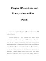
10.1 Installation and Robot Connection
The 8-mm bilateral robot trocars are placed pararectally and two 10-mm standard trocars (Versaport®, Ethi-
con) just anteromedial of the iliac spine (left). In procedures with only one assistant, the left-sided
standard 10-mm trocar may be replaced by a 5-mm multiuse trocar, which is positioned between the right-
sided robot trocar and the camera (right).
H. John ∙ M. T. Gettman
Chapter 10
145
Extraperitoneal Robotic Radical Prostatectomy
The 0° 3D endocamera is introduced (left). The abdominal wall is slightly lifted by the camera arm trocar
(“laparo-lift”).
The left arm is brought to the left robot trocar and attached (right).
The right arm is also connected and the instruments (bipolar forceps on the left side and round-tip scissors on
the right) are inserted under visual control (left). The bipolar cable is attached onto the forceps.
Before starting with the operation, always ensure the lower extremities are not compressed by the robotic arms
(right).
Chapter 10
H. John ∙ M. T. Gettman
10
146
The instruments allow wrist-like instrument movement (Endo-wrist®-technology). We use the bipolar
hemostatic forceps (a), a round-tip scissors (b) and two needle holders (c).
The table-side assistants are comfortably installed (left). They assist with an aspirator (right 10-mm trocar),
laparoscopic grasper, laparoscopic scissors and clip appliers.
The console surgeon leaves the operating table after port placement and is not sterile scrubbed during
radical prostatectomy (right).
Chapter 10
147
Extraperitoneal Robotic Radical Prostatectomy
The entire radical prostatectomy is performed by the operating urologist from the remote console (left).
He controls the robotic arms at the console (camera, working channels, additional fourth arm if installed).
The console surgeon controls the interchangeable instruments attached to the two working robotic arms
(right). They are felt as direct extensions of his arms and fingers.
10.2 Robotic Radical Extraperitoneal
Prostatectomy
If the preperitoneal space is completely developed, the anterior prostatic surface and the endopelvic fascia are
exposed and the fatty tissue overlying these structures is gently swept away.
Chapter 10
H. John ∙ M. T. Gettman
10
148
If the endopelvic fascia is freed from the fatty tissue, it is incised from the prostatovesical junction to the apex
of the prostate. Fibers of the levator ani muscle are swept off laterally until the entire lateral aspect of the pros-
tate is visible.
The puboprostatic ligaments are incised to expose the prostatic apex and the urethra.
Chapter 10
149
Extraperitoneal Robotic Radical Prostatectomy
Fibers of the rhabdosphincter are swept distally to the pelvic floor.
Control of the dorsal vein plexus is achieved by a simple or a figure-of-eight ligation. For this we use a
0 Vicryl suture with a slightly straightened MH+ needle.
Radical prostatectomy is performed in a descending fashion starting with the incision of the ventral bladder
neck. If necessary, the bladder neck can easily be identified by gentle traction on the catheter. The anterior
bladder neck is separated from the prostate by blunt and sharp dissection.
Chapter 10
H. John ∙ M. T. Gettman
10
150
As soon as the urethra is opened, the Foley catheter is grasped by the assistant. Upward traction on the cath-
eter permits the prostate to likewise be rotated upwards and ventrally, thereby optimizing exposure of the
dorsal structures.
The dorsal bladder neck is incised and the dissection continues in strictly posterior direction until the vas
deferens become visible.
Chapter 10
151
Extraperitoneal Robotic Radical Prostatectomy
The vasa deferentia are dissected and the seminal vesicles exposed.
We cut the seminal vesicles leaving their tips in place if PSA is <10 ng/ml and Gleason score <7, in order not to
injure the neurovascular bundles, which pass in very close proximity to the seminal vesicle tips and are more
likely to be damaged by the added tension that has to be exerted during full dissection of the seminal vesicles
[8, 9].
Chapter 10
H. John ∙ M. T. Gettman
10
152
The fascia of Denonvilliers is then opened.
The posterior prostate surface is lifted from the perirectal fat.