Evidence-Based Imaging - part 9 docx
Bạn đang xem bản rút gọn của tài liệu. Xem và tải ngay bản đầy đủ của tài liệu tại đây (1.19 MB, 60 trang )
lence of appendicitis identified by pathologic examination following
surgery or clinical follow-up was 25%. The diagnostic sensitivity of attend-
ing physicians was 95%, and the specificity was 98%. This yielded a posi-
tive predictive value of 94% and a negative predicative value of 98.3%.
These data are similar to other, retrospective, evaluations of CT in the pedi-
atric population (45,61), and correlate with a nonconsecutive prospective
study of CT (60). However, the limited number of children in this trial and
the lack of a direct comparison to graded compression ultrasound preclude
definitive comparison of CT versus ultrasound as the primary imaging
exam in the pediatric population.
The desire to increase the accuracy of imaging yet limit the radiation
exposure has led investigators to examine combinations of CT and graded
compression ultrasound exam. Two prospective studies examined the com-
bination of graded compression ultrasound as the initial imaging, followed
by CT study if the ultrasound exam was equivocal or failed to match the
clinical presentation (62,63). Another randomized trial compared CT and
ultrasound versus ultrasound alone in a pediatric population (59). These
trials enrolled 585 patients, and had a prevalence of appendicitis ranging
from 23% to 43%, with a pooled prevalence of 39%. The sensitivity of these
protocols varied from 77% to 97%, with a pooled sensitivity of 95% (95%
CI, 83–100%). The range of specificity was 89% to 99%, with a pooled result
of 93% (95% CI, 87–97%). As would be expected, these protocols demon-
strated a greater sensitivity when the combined ultrasound followed by CT
test results were considered than when the same series of ultrasound data
was considered alone. This increased sensitivity, however, was achieved
with the drawback of a lower overall specificity. The single randomized trial
demonstrated similar results, with CT and ultrasound combined demon-
strating a higher sensitivity than ultrasound alone; however, the sensitivi-
ties of the two groups were not found to be statistically different (59). The
positive and negative predictive values at the pooled prevalence of appen-
dicitis were 97% (range, 87% to 96%) and 88% (range, 93% to 99%), respec-
tively. The positive likelihood ratio of CT followed by graded ultrasound
was found to be 13.03, with a negative likelihood ratio of 0.06.
As with the studies of imaging in adult appendicitis, the trials examin-
ing imaging and pediatric appendicitis suffer from a number of potential
limitations, including the use of different reference standards with the
choice of reference standard determined by the imaging result. Thus, the
sensitivity and specificity for imaging may be falsely inflated (40). In addi-
tion, many included trials conducted imaging only after explicitly exclud-
ing patients with a typical disease presentation who underwent immediate
appendectomy (51–53,57,60,63,64). Since the diagnosis of “typical” appen-
dicitis is made by individual clinical judgment, the resulting study popu-
lations may not be strictly comparable.
As with imaging of appendicitis in adults, there has been conflicting data
regarding the effect of imaging on the rate of finding a normal appendix
by pathology following appendectomy. Some retrospective studies have
found a decrease in the rate of negative appendectomy (41,65,66). Other
studies, however, come to the opposite conclusion (67–70). All of these ret-
rospective examinations were potentially limited by sample bias and ver-
ification bias. Given these conflicting results, it is unclear if the impact of
imaging on the rate of negative appendectomies can be adequately deter-
mined outside of the performance of a randomized trial.
464 C.C. Blackmore et al.
The data examining the cost impact of imaging in pediatric patients with
suspected appendicitis are limited. A single prospective cohort trial has
examined the cost of a protocol of ultrasound followed by CT exam if indi-
cated (65). This trial, from the hospital point of view, was conducted using
the same cohort of 139 patients as was used to determine the overall sensi-
tivity of the protocol (63). This trial found that the overall cost was
decreased by $565 per patient using the protocol. However, this calculation
assumes that the decrease in negative appendectomy can be replicated
outside of the research setting. As has been noted previously, there is not a
consensus that imaging has decreased the rate of negative appendectomies.
III. What Is the Accuracy of Imaging for Diagnosing
Small Bowel Obstruction?
Summary of Evidence: Computed tomography and ultrasound have higher
sensitivity and specificity than conventional plain film abdominal imaging
for diagnosing small bowel obstruction (moderate evidence) (Table 25.3).
Computed tomography has a higher sensitivity in the detection of small
bowel obstruction than ultrasound examination (limited evidence) (Table
25.3).
Supporting Evidence: Four identified series, representing 199 patients, have
prospectively examined the efficacy of conventional abdominal imaging in
comparison to another imaging modality (38,71–73). No prospective trials
examining conventional radiography outside of a comparison study were
identified. The pooled sensitivity and specificity of conventional radiogra-
phy were 65% (95% CI, 42–88%) and 75% (95% CI, 58–92%), respectively.
If the prevalence of small bowel obstruction in those referred to imaging
is similar to the pooled prevalence found in this review (68%), the positive
predictive value of conventional radiography is 85% and the negative pre-
dictive value is 50%. In direct comparison trials, conventional plain film
examination was found to be less sensitive and specific in the diagnosis of
small bowel obstruction than ultrasound (38,71) or magnetic resonance
imaging (MRI) (72). When directly compared to CT examination, conven-
tional radiography was found to be both less specific and less sensitive in
one study (71), and to have similar specificity, but lower sensitivity in
another (73).
The reliability of ultrasound examination of patients with suspected
small bowel obstruction has been examined in at least four prospective
trials, representing 306 total exams (38,71,74,75). The pooled sensitivity and
specificity of ultrasound examination were 92% (95% CI, 87–96%) and 95%
(95% CI, 87–100%), respectively. These test characteristics, evaluated with
a prevalence of obstruction of 68%, yield a positive predictive value of 98%
and a negative predicative value of 84%.
A single, small (n = 32) prospective trial has compared ultrasound exam-
ination to CT for evaluation of this patient population, and found that
ultrasound has lower sensitivity than CT exam in detecting bowel obstruc-
tion (71). This study did not find any difference in specificity between ultra-
sound and CT; however, this work was limited in that only two of 32
patients were not diagnosed with bowel obstruction.
The test characteristics of CT examination have the most prospective data
in this area, with a total of seven studies representing 365 patients identi-
Chapter 25 Imaging in Acute Abdominal Pain 465
fied in the literature (71,72,76–80). The sensitivity of CT exam ranged from
71% to 100%, with a pooled sensitivity of 94% (95% CI, 86–100%). The speci-
ficity of CT exam was found to range from 57% to 100%, with a pooled result
of 78% (95% CI, 63–93%). In a population referred for radiologic imaging
with a prevalence of small bowel obstruction of 68%, this would result in a
positive predictive value of 90% and a negative predictive value of 86%.
Two small investigatory studies have examined the possibility of utiliz-
ing specialized MRI protocols to detect small bowel obstruction (72,80).
These two trials, with a total sample size of 51 patients, suggest that MRI
has a high sensitivity (range, 93% to 95%) and a high specificity (100%).
One study found that MRI had a higher sensitivity and specificity than CT
exam; however, this trial was limited in that only 16 patients underwent
both radiographic examinations (80).
All of the studies of imaging in patients with suspected small bowel
obstruction demonstrate some common limitations. There is potential ver-
ification bias, as the imaging exams had a direct impact on the type of
outcome verification that the patient was likely to receive. In addition,
sample sizes were uniformly small in the eligible studies, with no study
enrolling over 100 patients.
IV. What Is the Accuracy of Computed Tomography for
Detecting Small Bowel Ischemia?
Summary of Evidence: Computed tomography examination of patients
with suspected small bowel is highly sensitive and specific in detecting
small bowel ischemia (moderate evidence) (Table 25.3).
Supporting Evidence: Detecting small bowel ischemia is important due to
changes in the management of patients with suspected small bowel
obstruction. While surgical tradition has dictated “never let the sun set or
rise” on a small bowel obstruction, studies have suggested that up to 69%
of patients may be safely observed and managed nonoperatively (81–83).
The determination of bowel strangulation or ischemia is important in can-
didates for nonoperative management, as bowel ischemia is considered an
indication for initial operative management. However, patient history,
physical signs, and laboratory data are neither sufficiently sensitive nor
specific to satisfactorily separate patients with and without small bowel
ischemia (84,85).
Computed tomography signs such as increased or decreased enhance-
ment of the bowel wall, a “target” sign, closed loop bowel configuration,
bowel wall thickening, increased mesenteric fluid, congestion of mesen-
teric veins, and a “serrated beak” sign have all been retrospectively
described as indicating small bowel ischemia (86,87).
Five studies, representing 399 CT exams, have prospectively examined
the diagnostic accuracy of CT in detecting small bowel ischemia
(76–78,88,89). These studies have demonstrated a high sensitivity in detect-
ing small bowel ischemia, ranging from 83% to 100%, with a pooled result
of 95% sensitivity (95% CI, 88–100%). The demonstrated specificity at this
high level of sensitivity ranged from 61% to 100%, with a pooled specificity
of 90% (95% CI, 78–100%). When these results are evaluated at the pooled
prevalence of small bowel ischemia found in these studies (24%), the pos-
466 C.C. Blackmore et al.
itive predictive value of CT in predicting bowel ischemia due to small bowel
obstruction was found to be 76% and the negative predictive value 98%.
These results indicate that, at least in the research setting, a patient with
a negative CT exam is highly unlikely to be suffering from intestinal
ischemia due to bowel obstruction. However, it should be acknowledged
that the studies identified did not examine changes in overall patient
outcome with CT exam. There is limited evidence that CT exam influences
patient management. A single prospective study of 57 patients found
that when surgeons were required to state management plans before and
after CT examination, 23% of patients had a change in plan due to the CT
findings (90).
All of the studies examining CT imaging of small bowel ischemia due
to bowel obstruction are limited by verification bias and small individual
study sample size. In addition, some trials were limited in that only
patients with initial CT findings of small bowel obstruction were enrolled
in these trials, possibly selecting for a patient population with increased
probability for CT findings (88,89). However, similar results were obtained
in trials not limited to this patient population (76–78).
V. What Is the Accuracy of Imaging for Acute
Colonic Diverticulitis?
Summary of Evidence: Computed tomography demonstrates a higher sen-
sitivity and specificity in detecting acute colonic diverticulitis than graded
compression ultrasound (moderate evidence) (Table 25.4).
The data regarding the relative sensitivity and specificity of CT com-
pared with contrast enema radiography is limited.
Supporting Evidence: The radiographic imaging exam with the longest
history of use in the diagnosis of acute colonic diverticulitis is a contrast
enema in conjunction with conventional radiography (14). The accuracy of
this exam has been examined by two small (n = 86 and n = 38) prospective
trials as a comparison to CT exam (91,92). Sensitivity of contrast enema in
detection of acute diverticulitis ranged between 80% and 82%, while the
specificity ranged between 80% and 100%. When these test characteristics
are applied to a patient population with the prevalence of diverticulitis
equivalent to the pooled prevalence in the eligible studies of imaging and
diverticular disease (50%), the positive predictive value of contrast enema
was found to be 84%, and the negative predictive value 82%. Both of these
studies were performed to prospectively compare CT and contrast enema
in patients with suspected acute diverticulitis. The study, by Stefansson et
al. (92) in 1990, found that CT had a lower sensitivity but higher specificity
than contrast enema exam. However, another examination of this topic by
Cho et al. (91) determined that CT was more sensitive than contrast enema,
but that no difference was found in the imaging modalities’ specificities.
Both studies were potentially limited due to small sample size and verifi-
cation bias. In addition, the study by Cho et al. was limited by a failure to
blind the image interpreters to the outcome of the other imaging result.
Due to this limited, conflicting data, no conclusion can be made regarding
the more accurate exam modality for detecting acute diverticulitis. Two
more studies looked at CT without direct comparison to radiography
Chapter 25 Imaging in Acute Abdominal Pain 467
(93,94), and these four studies (91–94) include 412 subjects, and indicate
that CT is highly specific, with a pooled specificity of 99% (95% CI,
98–100%). The pooled sensitivity of CT was found to be 89% (95% CI,
78%–100%), resulting in a positive predictive value of 99% and a negative
predictive value of 90%. No prospective studies comparing ultrasonogra-
phy and CT examinations were identified.
Ultrasound examination has been proposed in cases of suspected acute
diverticulitis due to its cross-sectional capability, lack of ionizing radiation,
and wide availability (15,35). Four eligible prospective trials were identi-
fied, consisting of 571 imaging exams (95–98). The pooled sensitivity and
specificity were found to be 91% (95% CI, 82%–100%) and 92% (95% CI,
82–100%), respectively, resulting in a positive predictive and negative pre-
dictive value of 92% and 91%, respectively. No eligible studies performed
a comparison between sonography and other imaging modalities. As with
other investigations in this area, all the identified studies were limited by
verification bias.
VI. What Is the Accuracy of Computed Tomography
in Predicting the Success of Conservative
Management in Patients with Suspected Acute
Colonic Diverticulitis?
Summary of Evidence: Patients judged to have severe diverticular disease
on CT are more likely to require initial surgical management and to sec-
ondarily experience relapse, persistence, sigmoid stenosis, and fistula or
abscess formation (limited evidence).
Supporting Evidence: A single study by Ambrosetti et al. (99) investigated
the accuracy of CT in predicting patient management outcome during the
initial episode of diverticulitis (medical versus surgical therapy) and like-
lihood of relapse of diverticulitis following initially successful medical
therapy. This investigation of 542 patients with a positive imaging diag-
nosis of diverticulitis found that a significantly higher proportion of those
judged to have severe diverticulitis on CT examination (26%) went on to
require surgical management during the initial hospitalization, compared
to 4% of those judged to have mild diverticulitis. In addition, patients con-
sidered to have severe diverticulitis by CT exam were more likely to
acquire a secondary complication (relapse, persistence, sigmoid stenosis,
fistula formation, or abscess persistence) after the initial hospitalization,
with secondary complication rates of 36% and 17% for the severe and mod-
erate groups, respectively. This study only enrolled those patients with
positive imaging results; therefore, it is unknown how accurately imaging
predicts patient outcome in those with negative exams. This study was
potentially limited by a lack of blinding and possible verification bias.
Future Research
• The data regarding the effect of imaging on negative appendectomy rate
are in conflict. Resolution of this question is critical to determining the
effect of imaging on patient outcome.
468 C.C. Blackmore et al.
• While studies have demonstrated that CT has a high accuracy in the
detection of ischemia in patients with suspected small bowel obstruc-
tion, no investigation has yet determined the impact of CT on overall
patient outcome.
• The ability of imaging to differentiate medical from surgical causes
of abdominal pain and to influence patient management is not well
established.
• Relatively little is known regarding the overall cost and cost-effective-
ness of imaging for the set of conditions that make up the acute
abdomen.
Take-Home Tables
Chapter 25 Imaging in Acute Abdominal Pain 469
Table 25.2. Sensitivity and specificity of imaging in patients with
suspected acute appendicitis
Positive Negative
Sensitivity Specificity predictive predictive
(%) (%) value (%)
1
value (%)
1
Adults
2
Ultrasound 86 81 81 86
CT 94 95 95 95
Pediatric
Ultrasound
3
92 97 88 98
CT
4
95 98 92 99
Ultrasound 95 93 77 99
followed by CT
5
1
Calculated utilizing a prevalence of appendicitis of 48% and 20%, the mean prevalence of
appendicitis in the adult and pediatric trials, respectively.
2
From Terasawa (39).
3
Derived from references 51–58 and 64.
4
From reference 60.
5
From references 59, 62, and 63.
Table 25.3. Sensitivity and specificity of imaging in patients with sus-
pected small bowel obstruction
Positive Negative
Sensitivity Specificity predictive predictive
Modality (%) (%) value (%)
1
value (%)
1
Detection of
obstruction
Plain film
2
65 75 85 50
Ultrasound
3
92 95 98 84
CT
4
94 78 90 86
Detection of
ischemia
CT 95 90 76 98
1
Calculated utilizing a prevalence of small bowel obstruction of 68% of those imaged and a
prevalence of small bowel ischemia of 25%; these were the pooled prevalence found in the eli-
gible studies.
2
Adapted from references 38 and 71–73.
3
Adapted from references 38, 71, and 74.
4
Adapted from references 71, 72, and 76–80.
Imaging Case Studies
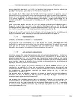
Case 1
A 67-year-old man with a history of diabetes and hypertension presented
to the ED with a 2-day history of central abdominal pain migrating to the
bilateral lower quadrants, nausea, and constipation (Fig. 25.1). In the emer-
gency department he exhibited abdominal tenderness, leukocytosis, and
neutrophilia.
A CT scan with intravenous and oral contrast demonstrated an enlarged
appendix (11mm in diameter) with associated periappendicular fat strand-
ing. Following the positive CT examination, the probability of confirmed
appendicitis (positive predictive value) rises to 95%, as opposed to the 48%
probability found in those who are referred for imaging. The diagnosis of
appendicitis was confirmed with pathologic examination of the vermiform
appendix removed at surgery.
470 C.C. Blackmore et al.
Table 25.4. Sensitivity and specificity of imaging in patients with
suspected acute colonic diverticulitis
Positive Negative
Sensitivity Specificity predictive predictive
Modality (%) (%) value (%)
1
value (%)
1
Contrast enema
2
81 85 84 82
Ultrasound
3
91 92 92 91
CT
4
89 99 99 90
1
Calculated utilizing a prevalence of diverticulitis of 50%, a prevalence equal to the pooled
prevalence of the eligible studies.
2
Adapted from references 91 and 92.
3
Adapted from references 95, 96, 98, and 100.
4
Adapted from references 91–94.
Figure 25.1. A: Enlarged appendix in sagittal plane. B: Enlarged appendix in transverse plane.
A
B
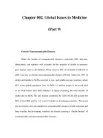
Case 2
A 70-year-old woman presented to the ED with a 2-day history of abdom-
inal pain, nausea, and vomiting. The patient has a history of abdominal
surgeries, including repair of an anterior abdominal wall hernia (Fig. 25.2).
An abdominal and pelvic CT examination with intravenous and oral
contrast revealed multiple dilated loops of jejunum with decompressed
ileum distally. There was no evidence of bowel wall ischemia on the exam-
ination. The patient underwent surgical decompression of small bowel
obstruction and recovered without complication.
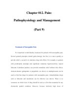
Case 3
A 39-year-old woman presented to the ED with a 3-day history of left lower
quadrant abdominal pain, fevers, chills, and vomiting, as well as leukocy-
tosis. The studies in this chapter suggest a clinical suspicion of divertic-
ulitis, as in this case, is accurate approximately 50% of the time (Fig. 25.3).
Chapter 25 Imaging in Acute Abdominal Pain 471
Figure 25.2. Small bowel obstruction.
Figure 25.3. Diverticulitis with abscess
formation.
Computed tomography revealed multiple diverticula and bowel wall
thickening in the sigmoid colon, with fat stranding in the mesocolon, and
an extraperitoneal abscess. Under CT guidance a percutaneous drainage
catheter was placed into the abscess, with subsequent aspiration of 40cc of
purulent material. The patient recovered and was discharged 72 hours after
drainage catheter placement.
Suggested Protocols
Appendicitis and Bowel Obstruction Protocol
Patient preparation: 1000mL oral contrast, drink over a 90-minute period.
Give rectal contrast if patient is unable to tolerate oral contrast.
Intravenous (IV) contrast: 150cc at 3.0 cc/second.
Imaging: venous phase (60-second scan delay), dome of the diaphragm to
ischial tuberosities, 2.5-mm detector collimation.
Diverticulitis Protocol
Patient preparation: 1000 to 1500mL rectal contrast instilled via soft rectal
tube.
IV contrast: 150cc at 3.0 cc/second.
Imaging: venous phase (60-second scan delay), dome of the diaphragm to
ischial tuberosities, 2.5-mm detector collimation.
References
1. Tsushima Y, et al. Clin Radiol 2002;57(6):507–513.
2. Urban BA, Fishman EK. Radiographics 2000;20(3):725–749.
3. Ann Emerg Med 1994;23(4):906–922.
4. Martin RF, Rossi RL. Surg Clin North Am 1997;77(6):1227–1243.
5. Adams ID, et al. Br Med J (Clin Res Ed) 1986;293(6550):800–804.
6. Contran R, Kumar V, Collins T, Robbins S. Robbins Pathologic Basis of Disease,
6th ed. Boston: WB Saunders, 1999:1425.
7. Goldman L. Cecil Textbook of Medicine, 21st ed. Philadelphia: WB Saunders,
2000:729–731.
8. Frank JL, et al. Surg Endosc 1998;12(3):274 –275.
9. Townisend CM. Sabiston Textbook of Surgery, 16th ed. 919–920.
10. Megibow AJ, et al. Radiology 1991;180(2):313–318.
11. Shih SC, et al. World J Gastroenterol 2003;9(3):603–605.
12. Miller G, et al. Br J Surg 2000;87(9):1240–1247.
13. Fevang BT, et al. Ann Surg 2004;240(2):193–201.
14. Parks TG, et al. Br Med J 1970;2(702):136–138.
15. Ferzoco LB, Raptopoulos V, Silen W. N Engl J Med 1998;338(21):1521–1526.
16. Diner WC, Barnhard HJ. Semin Roentgenol 1973;8(4):415–431.
17. Burkitt DP, Walker AR, Painter NS. JAMA 1974;229(8):1068–1074.
18. Hinchey EJ, Schaal PG, Richards GK. Adv Surg 1978;12:85–109.
19. McCaig LF, Stussman BJ. Adv Data 2002;1–20.
20. Advanced data from Vital and Health Statistics. Hyattsville, MD: National
Center for Health Statistics, 2004:340.
21. Powers RD, Guertler AT. Am J Emerg Med 1995;13(3):301–303.
22. Brewer BJ, et al. Am J Surg 1976;131(2):219–223.
23. Lukens TW, Emerman C, Effron D. Ann Emerg Med 1993;22(4):690–696.
24. Jess P, et al. Am J Surg 1982;144(3):338–340.
472 C.C. Blackmore et al.
25. Ann Emerg Med 2000;36(4):406–415.
26. Addiss DG, et al. Am J Epidemiol 1990;132(5):910–925.
27. Al-Omran M, Mamdani M, McLeod RS. Can J Surg 2003;46(4):263–268.
28. Korner H, et al. World J Surg 1997;21(3):313–317.
29. HCUPnet 2002 Statistics [Internet]. Rockville, MD: Agency for Healthcare
Research and Quality. [Cited 5/20/05]. Available from: />HCUPnet.asp
30. Tito SSM. Intestinal obstruction. In: Zuidema GD, ed. Surgery of the Alimen-
tary Tract, 5th ed. New York: WB Saunders, 2001.
31. Pradel JA, et al. Radiology 1997;205(2):503–512.
32. Kircher MF, et al. AJR 2002;178(6):1313–1318.
33. Rhea JT. Emerg Radiol 2000;7(4):237–244.
34. Brengman ML, Otchy DP. Dis Colon Rectum 1998;41(8):1023–1028.
35. Halligan S, Saunders B. Best Pract Res Clin Gastroenterol 2002;16(4):595–610.
36. Gresenz CR, Studdert DM. Ann Emerg Med 2004;43(2):155–162.
37. Flum DR, Koepsell T. Arch Surg 2002;137(7):799–804; discussion 804.
38. Ogata M, Mateer JR, Condon RE. Ann Surg 1996;223(3):237–241.
39. Terasawa T, et al. Ann Intern Med 2004;141:537–546.
40. Whiting P, et al. Ann Intern Med 2004;3(140):189–202.
41. Rao PM, et al. Ann Surg 1999;229(3):344–349.
42. Brandt MM, Wahl WL. Am Surg 2003;69(9):727–731.
43. Horton M, Counter SF, Florence M, Hart M. Am J Surg 2000;179(5):379–381.
44. Weyant MJ, et al. Surgery 2000;128(2):145–152.
45. Balthazar EJ, Rofsky NM, Zucker R. Am J Gastroenterol 1998;93(5):768–771.
46. Franke C, et al. World J Surg 1999;23(2):141–146.
47. Perez J, et al. Am J Surg 2003;185(3):194–197.
48. Lee SL, Walsh AJ, Ho HS. Arch Surg 2001;136(5):556–562.
49. Flum DR, et al. JAMA 2001;286(14):1748–1753.
50. Rao PM, et al. N Engl J Med 1998;338(3):141–146.
51. Rice HE, et al. J Pediatr Surg 1999;34(5):754–758; discussion 758–759.
52. Baldisserotto M, Marchiori E. AJR 2000;175(5):1387–1392.
53. Lessin MS, et al. Am J Surg 1999;177(3):193–196.
54. Hahn HB, et al. Pediatr Radiol 1998;28(3):127–151.
55. Vignault F, et al. Radiology 1990;176(2):501–504.
56. Sivit CJ, et al. Radiology 1992;182(3):723–726.
57. Quillin SP, Siegel MJ. Radiology 1994;191(2):557–560.
58. Schulte B, et al. Eur J Ultrasound 1998;8(3):177–182.
59. Kaiser S, Frenckner B, Jorulf HK. Radiology 2002;223(3):633–638.
60. Lowe LH, et al. Radiology 2001;221(3):755–759.
61. Mullins ME, et al. AJR 2001;176(1):37–41.
62. Teo EL, et al. Singapore Med J 2000;41(8):387–392.
63. Garcia Pena BM, et al. JAMA 1999;282(11):1041–1046.
64. Ramachandran P, et al. J Pediatr Surg 1996;31(1):164–167; discussion 167–169.
65. Pena BM, et al. Pediatrics 2000;106(4):672–676.
66. Applegate KE, et al. Radiology 2001;220(1):103–107.
67. Karakas SP, et al. Pediatr Radiol 2000;30(2):94–98.
68. Martin AE, et al. J Pediatr Surg 2004;39(6):886–890; discussion 886–890.
69. Partrick DA, et al. J Pediatr Surg 2003;38(5):659–662.
70. Bendeck SE, et al. Radiology 2002;225(1):131–136.
71. Suri S, et al. Acta Radiol 1999;40:422–428.
72. Matsuoka H, et al. Am J Surg 2002;183(6):614–617(6):614–617.
73. Frager D, et al. AJR 1994;162:37–41.
74. Czechowski J. Acta Radiol 1996;37:186–189.
75. Schmutz GR, et al. Eur Radiol 1997;7(7):1054–1058.
76. Frager D, et al. AJR 1996;166:67–71.
77. Balthazar EJ, Liebeskind ME, Macari M. Radiology 1997;205:519–522.
78. Obuz F, et al. Eur J Radiol 2003;48:299–304.
Chapter 25 Imaging in Acute Abdominal Pain 473
79. Peck JJ, Milleson T, Phelan J. Am J Surg 1999;177:375–378.
80. Beall DP, et al. Clin Radiol 2002;57(8):719–724.
81. Seror D, et al. Am J Surg 1993;165(1):121–125; discussion 125–126.
82. Fevang BT, et al. Eur J Surg 2002;168(8–9):475–481.
83. Cox MR, et al. Aust N Z J Surg 1993;63(11):848–852.
84. Deutsch AA, et al. Postgrad Med J 1989;65(765):463–467.
85. Sarr MG, Bulkley GB, Zuidema GD. Am J Surg 1983;145(1):176–182.
86. Balthazar EJ. AJR 1994;163(5):1260–1261.
87. Ha HK, et al. Radiology 1997;204(2):507–512.
88. Zalcman M, et al. AJR 2000;175:1601–1607.
89. Donckier V, et al. Br J Surg 1998;85:1071–1074.
90. Taourel PG, et al. AJR 1995;165:1187–1192.
91. Cho KC, et al. Radiology 1990;176(1):111–115.
92. Stefansson T, et al. Acta Radiol 1997;38(2):313–319.
93. Rao PM, Rhea JT. Radiology 1998;209(3):775–779.
94. Werner A, et al. Eur Radiol 2003;13(12):2596–2603.
95. Verbanck J, et al. J Clin Ultrasound 1989;17(9):661–666.
96. Zielke A, et al. Br J Surg 1997;84(3):385–388.
97. Hollerweger A, et al. AJR 2000;175(4):1155–1160.
98. Schwerk WB, Schwarz S, Rothmund M. Dis Colon Rectum 1992;35(11):
1077–1084.
99. Ambrosetti P, Becker C, Terrier F. Eur Radiol 2002;12:1145–1149.
100. Hollerweger A, et al. Eur Radiol 2001;11(10):1956–1963.
474 C.C. Blackmore et al.
26
Intussusception in Children:
Diagnostic Imaging and Treatment
Kimberly E. Applegate
I. Diagnosis of intussusception: what are the clinical predictors? who
should undergo imaging?
A. What are the clinical predictors of intussusception?
B. What are the clinical predictors of reducibility and bowel
necrosis?
II. Which imaging should be performed?
A. What is the diagnostic performance of abdominal radiographs?
B. What is the diagnostic performance of sonography?
C. What are the sonographic predictors of reducibility and bowel
necrosis?
D. What are the pathologic lead points?
III. Treatment of intussusception: how should the enema be
performed?
A. Air vs. liquid enema
B. Rule of threes
C. Radiation dose
D. Alternative enema approaches
E. Fluoroscopy vs. sonography
F. Delayed repeat enema
G. Where should patients be treated?
H. What are the complications of enema therapy?
I. What are the surgical management and complications?
J. Cost-effectiveness analysis
IV. What is appropriate management in recurrent cases?
V. Special case: intussusception limited to the small bowel
VI. Special case: intussusception with a known lead point mass
475
Children with clinically suspected intussusception should undergo
enema reduction after surgical consultation. The only absolute con-
traindications to enema are signs of peritonitis on clinical exam or free
air on abdominal radiographs. Air enema has better overall reduction
Issues
Key Points
Definition and Pathophysiology
Intussusception is an acquired invagination of the bowel into itself, usually
involving both small and large bowel, within the peritoneal cavity. The
more proximal bowel that herniates into the more distal bowel is called the
intussusceptum and bowel that contains it is called the intussuscipiens. It
is an emergent condition where delay in diagnosis is not uncommon, and
leads to an increased risk of bowel perforation, obstruction, and necrosis.
There may be an accompanying pathologic lead point mass in approxi-
mately 5% of children (1). Intestinal intussusception may occur along the
entire length of the bowel from the duodenum to prolapse of intussus-
cepted bowel through the rectum. It can also range from classic clinical pre-
sentations to asymptomatic transient intussusception seen increasingly on
multichannel computed tomography (CT) studies of the abdomen for other
indications (2,3). Most cases are idiopathic in that the etiology of the intus-
susception is due to hypertrophied lymphoid tissue in the terminal ileum,
which results in ileocolic intussusception. Some reports have suggested a
viral etiology, most commonly adenovirus but also enterovirus, echovirus,
and human herpes virus 6 (4). The clinical signs and symptoms of intus-
susception are often nonspecific and overlap with those of gastroenteritis,
malrotation with volvulus, and, in older children, Henoch-Schönlein
purpura (HSP). The large majority of clinically symptomatic cases occur in
the infant and toddler, with a peak age of 5 to 9 months, although it has
been reported on prenatal imaging and may occur in children who present
without the typical clinical presentation of vomiting, bloody stools, palpa-
ble abdominal mass, and colicky abdominal pain (5). The classic triad of
colicky abdominal pain, vomiting, and bloody stools is present in only 7%
to 20% of children (6–8).
476 K.E. Applegate
rates than liquid enema but the outcome depends on the experience
of the radiologist (moderate evidence).
Barium should not be used due to the poorer outcomes in those chil-
dren who perforate (moderate evidence).
Ultrasound (US) is the primary imaging modality for initial diagno-
sis outside of the United States and for a growing majority of pedi-
atric radiologists in the U.S. Ultrasound also plays a role in the
evaluation of reducibility of intussusception, the presence of a lead
point mass, potential incomplete reduction after enema, and of intus-
susception limited to small bowel (limited evidence).
Abdominal radiographs have poor sensitivity for the detection of
intussusception but may serve to screen for other diagnoses in the dif-
ferential diagnosis, such as constipation, and for free peritoneal air.
For screening children with a low probability for intussusception,
sonography is the preferred screening test (limited evidence).
The use of delayed repeat enema for the reduction of intussusception
shows promise, but there are few data on the appropriate methods or
timing (limited evidence).
For recurrence of intussusception, including multiple recurrences,
enema is the preferred method for reduction (limited evidence).
Epidemiology
Intussusception is the most common cause of small bowel obstruction in
children and occurs in at least 56 children per 100,000 per year in the U.S.
(9). It is second only to pyloric stenosis as the most common cause of gas-
trointestinal tract obstruction in children. It occurs in boys more than girls
at a ratio of 3: 2. Some studies have reported associations with viruses, par-
ticularly adenovirus, although the lack of seasonality suggests more than
one pathogen (9). Intussusception occurs most commonly in infants
beyond the newborn period, with large series reporting 57% to 85% of cases
occurring before the age of 1 year (average 67% occur by age 1 year) (5).
Delay in diagnosis and treatment is not uncommon, making enema reduc-
tion less successful, bowel resection more likely, and death due to bowel
ischemia possible (1,5,10,11). There were 323 intussusception-associated
deaths in American infants reported to the Centers for Disease Control and
Prevention (CDC) between 1979 and 1997. In a review of administrative
discharge data of intussusception-associated hospitalizations and deaths
in the U.S., Parashar and colleagues (9) noted a peak age of 5 to 7 months
with two thirds of patients under age 1 year, no consistent seasonality, hos-
pitalization rates of approximately 56 per 100,000 children, and a general
trend toward fewer hospitalizations over the past two decades. The mor-
tality rates also decreased over this time period, from 6.4 per 1,000,000 to
2.3 per 1,000,000 live births. The authors also reported an increased risk of
intussusception-related deaths among infants whose mothers were <20
years old, unmarried, nonwhite, and had less than a grade 12 education.
The authors concluded that these data suggest that reduced access or delay
in seeking care contributed to the risk of death. They did not investigate
costs or rates of surgical versus enema reductions.
In another study comparing worldwide data, Meier and colleagues (11)
noted that the most important difference between industrialized and
developing countries’ outcomes was the delay in presentation for treat-
ment and consequent lower rates of enema reduction and higher rates of
surgical mortality (18%) from bowel necrosis.
Rotavirus Vaccine
Shortly after the first and only rotavirus vaccine was introduced in the U.S.
in 1998 for routine vaccination of infants at ages 2, 4, and 6 months, several
reports to the CDC suggested an association between the vaccine and
intussusception. This was noted particularly within 2 weeks after vaccina-
tion with the first dose. The vaccine was removed from the world market
in 1999 (12). Although controversial, subsequent investigations have not
found a higher rate of intussusception after rotavirus vaccination (13,14).
A new rotavirus vaccine is currently under development (15).
Overall Cost to Society
No data have been identified detailing the total cost to society of intus-
susception. Three recent surveys have documented practice patterns for
the evaluation of intussusception (3,16,17). In centers without pediatric
radiologists, the enema is the initial and often only imaging test performed
for both diagnosis and treatment. In contrast, at the 2004 Society for Pedi-
atric Radiology (SPR) annual meeting, a survey of pediatric radiologists
Chapter 26 Intussusception in Children 477
showed that 57% now use sonography for initial diagnosis prior to enema
(16). Overall, the total hospital cost for children with intussusception
treated with surgery is approximately four times that of those treated with
enema (18–20).
Goals
The goal of initial bowel imaging is early detection of intussusception to
enable enema reduction of the intussusception. Additional imaging studies
may be performed to further characterize indeterminate results. The ulti-
mate goal that radiologists should strive for is nonoperative reduction for
all children with idiopathic intussusception (approximately 95% cases).
Methodology
A Medline search was performed using PubMed (National Library of Med-
icine, Bethesda, Maryland) for original research publications discussing the
diagnostic performance and effectiveness of imaging strategies in intus-
susception. Clinical predictors of intussusception were also included in the
literature search. The search covered the period 1966 to June 2004. The
search strategy employed different combinations of the following terms:
(1) intussusception, (2) children, ages under 18 years, (3) diagnosis, and (4)
therapy or surgery or etiology. Additional articles were identified by review-
ing the reference lists of relevant papers, identifying appropriate authors,
and use of citation indices for MeSH terms. This review was limited to
human studies and the English-language literature. The author performed
an initial review of the titles and abstracts of the identified articles followed
by review of the full text in articles that were relevant.
I. Diagnosis of Intussusception: What Are the Clinical
Predictors? Who Should Undergo Imaging?
Summary of Evidence: At this point there are no reliable clinical prediction
models that can accurately identify all patients with intussusception
(limited evidence). Determination of which children should undergo
imaging, and which should not undergo imaging, has not been studied in
formal prospective trials.
Supporting Evidence
A. What Are the Clinical Predictors of Intussusception?
Ideally, children with intussusception should be diagnosed early to avoid
bowel necrosis and surgery. Yet this goal remains elusive. One report found
that only 50% of children were correctly diagnosed at initial presentation to
a health care provider (20). The classic triad of colicky abdominal pain (58%
to 100% cases), vomiting (up to 85% cases), and bloody stools is present in
only 7% to 20% of children (6,8,22). Guaiac-positive stool is present in 75%
of children with intussusception (8,23). Kuppermann and colleagues (24)
published a cross-sectional study that evaluated the clinical factors that
might predict intussusception in 115 children (limited evidence). Using mul-
tivariate logistic regression and bootstrap sample analysis, they found that
478 K.E. Applegate
the presence of highly suggestive abdominal radiographs, rectal bleeding,
and male sex were independent predictors of intussusception but also noted
that these factors were not specific. Harrington and colleagues (6) investi-
gated the positive and negative clinical predictors of intussusception in a
prospective cohort study (moderate evidence). They recorded signs and
symptoms in 245 children and correlated them with sonographic and enema
findings. Significant positive predictive factors for intussusception were the
presence of right upper quadrant mass, gross blood in stool, guaiac-positive
stool, and the triad of colicky abdominal pain, vomiting, and right upper
quadrant mass. They were unable to identify significant negative predic-
tors. Klein and colleagues (25) reviewed clinical history, physical exam, and
radiographic findings to develop a prediction model of children with pos-
sible intussusception (moderate evidence). Their univariate analysis iden-
tified several known factors associated with intussusception, including
vomiting, abdominal pain, palpable abdominal mass, guaiac-positive stool,
and rectal bleeding. However, they concluded that they were “unable to
develop a prediction model that would reliably identify all patients with the
diagnosis of intussusception. Previously identified predictors of intussus-
ception remain important in increasing suspicion of this important diagno-
sis. At this point there is no reliable prediction model that can accurately
identify all patients with intussusception.”
B. What Are the Clinical Predictors of Reducibility and
Bowel Necrosis?
The most important factor that decreases the reduction rate of enema is a
longer duration of symptoms. This finding is supported by multiple case
series. A significant delay is typically 48 hours, but some reports suggest
24 or 72 hours, as either one of several factors or the single factor predict-
ing unsuccessful enema reduction (5,26). Other factors associated with
lower reduction rates include age less than 3 months, dehydration, small
bowel obstruction, and intussusception encountered in the rectum (25%
reduction rate) (3,21,22,26,27) (limited evidence).
II. Which Imaging Should Be Performed?
Summary of Evidence: Ultrasound has higher accuracy in the diagnosis
of intussusception than plain radiographs. Ultrasound also has higher
diagnostic accuracy in identifying pathologic lead points than plain radi-
ographs or enema. The role of ultrasound findings in predicting success of
reduction is not well known with available literature. Given current evi-
dence, the diagnostic approach should include (1) abdominal radiographs
if concern for other diagnoses or for perforation; (2) sonography for diag-
nosis or exclusion of intussusception; (3) if positive, a surgical consult
should be obtained prior to the enema reduction attempt; and (4) air enema
reduction (or if no experience with the air technique, liquid enema) (mod-
erate evidence).
Supporting Evidence
A. What Is the Diagnostic Performance of Abdominal Radiographs?
The presence of a curvilinear mass within the course of the colon (the
crescent sign), particularly in the transverse colon just beyond the hepatic
Chapter 26 Intussusception in Children 479
flexure, is a nearly pathognomonic sign of intussusception. The absence of
bowel gas in the ascending colon is one of the most specific sign of intus-
susception on radiographs (28). However, small bowel or sigmoid colon
gas located in the right abdomen on radiographs may mimic ascending
colon or cecal gas. Radiographs have low sensitivity and specificity, even
when viewed by experienced pediatric radiologists (28,29) (limited evi-
dence). Sargent and colleagues (27) reported a 45% sensitivity in 60 chil-
dren when evaluated prospectively by pediatric radiologists, using the
enema as the reference standard (Table 26.1). Others report similar poor
sensitivity in the detection of intussusception (5). In a survey of the SPR
2004 attendees, Daneman (16) found that 79% obtain radiographs, but this
practice may not be under the control of radiologists. Only 10% of pedi-
atric radiologists in this survey preferred radiographs for the diagnosis.
B. What Is the Diagnostic Performance of Sonography?
Intussusception can be reliably diagnosed when a donut, target, or
pseudokidney sign is seen using linear transducer sonography (30–33). The
optimal US technique in this population is well described (31–35). There
are no known contraindications or complications resulting from US for this
purpose. Ultrasound also plays a role in the evaluation of reducibility of
the intussusception, the presence of a pathologic lead point (PLP) mass,
and intussusception limited to small bowel, in diagnosing or excluding
residual intussusception after enema, and in identifying alternative
diagnoses (6,32,34,35) (limited evidence). In a 2004 survey, 57% of North
American pediatric radiologists reported the use of sonography to diag-
nose intussusception as compared to 93% of European pediatric radiolo-
gists in a 1999 survey (16,36).
Sonography screening in children has been suggested to reduce cost,
radiation exposure, and both patient and parental anxiety/discomfort with
enema (35) (limited evidence). Published series from single institutions
suggest high accuracy, approaching 100% in experienced hands, with sen-
sitivity of 98% to 100% and specificity of 88% to 100% (6,32,37,38) (limited
evidence) (Table 26.1). Eshed and colleagues (39) found similar abilities in
sonographic diagnosis of intussusception for staff radiologists as well as
senior and junior radiology residents: sensitivity and specificity were 85%
and 98% for staff radiologists, 75% and 96% for senior residents, and 83%
and 97% for junior residents, respectively. Given that the theoretical cost-
effectiveness of sonography is dependent on the prevalence of intussus-
ception, optimization of imaging will require stratification of subjects into
different levels of probability of intussusception (40). However, data are
lacking for such stratification. Henrikson and colleagues (35) noted a trend
480 K.E. Applegate
Table 26.1. Summary of sensitivity and specificity of diagnostic imaging
for intussusception
Test Sensitivity (%) Specificity (%)
Abdominal radiographs
1
45 45
Ultasound
2
98–100 88–100
Enema
3
100 100
1
Based on references 5 and 28.
2
Based on references 6, 32, 37 and 38.
3
Approximate levels for ileocolic intussusception (does not include intussusception limited to
small bowel) (26).
of decreased prevalence of intussusception (22%) in those children referred
for enema and began sonographic screening (limited evidence). In their
small series of 38 children, they were able to avoid 19 enemas in those with
negative sonography, resulting in savings in both radiation exposure (an
average of 8.2mGy for negative enemas) and hospital charges. Future cost-
effectiveness modeling research is needed to define the population that
should undergo sonography.
C. What Are the Sonographic Predictors of Reducibility
and Bowel Necrosis?
Del-Pozo and colleagues (41) performed sonography in 145 children with
intussusception and found that fluid seen inside the intussusception rep-
resented trapped peritoneal fluid and was associated with significantly
fewer reductions on enema and bowel ischemia at surgery (42) (limited
evidence).
Some US reports have noted that thicker bowel wall was associated with
fewer enema reductions (32,43) but others did not find this association (42).
Lack of color Doppler signal in the intussuscepted bowel wall suggested
bowel ischemia in several small series (44–46). Free intraperitoneal fluid in
small or moderate amounts is present in approximately half of children
with intussusception and is not a contraindication for enema (33). There
are conflicting reports that free peritoneal fluid is associated with fewer
reductions (5,22,26,34,47). Some descriptive studies report that the pres-
ence of lymph nodes trapped in the intussusception is associated with
fewer reductions (34,48). For these US findings, due to the conflicting
reports or small series, the evidence is inconclusive.
D. What Are the Pathologic Lead Points?
Approximately 5% to 6% of intussusceptions in children are caused by
PLPs, which are due to either focal masses or diffuse bowel wall abnor-
mality. The most common focal PLPs are (in decreasing order of incidence)
Meckel’s diverticulum, duplication cyst, polyp, and lymphoma (1,5,49)
(limited evidence). Diffuse PLPs are most commonly associated with cystic
fibrosis or HSP. Although the common teaching remains that focal PLPs
are more common in older children, this is somewhat misleading. The rel-
ative prevalence of PLP with intussusception is higher in children over the
age of 3 years, particularly for lymphoma. However, the absolute number
of PLPs in infants versus older children is approximately equal (1).
The detection of lead points by imaging remains problematic, although
US is the noninvasive standard of reference 66% of PLPs may be identified
on US (50) and 40% of PLPs may be diagnosed by liquid enema (5). Air
enema has a lower rate of detection of PLP of 11%–29% (50,51), so that
some researchers suggest that US be used afterward to search for PLP (3)
(limited evidence).
III. Treatment of Intussusception: How Should the Enema
Be Performed?
Summary of Evidence: The air enema is considered superior at reduction,
cleaner (based on appearance of peritoneal cavity at surgery when perfo-
ration occurs), safer, and faster, with less radiation when compared to
Chapter 26 Intussusception in Children 481
liquid enema (23,52–56) (moderate evidence). The recurrence rates for air
versus liquid enema reductions do not differ (both are approximately 10%).
The rule of threes that is used to guide liquid enema technique is supported
by limited evidence. Barium is no longer the liquid contrast medium of
choice due to the risk of barium peritonitis, infection, and adhesions when
perforation occurs during the enema (23,47,53,57). Neither sedation nor
medications increase the enema success rate (limited evidence). Direct
comparison of reduction with fluoroscopy versus ultrasound has not been
studied (insufficient evidence).
Supporting Evidence: There are multiple investigations of success rates for
enema reduction, although most are retrospective. Seventy-one published
studies of this question were largely level III (limited evidence) investiga-
tions consisting of unselected but often consecutive case series. The
average reduction rate for these 71 published studies was 74%. In 19 series
with at least 150 children each, retrospective analysis demonstrated reduc-
tion rates averaging 80%, range 53% to 96% (26) (Table 26.2). The two
largest series from China, using air enema in 6396 and 9028 children,
reported reduction rates of 95% and 92% (54,55) (limited evidence).
However, while the air enema may be preferred in experienced hands, the
liquid enema is also safe and effective. The air enema technique is well
described in the literature (54,56,58). Briefly, the enema tip should be
placed within the child’s rectum and taped in place with abundant tape.
The child is placed in a prone position to allow the radiologist or assistant
to squeeze the buttocks closed and prevent air from leaking. Air is rapidly
insufflated into the colon under fluoroscopic observation. Once the intus-
susception is encountered, its reduction is followed fluoroscopically until
it is completely reduced. Air should flow freely from the cecum into the
distal small bowel loops to signify complete reduction. One critical safety
issue is to keep air pressure below a maximum limit of 120mm Hg
(although higher pressures occur when the patient performs a Valsalva
maneuver) to avoid the risk of perforation (23,47,56).
A. Air vs. Liquid Enema
There are only two randomized controlled trials of the reduction rates of
air versus liquid enema (60) (moderate evidence). The 1993 study by Meyer
et al. enrolled 101 children and found similar success rates of 76% for air
and 63% for liquid enema. However, the trial used sedation and had lower
reduction rates than those not using sedation (25). The authors abandoned
482 K.E. Applegate
Table 26.2. Summary of published intussusception enema reduction rates and perforation rates
All studies Studies with cases >150
No. of Wt. mean No. of Wt. mean
Rates studies Mean (SD) (SD) studies Mean (SD) (SD)
Reduction (%) 71 74.1 (16.8) 87.3 (12) 19 79.6 (12.5) 89.5 (9.3)
Perforation (%) 66 0.8 (1.4) 0.3 (0.7) 18 0.6 (0.8) 0.2 (0.4)
Note: Summary data include a weighted average measure of reduction and perforation rates based on publications with at
least 150 pediatric cases. The enema techniques varied and included air versus liquid media, with sonographic or fluoro-
scopic guidance.
Wt. mean, weighted mean; SD, standard deviation.
Source: Adapted from Daneman and Navarro (26).
the use of sedation after this study. The use of sedation may reduce the
intraabdominal pressure children create by the Valsalva maneuver and is
reported to improve reducibility at enema (47,56). In contrast, Hadidi et al.
found significantly higher reduction rates using air (90%) versus barium
(70%) in 100 children (61). More recent reports of air reduction show better
results than liquid enema reduction (26). The superior air enema results
may be due to the level of experience of those who use air reduction
techniques as well as the presence of higher intraluminal pressure for air
as compared to standard hydrostatic reduction (62,63).
In a 1991 survey of American pediatric radiology chairs, Meyer et al. (17)
found that only 24% were using air enema but 64% used barium and 12%
water-soluble contrast, as compared to 35% of international pediatric radi-
ologists who used air enema (59). More recently, 65% of American pedi-
atric radiologists now use air enema, 33% use liquid enema (water-soluble
contrast or barium), and 3% use liquid enema with sonographic guidance
(16). Some pediatric radiologists use air for children older than 3 months,
but for younger infants, especially neonates, they prefer liquid contrast due
to the greater differential diagnosis in this group (26).
All children should have a surgical consultation prior to enema (1) to
assess for peritoneal signs precluding enema, (2) to identify children who
cannot be reduced with enema or who are found to have perforation,
and (3) for postreduction management. Prior to enema reduction, dehydra-
tion should be treated with intravenous fluid resuscitation. Children with
evidence of peritonitis, shock, sepsis, or free air on abdominal radiographs
are not candidates for enema. Radiologists should achieve enema reduction
rates of at least 80% and up to 95% (moderate evidence). Several reports esti-
mate that the rate of spontaneous reduction based on sonographic or enema
diagnosis prior to surgery is 10% (1,3,22,43) (limited evidence).
Bratton and colleagues (18) suggest that more experienced radiologists
and caregivers at children’s hospitals decrease the risk of surgical reduc-
tion, length of hospital stay, and cost of care (moderate evidence). Surgical
management is performed when the patient is too unstable (shock, dehy-
dration, sepsis) for enema reduction, when the enema is unsuccessful, or
when PLP is diagnosed.
B. The Rule of Threes
A general guideline to the liquid enema technique, often taught to radiol-
ogy residents, is the rule of threes: three attempts of 3 minutes’ duration,
with the liquid enema bag at 3 feet above the fluoroscopy table. There is
little evidence to support this rule, particularly regarding the height of the
enema bag (26,64). Many experienced pediatric radiologists alter this
general guide in response to the clinical status of the patient and the move-
ment of the intussusceptum mass achieved with the initial enema (22,64).
For example, if the intussusception is partially reduced to where it most
frequently hangs up, at the ileocecal valve, some radiologists will make
further or longer attempts or raise the enema bag above 3 feet. The exam
is tailored to the patient and performed in conjunction with the surgeon
involved.
C. Radiation Dose
The dose deposited depends on a number of factors, including the type of
fluoroscopy equipment, the use of pulsed fluoroscopy, and the fluoroscopy
Chapter 26 Intussusception in Children 483
time (1,47). A 1993 study reported a mean effective dose of 55mSv for enema
reduction of an intussusception (65). Experienced pediatric radiologists
using air enema averaged 95 seconds of fluoroscopy time to reduce an
intussusception and 42 seconds to exclude one in a child without intus-
susception (56). Air enema radiation doses average one-third to one-half
less than the dose for liquid enema (47). A 2003 report showed the average
radiation dose saved was estimated at 8.2mSv (820 mR) (the average dose
for negative enema) per patient (35).
D. Alternative Enema Approaches
A number of different approaches have been described to try to improve
intussusception reduction on enema that include sedation, anesthesia, use
of glucagon, manual palpation, and delayed repeat enema. In the past,
sedation, and sometimes anesthesia, were commonly used to improve
reduction rates, but case series showed no improvement (17,66,67) (limited
evidence). In a 1991 survey Meyer (17) found only 10% of respondents used
sedation either always or almost always, as compared to 54% of interna-
tional pediatric radiologists, and those using sedation reported lower
reduction rates (59). Therefore, few pediatric radiologists currently use
sedation in the U.S. Glucagon was shown not to improve enema reduction
rates in one study (68) and is no longer used (17). The use of manual pal-
pation has been suggested to improve intussusception reduction at enema
but has not been systematically studied (47,69). One study by Grasso et al.
(69) reported a reduction rate of 76% when manual palpation was used,
less than the average of 80% in large series.
E. Fluoroscopy vs. Sonography
In the West, fluoroscopy is almost always used during enema reduction.
In the East, and in a few European centers, reports on the use of sonogra-
phy with either water (70–76) or air (77–79) reduction show reduction rates
as high as or higher than those in the West; however, the experience level
required for these techniques has not been studied nor has the ability of
sonography to detect perforations (limited evidence).
F. Delayed Repeat Enema
In the small percent of children who fail initial enema reduction, delayed
repeat enema may avoid the need for surgical reduction. The use of
delayed attempts at between 30 minutes and 19 hours after initial attempt
have shown promise in increasing the success of enema reductions (80–84)
(limited evidence). These four small series showed further reduction rates
of 50% to 82% by waiting at least 30 minutes prior to further attempts at
enema reduction. Further research to understand optimal timing and tech-
nique for delayed repeat enemas is needed. Daneman and Navarro (26),
with the largest reported experience to date, suggest a delay of 2 to 4 hours
until further research yields more rigorous guidelines. The child must
remain clinically stable and be appropriately monitored during this time
interval. Delayed enema should not be performed if the initial enema does
not move the intussusception at all (26,83).
484 K.E. Applegate
G. Where Should Patients Be Treated?
Bratton and colleagues (18) performed a retrospective cohort analysis of all
children hospitalized with intussusception in the state of Washington from
1987 through 1996 (moderate evidence). They investigated whether the risk
of surgical management for these children varied by hospital pediatric
caseload, measured by the annual number of pediatric hospital admis-
sions. By reviewing the discharge data of all 507 children, they found an
overall surgical reduction rate of 53%, with 20% undergoing bowel resec-
tion. Surgical reduction rates varied by pediatric caseload from 36% at hos-
pitals with large pediatric caseloads to 64% at hospitals with low pediatric
volumes, resulting in nearly twice the risk of surgical reduction. Children
who underwent surgery, versus enema reduction, had similar gender and
median age characteristics, but those who had bowel resection were more
likely to have coexisting conditions. The median cost of hospital care
for these children was $5724 for surgical reduction and $1184 for enema
reduction.
H. What Are the Complications of Enema Therapy?
The most important potential complication of enema is bowel perforation.
Sixty-six published studies of this question were largely level III (limited
evidence) investigations consisting of unselected but often consecutive
case series. The mean perforation rate was 0.8% (Table 26.2). In 18 case
series with at least 150 children each, perforation rates averaged 0.6%, with
a range of 0% to 1.6% (26). There were no statistically significant differ-
ences between air and liquid enema perforation rates (Table 26.3). When
these averages were weighted to reflect the sample size of each published
study, the perforation rates were even lower, at 0.3% for all 66 studies and
0.2% for the larger studies.
Ultimately, however, the risk of perforation depends on each radiolo-
gist’s patient population and technique. Though determination of clinical
predictors of perforation is complicated by a lack of prospective studies,
the one acknowledged key factor is symptom length greater than 48 hours.
Several reports in both pig models and children suggest that there may be
Chapter 26 Intussusception in Children 485
Table 26.3. Summary comparison of air versus liquid contrast enema reduction and perfora-
tion rates
All studies Studies with cases >150
No. of Mean Wt. mean No. of Mean (SD) Wt. mean
studies (SD) (SD) studies (SD)
Reduction Pneumatic 32 82.1 (11.9) 91.4 (5.2) 10 86.4 (6.3) 92.2 (3.3)
(%) Hydrostatic 39 67.5 (17.6) 69.1 (15.2) 9 72.1 (13.7) 70.0 (14.1)
p-value <0.001 0.009 < 0.001
<0.001
Perforation Pneumatic 31 1.0 (1.5) 0.3 (0.6) 11 0.8 (0.9) 0.2 (0.4)
(%) Hydrostatic 35 0.6 (1.4) 0.4 (1.0) 7 0.3 (0.6) 0.2 (0.4)
p-value 0.30 0.53 0.28 0.99
Note: While the liquid contrast media reduction rates are lower, a number of these studies are older than the newer air
enema reports. There was no significant difference in perforation rates.
P values are based on the t-test
Source: Adapted from Daneman and Navarro (26).
preexisting focal perforation in the necrotic intussuscipiens or, less com-
monly, the intussusceptum, that are rarely radiographically apparent as
free air (21,23,26,85–88) (moderate evidence). The most common site is at
or just proximal to the intussusception in the transverse colon (88). Perfo-
rations with air tend to be smaller than those with liquid enema although
the overall perforation rates are similar (23,86).
In 1989, Campbell (89) surveyed enema techniques and complications of
North American pediatric radiologists. Respondents’ combined experience
was 14,000 intussusception enemas. Although they did not report enema
reduction rates, the combined perforation rate was 0.39% (55/14,000), with
only one death. This study remains the basis for the risk of perforation that
is explained to parents for consent prior to enema reduction (one in 250 to
one in 300) (limited evidence).
Barium is no longer the liquid contrast medium of choice for reduction
of intussusception due to the risk of barium peritonitis, infection, and
adhesions when perforation occurs during the enema (23,47,53,58) (mod-
erate evidence). While iodinated contrast is now preferred and is consid-
ered a safer agent than barium, one should be aware that it may produce
fluid and electrolyte shifts if perforation occurs since contrast is absorbed
from the peritoneum.
One complication unique to air enema is the tension pneumoperi-
toneum. In an early report, two deaths occurred from this complication,
leading the proponents of air enema to advise having an 18-gauge needle
readily available in the fluoroscopy room for emergent decompression
(26,47,53). Although theoretically possible, there have been no reports of
air embolism.
I. What Are the Surgical Management and Complications?
Depending on the patient population, approximately 20% to 40% of
children who undergo surgical reduction of their intussusception require
bowel resection [20% (17); 30% to 40% (1)]. If we estimate that 20% of
children with intussusception will fail enema reduction and undergo sur-
gical reduction, then only 4% to 8% of all children will require bowel resec-
tion. Ideally, only this population and those with pathologic lead points
should need surgical intervention.
Short-term complications from laparotomy include infection and bowel
perforation. The long-term risk of small bowel obstruction from adhesions
is approximately 8% for neonates and 3% to 5% for those children older
than 1 month (90).
J. Cost-Effectiveness Analysis
There are no known rigorous economic analyses on diagnosis and treat-
ment strategies for intussusception, although one study evaluated the cost
savings of more aggressive enema reduction compared to surgical reduc-
tion (20). Stein and colleagues (20) analyzed single institution billing
records of 703 children with intussusception to compare government
Diagnosis-Related Groups (DRG) reimbursements of hospital care in Aus-
tralia (limited evidence). In 1993 Australian dollars, the government paid,
on average, $727 for enema reduction and $4514 for surgical reduction in
hospital care. With the broader indications for enema and the increased use
486 K.E. Applegate
of air, the authors noted decreased use of surgical reduction at their insti-
tution—in 1983, 65% children underwent surgical reduction, decreasing to
25% in 1992. Ironically, the authors noted that hospital profit, however, is
greater for surgical reductions.
IV. What Is Appropriate Management in Recurrent Cases?
Summary of Evidence: Intussusception recurrence rates average 10% in
large series, with a range of 5.4% to 15.4% (1,26,91), regardless of air versus
liquid enema technique (moderate evidence). The recurrence rates are
less than or equal to 5% when surgical reduction is performed, presum-
ably due to the development of adhesions (92). Repeat enema is both
safe and effective in recurrent intussusception (1,47,92,93) as long as the
child remains clinically stable (limited evidence). There is insufficient
evidence to support any particular approach beyond the performance of
the enema and referral to a surgeon for shared decision making with the
patient.
Supporting Evidence: Fifty percent of children who develop recurrent intus-
susception present within 48 hours, although recurrences have been
reported up to 18 months later (53) (limited evidence). No clear risk factors
are known for why some children have recurrences, although some have
focal PLP. In those with PLP, children with diffuse bowel abnormality such
as cystic fibrosis, HSP, or celiac disease may be treated with enema reduc-
tion more aggressively than those with focal PLPs.
The risk of PLP in children with recurrent intussusception is low. In one
large series of 763 children it was 7% (5/69) (53) only slightly higher than
the reported 5% to 6% incidence of PLP at first presentation of intussus-
ception (1) (insufficient evidence). No predictive clinical factors have been
identified for PLP in these children with recurrent intussusception. Reduc-
tion with air enema was possible in 95% of recurrences in the largest
reported experience (1,53) (limited evidence).
When there is concern about PLP, sonography may play an important
role and may detect 60%–66% of PLPs (1,45,92) (limited evidence). While
US will not detect all PLPs, the risk of missing a PLP without other signs
or symptoms to guide management is unlikely (49). Ein (92) reviewed 1200
intussusception cases covering 40 years’ experience at one institution to
analyze this risk. When the enema failed to detect lymphoma as a PLP, Ein
noted the presence of clinical signs of illness of greater than 1 week, patient
age greater than 3 years, weight loss, and palpable mass in all of these chil-
dren (limited evidence).
In a randomized, double-blind trial comparing 144 children who
received intramuscular corticosteroids versus 137 who received placebo
before air enema reduction, Lin and colleagues (4) reported significantly
fewer intussusception recurrences at 6 months (moderate evidence). In
both groups, the initial reduction rate was 85%. There were no recurrences
in the children who received dexamethasone, compared to 5% in the
placebo group. They hypothesized that steroids decreased the volume of
mesenteric adenopathy and lymphoid hyperplasia in the terminal ileum
and thus the risk of recurrence. However, further investigation of the risks
and benefits of this intervention is needed.
Chapter 26 Intussusception in Children 487
V. Special Case: Intussusception Limited to
the Small Bowel
With the increasing use of multidetector CT scanners, radiologists are
reporting the more frequent presence of small, asymptomatic small
bowel–small bowel intussusception (2,3,94) (limited evidence). These
intussusceptions are typically transient and, since the children are asymp-
tomatic, they are of no known clinical significance.
There is little evidence in the literature regarding the optimal diagnosis
and treatment of symptomatic intussusception limited to the small bowel.
Most authors agree, however, that the diagnosis is more difficult both clin-
ically and radiologically (1,22,27). Small bowel intussusceptions are
unlikely to have associated abdominal mass or rectal bleeding. Treatment
is virtually always surgical reduction. Special risk factors for small bowel
intussusception include the early postoperative period after either
intraperitoneal and retroperitoneal surgery, the presence of long enteric
feeding tubes, diffuse PLP (cystic fibrosis or HSP), and small bowel polyps
(1,27,95) (limited evidence).
VI. Special Case: Intussusception with a Known Lead
Point Mass
The optimal imaging approach to children with intussusception and
known PLP is unknown. However, Daneman (16) surveyed the SPR
members at their 2004 annual meeting and found that 76% of respondents
attempt reduction in these patients. Some surgeons may request enema
reduction in these children to partially reduce the intussusception and
perhaps decrease the laparotomy incision size (82). There is insufficient evi-
dence to support any particular approach beyond referral to a surgeon for
shared decision making with the patient and, if requested, the performance
of an enema (26,59,93).
Imaging Case Study
A 9-month-old boy presents to the emergency department with a 1-day
history of irritability, vomiting, and intermittent crying (Figs. 26.1 and
26.2).
Future Research
• Investigate the optimal technique and timing of delayed, repeat enema
reduction.
• Investigate the role of corticosteroids to decrease the rate of recurrence
in a prospective controlled trial.
• Perform cost-effectiveness analyses (CEA) of the role of US in the diag-
nosis of intussusception. This investigation would include this question:
At what disease prevalence or individual case probability is US cost-
effective prior to enema?
488 K.E. Applegate