báo cáo khoa học: " Surgical treatment of giant mesenteric fibromatosis presenting as a gastrointestinal stromal tumor: a case report" doc
Bạn đang xem bản rút gọn của tài liệu. Xem và tải ngay bản đầy đủ của tài liệu tại đây (353.4 KB, 4 trang )
CAS E REP O R T Open Access
Surgical treatment of giant mesenteric
fibromatosis presenting as a gastrointestinal
stromal tumor: a case report
Christos N Stoidis
1*
, Basileios G Spyropoulos
2
, Evangelos P Misiakos
2
, Christos K Fountzilas
3
, Panorea P Paraskeva
4
,
Constantine I Fotiadis
2
Abstract
Introduction: Intra-abdominal fibromatosis, usually located at the mesente ric level, is a locally invasive tumor of
fibrous origin, with no ability to metastasize, but a tendency to recur. Certain non-typical cases of intra-abdominal
fibromatosis with involvement of the bowel wall can be misdiagnosed because of their different biological
behavior.
Case presentation: We describe the case of a 64-year-old Caucasian man presenting with mesenteric fibromatosis
and involvement of the bowel wall, who was treated surgically. The macroscopic and microscopic appearance of
the lesion mimicked a gastrointestinal stromal tumor, a tumor with potential malignant behavior.
Conclusion: It is essential to make an early and correct diagnosis in such equivocal cases, so that the appropriate
treatment can be chosen and suitable patients admitted to clinical trials if appropriate. New and reliable criteria for
discriminating between intra-abdominal fibromatosis and gastrointestinal stromal tumor should be proposed and
established because novel sophisticated therapeutic strategies have been introduced in the international literature.
Introduction
Mesenteric desmoids account for less than 10% of
sporadic desmoid tumors and are particularly common
in patients with familial adenomatous polyposis (FAP)
[1]. Of these tumors, 70% are intra-abdominal, and most
of these involve the mesentery [1]. The association
between desmoi d tumors and FAP is particularly strong
in patients with Gardner’s syndrome [2]. Patients with
FAP and a family history of desmoid tumors have a 25%
chance of developing a desmoid tumor [2].
Gastrointestinal stromal tumors (GISTs) on the other
hand originate from gastrointestinal pacemaker Cajal
cells, which are the primary e ffectors controlling gut
motility [3]. GISTs may dev elop to a large size and
usually present with bleeding. Radiologically, the tumor
usually contains a central ulceration caused by necrosis
from outgrowth of its blood supply. GISTs may grow
into the organ lumen, remain entirely on the serosal
surface, or even become pedunculated within the
abdomina l cavity. Spread is by direct invasion or blood-
borne metastases. Computed tomography (CT) scans
provide useful information about the extent of extra-
organ spread.
The enigmatic biology and anatomic location of intra-
abdominal fibromatosis (IAF) highlight the need to dis-
criminate between these two diseases. IA F is benign and
exclusively locally aggressive, whereas GISTs present
with a risk of aggressive clinical behavior, depending on
location, diameter and number of mitoses; thus GISTs
are potentially malignant and may lead to distant metas-
tases. The fact that IAF and GISTs have different biolo-
gical behaviors makes treatment recommendations
difficult despite the recent introduction of new thera-
peutic st rategies. A signi ficant factor limiting any
attempts at generalizing management strategies is the
small number of cases available for analysis, reflecting
the relative rarity of the disease.
* Correspondence:
1
Department of Surgery, Athens Navy Hospital, 70 Deinokratous Street,
11521, Athens, Greece
Full list of author information is available at the end of the article
Stoidis et al. Journal of Medical Case Reports 2010, 4:314
/>JOURNAL OF MEDICAL
CASE REPORTS
© 2010 Stoidis et al; licensee BioMed Central Ltd. This is an Open Access article distributed under the terms of the Creative Commons
Attribution License (http://creativec ommons.org/licenses/by/2.0), which permits unr estricted use , distribution, and reproduction in
any medium, provided the original work is properly cited.
Case presentation
A 64-year-old Caucasian man was admitted to our hos-
pital with a ten- year-history of a mild diffus e abdominal
pain associated with anorexia. He reported no noticeable
weight loss or other symptoms. His family history was
unremarkable and he had no history of previous abdom-
inal surgery.
On physical examination, we noted a mass in the left
upper quadrant of the abdomen, which was mobile. A
CT scan of the abdomen reveale d a homogeneous, non-
enhancing mass, 70 × 100 mm in size, in the mesenteric
region near the small bowel, with a consistency suggest-
ing thick mucinous or proteinaceous material, which
possibly represented an intestinal wall tumor. There
were no other relevant findings.
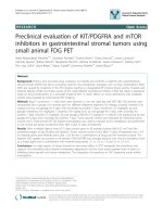
At laparotomy, a solid mass measuring 80 × 100 × 120
mm was identified at the root of the jejunal mesentery,
infiltrating the adipose tissue and bowel wall, and in
close association with the superior mesenteric and the
middle colic vessels (Figure 1). The mesentery contained
several large lymph nodes. A small amount of free peri-
toneal fluid was present. The mass was totally excised
with a loop of jejunum and without appar ent interfer-
ence with the blood supply to the bowel. However,
bowel ischemia did occur, and the patient required a
second laparotomy three days later. The ischemic injury
seemed to be secondary to venous obstruction. It was
necessary to resect an additional segment of small bowel
measuring 600 mm in length. A primary anastomosis
was performed.
Our patient recovered well, and was discharged from
the hospital one week after the second laparotomy. The
final tissue diagnosis showed spindle-shaped fibroblasts
with elongated nuclei and a benign appearance. The cut
surface was tan, whorled, and firm, without necrosis,
cystic change, o r hemorrhage. Microscopy showed
loosely arranged spindle cells with bland, oval nuclei
and minimal cytoplasm (Figure 2). There were also
plump spindle cells with tapering ends, with oval, vesi-
cular nuclei and moderate amounts of eosinophilic cyto-
plasm. There were many t hin-walled vessels of varying
caliber. There were no cells with epithelioid features,
and any inflammatory cells, calcification, osseous meta-
plasia, necrosis, or mitoses. The tumor had infiltrated
the muscularis propria and had non-infiltrating margins.
The tumor cells were negative for antibodies to CD117,
S100, CD34, and smooth muscle actin, a nd positive for
desmin. The sm all sample of peritoneal fluid was free of
malignant cells.
A diagnosis of fibromatosis of the jejunum and
mesentery was made. Six months after resection, a CT
scan of the abdomen showed no evidence of resid ual or
recurrent tumor. Our patient had no evidence of disease
at follow-up 16 months after surgery. However, the clin-
ical data and radiologic findings gave rise to a diagnostic
dilemma: was the lesion an intermediate or high-risk
malignant GIST originating from the bowel wall and
spreading to the mesentery, or was this a benign lesion
such as IAF o riginating from the m esentery and infil-
trating the bowel wall?
Discussion
Intra-abdominal spindle cell lesions are uncommon, and
often present a diagnostic chall enge [4]. The differential
diagnosis of a bland spindle cell tumor involving the
gastrointestinal (GI) tract and the mesentery includes
GIST, fibromatosis, and inflammatory myofibroblastic
tumor [5].
IAF is the most common primary mesenteric tumor
with spindle cell morphology [6]. Its biological behavior
is intermediate between benign fibrous tissue prolifera-
tion and fibrosarcoma. Fibromatosis characteristically
is locally invasive and tends to recur, but does not
Figure 1 Surgical preparation. The resected tumor measured
120 × 80 × 100 mm.
Figure 2 Spindle cells growing in sweeping fascicles, with
eosinophilic cytoplasm.
Stoidis et al. Journal of Medical Case Reports 2010, 4:314
/>Page 2 of 4
metastasize. Although mesenteric desmoid tumors tend
to be aggressive, there is cons iderabl e variability in their
growth rate during the course of the disease. In fact, the
biology of intra-abdominal desmoid may be character-
ized by initial rapid growth, followed by stability or even
regression. However, mesenteric desmoid, by virtue of
its relationship to vital structures and its ability to infil-
trate adjacent organs, may cause important complica-
tions, including intestinal obstruction, ischemia and
perforation, hydronephrosis, and even aortic rupture
[7,8]. Despite these complications, the overall ten-year-
survival for patients with intra-abdominal desmoids can
be as high as 60 to 70% [9].
In contrast to abdominal wall desmoids, surgery for
intra-abdominal desmoid tumors is much more danger-
ous, and is associated with increased morbidity and
mortality, mainly due to h emorrhagic complications or
extensive enterectomy (cause d by small bowel involve-
ment or involvement of the base of the mesentery and
major portions of the mesenteric blood supply) requir-
ing long-term parenteral nutrition [10]. Additionally, a
large percentage of patients with int ra-abdominal des-
moid tumors have unresectable disease [11]. In these
cases, expectant treatment is favored, and biopsy should
be preferred to excision. If clinical obstruction warrants
operative therapy, then bypass, a s opposed to major
resection, is indicated, followed by a trial of medical
therapy. Small bowel obstruction can occur in nearly
half of patients with intra-abdominal desmoid tumors,
and in 70% of these, diffuse, dense fibrotic adhesions are
responsible [12]. Debulking has no place as a therapeu-
tic measure, as it almost invariably leads to more aggres-
sive and infiltrative desmoid growth [12]. Most authors
consider surgery a reasonable first-line treatment for
abdominalwalltumors,butitshouldbeusedasalast
resort for intra-abdominal desmoid tumors, and only in
specific circumstances (for example, when tumors cause
major complications, do not involve vital organs and
vessels on preoperative imaging, after failure of systemic
pharmacologic therapy, or when surgery is the only pos-
sible therapeutic option, such as in the case of a rapidly
growing tumor) [13]. Radical (free margin) excision
offers the best chance for cure and of avoiding local
recurrence [14]. Unfortunately, radical surgery is not
always a straightforward procedure because of the extent
and invasiveness of the tumor. Therefore, very high
rates of recurrence (up to 88%) should be expected [14].
Given the high likelihood of recurrence and prolonged
survival even in the setting of advanced disease, some
authors have suggested that a trial of watchful waiting
along with minimally toxic agents such as s ulindac and
anti-estrogen therapy may be the best strategy, particu-
larly in patients w ith minimal symptoms [15]. For
clearly inoperable cases, cytotoxic chemotherapy,
especially doxorubicin-based or low-dose vinblastin and
methotrexate has been proposed [12].
The location of a desmoid tumor within the mesen-
tery of the small bowel may complicate the management
of patients with FAP and interfere with surgical strategy,
preventing proctectomy or ilial pouch-anal anastomosis
(IP AA), at least in some patients [15]. It may also make
a diverting ileostomy impossible. Moreover, in this
group of patients, recurrence rate after complete resec-
tion is particularly high [16]. It has been suggested that
this increased risk may be due, at least in part, to the
added manipulations of the mesentery during IPAA.
Management of desmoid tumors in patients with FAP is
further complicated by the fact that clinical course of
the disease in this group is particularly variable and
unpredictable, and may be disastrous. In a few highly
selected patients with extensive des moid tumors invol-
ving the mesentery, intestinal transplan tation has been
performed [17].
It is generally easy to diagnose primary IAF in its clas-
sic presentation as a mesenteric mass, because of its dis-
tinctive gross and microscopic features [18]. However,
when it presents primarily as an intestinal wall tumor,
as in our patient, the diagnosis of GIST must be s er-
iously considered. For our patient, we considered GIST
an unlikely diagnosis because of the whorled appearance
on the cut surface of the tumor, and the histolo gic eva-
luation confirmed this, hence the diagnosis of fibroma-
tosis of the jejunum and mesentery was established.
Histological features characteristic of GIST include the
presence of spindle or epithelioid cells with variable
architecture, mitotic activity and nuclear atypia, and
myxoid or hyalinized stroma. Necrosis and hemorrhage
can also be seen. By contrast, IAFs are characterized by
a spatially homogeneous proliferation of wavy spindle
cells without atypia, associated with collagen deposition
(often of the keloidal type) and an infiltrative border.
These features are sufficiently characteristic of mesen-
teric fibromatosis to allow distinction from GIST on the
basis of routinely stained sections in most cases. In the
few equivocal ca ses, immunohistochemical analysis can
provide more precise diagnosis.
The distinction between these neoplasms is very
important because they have different biological beha-
viors, and there are important clinical implications for
the patient. Complete excision of the tumor with a mar-
gin of uninvolved tissue (when this is possible) is the
most effective treatment yet described, and it may be
followed by other therapies if GIST is diagnosed. After
the more radical (free margin) resections required for
GISTs, the five-year survival rate is approximately 45%,
whereas for metastatic disease it drops to 20% [19]. The
tumor is resistant to radiotherapy, whereas radiation is
controversial for intra-abdominal desmoid tumors
Stoidis et al. Journal of Medical Case Reports 2010, 4:314
/>Page 3 of 4
[18,19]. Imatinib mesylate is an effective systemic agent
and is indicated in patients with KIT (CD117)-positive
gastrointestinal stromal tumors that cannot be surgically
removed, and/or have spread to other parts of the body.
Recent research indicated it may also be useful as part
of the post-surgery adjuvant therapy for adult patients
who have had their GISTs completely removed. The
role of imatinib in the management of abdominal des-
moid tumors remains unproven [20].
Conclusion
The optimum treat ment protocol for desmoids tumors
has not yet been established and, in many cases, a multi-
disciplinary approach including surgery, chemotherapy,
and radiation therapy is required. The rarity of cases in
even major oncological centers has traditionally limited
the ability to study this disease. The notion that a speci-
fic genotype can predict the development of an aggres-
sive desmoid tumor in a given patient could prove to be
valuable in allowing appropriate patient selection for
early therapy or even a chemopreventive strategy. Sev-
eral novel pharmacologic and biologic treatment
approaches are actively being developed, although long-
term follow-up is needed for their substantiation.
The aim of this report is to stress the importance of
correctly characterizing a tumor localized in the bowel
wall and infiltrating the mesentery to plan the appropri-
ate treatment because tumor diagnoses based on immu-
nohistochemical staining or traditional histologic criteria
alone are not specific enough.
Consent
Written informed consent was obtained from the patient
for publication of this case report and accompanying
images. A copy of the written consent is available for
review by the Editor-in-Chief of this journal.
Acknowledgements
The authors state that there was no extra-institutional funding. We thank
Ilias A. Kouerinis, who was a major contributor in composing the manuscript.
Author details
1
Department of Surgery, Athens Navy Hospital, 70 Deinokratous Street,
11521, Athens, Greece.
2
Third Department of Surgery, University of Athens
Medical School, Attikon University Hospital, 1 Rimini Street, 12462, Chaidari,
Greece.
3
Department of Internal Medicine, Athens Navy Hospital, 70
Deinokratous Street, 11521, Athens, Greece.
4
Second Department of
Propaedeutic Surgery, University of Athens Medical School, Laikon General
Hospital, 17 Agiou Thoma Street, 11527, Athens, Greece.
Authors’ contributions
CIF was the patient’s surgeon, and was involved in drafting the manuscript
and critically revising it for important intellectual content. EPM, CNS, BGS,
PPP and CKF have made contributions to conception and design. CNS
contributed to the analysis and interpretation of data and wrote the paper.
All authors read and approved the final manuscript. All authors contributed
equally to the final draft of the manuscript. CIF has given the final approval
of the version to be published.
Competing interests
The authors declare that they have no competing interests.
Received: 5 April 2010 Accepted: 23 September 2010
Published: 23 September 2010
References
1. Okuno S: The enigma of desmoid tumors. Curr Treat Options Oncol 2006,
7:438-43.
2. Church J, Lynch C, Neary P, LaGuardia L, Elayi EL: A desmoid tumor-
staging system separates patients with intra-abdominal, familial
adenomatous polyposis-associated desmoid disease by behavior and
prognosis. Dis Colon Rectum 2008, 51:897-901.
3. Connolly EM, Gaffney E, Reynolds JV: Gastrointestinal stromal tumours. Br
JSurg2003, 90:1178-86.
4. Al-Nafussi A, Wong NACS: Intra-abdominal spindle cell lesions: a review
and practical aids to diagnosis. Histopathol 2001, 38:387-402.
5. Colombo P, Rahal D, Grizzi F, Quagliuolo V, Roncalli M: Localized intra-
abdominal fibromatosis of the small bowel mimicking a gastrointestinal
stromal tumor: a case report. World J Gastroenterol 2005, 11:5226-5228.
6. Rodriguez JA, Guarda LA, Rosai J: Mesenteric fibromatosis with
involvement of the gastrointestinal tract. A GIST simulator: a study of 25
cases. Am J Clin Pathol 2004, 121:93-8.
7. Holubar S, Dwivedi AJ, O’Connor J: Giant mesenteric fibromatosis
presenting as small bowel obstruction. Am Surg 2006, 72:427-9.
8. Collins D, Myers E, Kavanagh D, Lennon G, McDermott E: Mesenteric
desmoid tumor causing ureteric obstruction. International J Urol 2008,
15:261-262.
9. Dozois EJ, Dozois RR: Desmoid disease. In Mayo Clinic Gastrointestinal
Surgery. Edited by: Kelly KA, Sarr MG, Hinder RA. Saunders Eds: Philadelphia;
2004:563-565.
10. Pai SA, Zaveri SS: Intra-abdominal fibromatosis of the jejunum and
mesentery. J Clin Pathol 2004, 57:1119.
11. Schlemmer M: Desmoid tumors and deep fibromatoses. Hematol Oncol
Clin N Amer 2005, 19:565-71.
12. Latchford AR, Sturt NJH, Neale K, Rogers PA, Phillips RK: A ten-year-review
of surgery for desmoid disease associated with familial adenomatous
polyposis. Br J Surg 2006, 93:1258-64.
13. Rampone B, Pedrazzani C, Marrelli D, Pinto E, Roviello F: Updates on
abdominal desmoid tumors. World J Gastroenterol 2007, 13:5985-5988.
14. Melis M, Zager JS, Sondak VK: Multimodality management of desmoid
tumors: how important is a negative surgical margin? J Surg Oncol 2008,
98:594-602.
15. Sturt JNH, Clark SK:
Current ideas in desmoid tumors. Familial Cancer 2006,
5:275-85.
16. Tamura K, Tani M, Kinoshita H, Yamaye H: Mesenteric desmoids tumor of
the interposed jejunal pouch after total gastrectomy. World J Surg Oncol
2006, 4:27-31.
17. Chatzipetrou MA, Tzakis AG, Pinna AD, Kato T, Misiakos EP, Tsaroucha AK,
Weppler D, Ruiz P, Berho M, Fishbein T, Conn HO, Ricordi C: Intestinal
transplantation for the treatment of desmoid tumors associated with
familial adenomatous polyposis. Surgery 2001, 129:277-81.
18. Sakorafas GH, Nissotakis C, Peros G: Abdominal desmoid tumors. Surg
Oncol 2007, 16:131-142.
19. Yantiss RK, Spiro IJ, Compton CC, Rosemberg AE: Gastrointestinal stromal
tumor versus intra-abdominal fibromatosis of the bowel wall. Am J Surg
Pathol 2000, 24:947-957.
20. Mace J, Biermann JS, Sondak V: Response of extraabdominal desmoid
tumors to therapy with imatinib mesylate. Cancer 2002, 11:2373-2379.
doi:10.1186/1752-1947-4-314
Cite this article as: Stoidis et al.: Surgical treatment of giant mesenteric
fibromatosis presenting as a gastrointestinal stromal tumor: a case
report. Journal of Medical Case Reports 2010 4:314.
Stoidis et al. Journal of Medical Case Reports 2010, 4:314
/>Page 4 of 4