báo cáo khoa học: " Endovascular covered stenting for the management of post-percutaneous nephrolithotomy renal pseudoaneurysm: a case report" doc
Bạn đang xem bản rút gọn của tài liệu. Xem và tải ngay bản đầy đủ của tài liệu tại đây (1.29 MB, 4 trang )
CAS E REP O R T Open Access
Endovascular covered stenting for the
management of post-percutaneous
nephrolithotomy renal pseudoaneurysm:
a case report
Prodromos Philippou, Konstantinos Moraitis, Tamer El-Husseiny, Hassan Wazait, Junaid Masood, Noor Buchholz
*
Abstract
Introduction: Intrarenal pseudoaneurysm is a rare, yet clinically significa nt, complication of percutaneous
nephrolithotomy. A high index of clinical suspicion is necessary in order to recognize pseudoaneurysm as the
cause of delayed bleeding after percutaneous nephrolithotomy and angiography confirms the diagnosis which
allows endovascular management.
Case presentation: We present a case of a 65-year old Caucasian woman who underwent percutaneous
nephrolithotomy in the supine position for a two centimetre renal calculus. The postoperative course was
complicated by persistent bleeding due to a renal pseudoaneurysm. The vascular lesion was successfully managed
by endovascular exclusion through the use of a covered stent graft. We report the first successful use of this
method for the management of iatrogenic pseudoan eurysm in a branch of the left renal artery and we focus on
the imaging findings, technical details, advantages and limitations of this technique.
Conclusion: As a result of its high efficacy, interventional radiology has largely replaced open surgery for the
management of renal pseudoaneurysm related to percutaneous nephrolithotomy. Recent technical advancements
have allowed the use of covered stent grafts as an alternative to embolisation for the angiographic management
of visceral artery pseudoaneurysm located in other organs. This novel technique allows the endovascular exclusion
of the pseudoaneurysm, without compromising arterial supply to the end-structures - an advantage of critical
importance in organs supplied by segmental arteries - in the absence of collateral vasculature, such as the kidney.
Introduction
Renalpseudoaneurysm(PA)is a rare, yet clinically sig-
nificant, cause of delayed bleeding following percuta-
neous nephrolithotomy (PCNL). According to the
current literature, the reported incidence of intrarenal
PA following PCNL is low (0.6%-1%) [1,2]. A high index
of clini cal suspicion is of the utmo st importance, while
angiography is usually neces sary in order to identify the
source of bleeding and treat the vascular injury. Angio-
grap hic management - usually by superselective emboli-
sation of the injured vessel - has success rates that
exceed 90% and has largely replaced the need for open
surgery [2,3]. We report a unique case of a PCNL-
related renal PA which was successfully managed by a
covered stent graft exclusion, a technique that was
recently developed for the management of PAs located
elsewhere.
Case presentation
A 65-year old Caucasian woman, with a two centimetre
calculus located in the pelvis of the left kidney (Figure
1), underwent supine PCNL. Percutaneous access was
achieved through the middle calyx: the procedure was
uneventful and the intra-operative blo od loss was mini-
mal. At the end of the procedure, stone-free status was
achieved and a Mal ekot-type 22 Fr nephrostomy tube
was left in situ.
* Correspondence:
Department of Urology, Barts and The London NHS Trust, Smithfield,
London EC1A 7BE, UK
Philippou et al. Journal of Medical Case Reports 2010, 4:316
/>JOURNAL OF MEDICAL
CASE REPORTS
© 2010 Philippou et al; licensee BioMed Cent ral Ltd. This is an Open Access article distributed unde r the terms of the Creative
Commons Attribution License ( es/by/2.0), which permits unrestricted use, distribution, and
reproduction in any medium , provided the original work is properly cited.
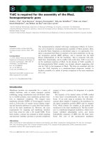
On the fourth post-operative day, the patient devel-
oped gross hematuria and severe pain of the left loin. A
significant drop in the hemoglobin level was noted
(from 9.5 g/dL to 6.8 g/dL) but she remained hemody-
namically stable and the coagulation parameters were
within normal limits. She was initiall y treated conserva-
tively with bed rest and transfusions but gross hema-
turia persisted. An abdominal computed tomography
(CT) scan (Figure 2) revealed the presence of a large
left perinephric hematoma with active extravasation of
contrast. An urgent selective left renal angiogram was
arranged in order to achieve endovascular control of
the bleeding vessel.
Access was achieved through the right common
femoral artery and selective catheterisation of the left
renal a rtery was performed. On angiography, a PA was
noted, arising from a branch of the posterior division of
the left renal artery, with active extravasation of contrast
(Figure 3). Selective catheterisation and embolisation of
the bleeding branch was technically not feasible. Emboli-
sation of a more proximal arterial branch was consid-
ered inappropriate due to the associated risk of more
extensive renal parenchymal ischemia. In order to over-
come these limitations, a 6 mm × 19 mm self-expand-
able Fluency covered stent™ (Bard, New Jersey, USA)
was advanced over a guidewire and deployed to achieve
endovascular exclusion of the PA. A control angiogram
at the end of the procedure revealed the absence of opa-
cification of the PA, with the appropriate preservation of
renal parenchymal perfusion (Figure 4).
Twenty-four hours later, hematuria ceased and the
patient remained hemodynamically stable. An abdominal
CT angiogram was performed in order to enable us to
evaluate the result of the endovascular manipulation.
Uniform global enhancement of the renal parenchyma
was noted. There were no signs of active contrast extra-
vasation or opacification of an aneurysmal cavity.
The woman was discharged 48 hours later and a plain
abdominal X-ray film, which was done six weeks later,
confirmed stone-f ree status and the presence of a cov-
ered stent graft in the anatomic location corresponding
to the left kidney (Figure 5).
Figure 1 Comput ed tomography of the kidneys, uret er and
bladder, prior to percutaneous nephrolithotomy. Note a two
centimetre stone located at the left renal pelvis.
Figure 2 Abdominal computed tomography scan prior to
angiography. A large left perinephric haematoma with active
extravasation of contrast is identified (arrows).
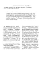
Figure 3 Selective angiogram of the left renal artery.A
pseudoaneurysm is noted, arising from a branch of the posterior
division of the left renal artery, with active extravasation of contrast
(dotted circle). Selective catheterisation and embolisation of the
bleeding branch was technically not feasible as advancement of the
endovascular catheter was not possible beyond the main stem of
the posterior branch of the renal artery (arrow).
Philippou et al. Journal of Medical Case Reports 2010, 4:316
/>Page 2 of 4
Discussion
Percutaneous access to the upper urinary tract was first
described in 1955, while PCNL was introduced 20 years
later [4]. Since then, PCNL has undergone many refine-
ments and is considered to be the current method of
choice for the manage ment of large, or otherwise com-
plex, renal stone disease [4,5]. Despite being a mini-
mally-invasi ve technique, PCNL is associated with
clinically-significant bleeding, with transfusion rates in
contemporary literature between 5%-18% [6].
Major vascular complications caused by vessel injury
during PCNL - namely, PAs or arteriovenous fistula -
usually present as delayed postoperati ve bleeding after a
mean delay o f eight days [1]. The percutaneous tract
disrupts the normal vessel wall and a PA is formed from
the tissues surrounding the high-pressure arterial
system, resulting in recanalisation between the intravas-
cular and extr avascular space that produces a pulsating ,
encapsulated hematoma. The PA may eventually grow
and become unstable, with erosion into the pelvicaliceal
system or the p erinephric tissue [7]. Srivas tava et al.
identified stone burden as a significant predictor of
severe vascular injuries aft er PCNL [1] and this has now
been reproduced by others [2]. Surgical experience was
also identified as a significant predictor for clinically sig-
nificant PCNL-related vascular injuries [ 2]. Post-PCNL
PA is usually located in the peripheral arteries. In our
case the tract was done by using the standard technique.
The segment al artery was slightly atypically located and
the Amplatz sheath and, later, the large bore nephrost-
omy catheter left in situ might have temporarily tampo-
naded the bleeding. This might explain the ab sence of
significant bleeding intraoperatively and immediately
postoperatively.
The diagnosis o f intrarenal P A is challenging. Angio-
graphy has emerged as the standard but multiple non-
invasive tests, such as renalultrasound,intravenous
pyelography, contrast-enhanced CT scanning (with three
dimensional reconstruction), magnetic resonance
imaging and renal scintigraphy, have been used with
moderate success in diagnosing renal artery pseudoa-
neurysm [8]. The advantages of angiography in this set-
ting include high sensitivity in identifying the PA (which
usually appears as a round or oval structure arising from
the main renal artery or one of its branches) and the
potential to achieve simultaneous endovascular manage-
ment of these lesions, with success rates exceeding 90%
[3]. Superselective embolisation is highly efficient in
achieving PA occlusion through th e injection of a perma-
nent agent at the fistulous point. Materials such as etha-
nol, gel foam particles and N-butyl-2-cyanoacrylate
[3,7,8] have been successfully used for embolisation.
However, embolisation for the management of PA does
have some shortcomings, such as possible reflux of
embolicmaterialintothenormalproximalvesselifthe
distal branch has not been select ively cannulated and the
risk of more generalised ischemia resulting from throm-
bosis of a main feeding branch [9].
In order to overcome these limitations, covered stent-
grafts have been used for the treatment of PA located in
branches of visceral arteries, such as the hepatic and sple-
nic artery [9,10]. To date, a total of 17 cases of visceral
artery PAs managed by en dovascular covered stenting
have been describe d in the medical literature [10]. How-
ever, our case represents the first report on the successful
use of this method for the management of an iatrogenic
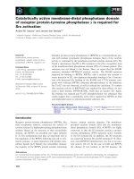
Figure 4 Control angiogram at the end of the procedure.A6×
19 mm self-expandable fluency covered stent was advanced over a
guidewire and deployed in order to achieve endovascular exclusion
of the pseudoaneurysm (PA; arrows). The control angiogram at the
end of the procedure revealed the absence of opacification of the
PA, with appropriate preservation of renal parenchymal perfusion.
Figure 5 A plain abdominal X-ray film. Six weeks postoperatively,
a plain abdominal X-ray confirmed stone-free status and the
presence of a covered stent graft in the anatomic location
corresponding to the left kidney (arrow).
Philippou et al. Journal of Medical Case Reports 2010, 4:316
/>Page 3 of 4
PA in a branch of the renal artery This technique allows
for the endovascular exclusion of a PA without compro-
mis ing blood flow to the end-s tructures, an advantag e of
critical importance in organs supplied by segmental end-
arteries in the absence of collatera l vasculature, such as
the kidney. The Fluency™ device (Bard, New Jersey, USA)
is a carbon coated, expanded polytetrafluoroethylene
(PTFE) encapsulated nitinol stent which has two mm of
bare metal exposed at each end [11].
An important limiting factor in the use of covered
stentsisthesizeandrigidityoftheavailablesystems.
Covered stents are reserved for lesions located at major
arterial branches. They are usually used for arteries that
are more than six millimeters in diameter because of the
risk of thrombosis when used for smaller vessels [12].
These factors may preclude the use of this technique for
the management of lesions involving small-calibre and
tortuous renal v essels [12]. Currently, there is a lack of
long-term data that support the indiscriminant use of
this technique. Embolisation remains the gold standard
for the management of post-PCNL PA, especially for
lesions located at the distal branches of the re nal artery.
However, the s hort-term data regarding the use of the
technique for the management of visceral PA located
elsewhere are promising. Another issue of concern is the
possibility of stenosis at the ends of the stent or within
the stent. The use of stents covered with autogenous
material or drug-eluding stents may resolve this problem.
One of the aims of future research in this field should be
the improvement of the profile and longitudinal flexibility
of these stents which could facilitate their positioning
and deployment, even in complex vascular lesions.
Conclusion
Expanding worldwide experience has allowed PCNL to
become a significant technique with high stone clear-
ance rates and low morbidity. PCNL-related vascular
injuries are rare but life-threatening complications. The
advances of the endovascular technique have allowed
the successful treatment of the v ast majority of renal
PAs by embolisation, while covered stenting may
emerge as a highly effective and safe alternative, allow-
ing the repair of a PA, without compromising arterial
supply to the end-structures.
Consent
Written informed consent was obtained from the patient
for publication o f this case report and accompanying
images. A copy of the written consent is available for
review by the Editor-in-Chief of this journal.
Abbreviations
CT: computed tomography; PA: pseudoaneurysm; PCNL: percutaneous
nephrolithotomy.
Authors’ contributions
PP analyzed and interpreted the patient data, reviewed the literature and
was responsible for drafting the manuscript. KM made substantial
contributions to the conception, design and acquisition of data. TEH made
substantial contributions to the analysis and interpretation of data and
revised the study critically for important intellectual content. HW reviewed
the current literature and was responsible for the interpretation of the
imaging finding. JM made substantial contributions to the conception and
design of this study and revised it critically for important intellectual
content. NB reviewed the literature, made substantial contributions to the
conception and design of this study and revised it critically for important
intellectual content. All authors read and approved the final manuscript.
Competing interests
The authors declare that they have no competing interests.
Received: 29 March 2010 Accepted: 23 September 2010
Published: 23 September 2010
References
1. Srivastava A, Singh KJ, Suri A, Dubey D, Kumar A, Kapoor R, Mandhani A,
Jain S: Vascular complications after percutaneous nephrolithotomy: are
there any predictive factors? Urology 2005, 66:38-40.
2. El-Nahas AR, Shokeir AA, El-Assmy AM, Mohsen T, Shoma AM, Eraky I, El-
Kenawy MR, El-Kappany HA: Post-percutaneous nephrolithotomy
extensive haemorrhage: a study of risk factors. J Urol 2007, 177:576-579.
3. Martin X, Murat FJ, Feitosa LC, Rouvière O, Lyonnet D, Gelet A, Dubernard J:
Severe bleeding after nephrolithotomy: results of hyperselective
embolization. Eur Urol 2000, 37:136-139.
4. Skolarikos A, Alivizatos G, de la Rosette JJ: Percutaneous nephrolithotomy
and its legacy. Eur Urol 2005, 47:22-28.
5. Tiselius HG, Ackermann D, Alken P, Buck C, Conort P, Gallucci M: EAU
guidelines on urolithiasis. Arnhem: European Association of Urology 2008.
6. Michel MS, Trojan L, Rassweiler JJ: Complications in percutaneous
nephrolithotomy. Eur Urol 2007, 51:899-906.
7. Lee KL, Stoller ML: Minimizing and managing bleeding after
percutaneous nephrolithotomy. Curr Opin Urol 2007, 17:120-124.
8. Massulo-Aguiar MF, Campos CM, Rodrigues-Netto N Jr: Intrarenal
pseudoaneurysm after percutaneous nephrolithotomy.
Angiotomographic assessment and endovascular management. Int Braz J
Urol 2006, 32:440-442.
9. Rami P, Williams D, Forauer A, Cwikiel W: Stent-graft treatment of patients
with acute bleeding from hepatic artery branches. Cardiovasc Intervent
Radiol 2005, 28:153-158.
10. Pasklinsky G, Gasparis AP, Labropoulos N, Pagan J, Tassiopoulos AK,
Ferretti J, Ricotta JJ: Endovascular covered stenting for visceral artery
pseudoaneurysm rupture: report of 2 cases and a summary of the
disease process and treatment options. Vasc Endovascular Surg 2009,
42:601-606.
11. Dale JD, Dolmatch BL, Duch JM, Winder R, Davidson IJ: Expanded
polytetrafluoroethylene-covered stent treatment of angioplasty-related
extravasation during hemodialysis access intervention: technical and
180-day patency. J Vasc Interv Radiol 2010, 21:322-326.
12. Nosher JL, Chung J, Brevetti LS, Graham AM, Siegel RL: Visceral and renal
artery aneurysms: a pictorial essay on endovascular therapy.
Radiographics 2006, 26:1687-1704.
doi:10.1186/1752-1947-4-316
Cite this article as: Philippou et al.: Endovascular covered stenting for
the management of post-percutaneous nephrolithotomy renal
pseudoaneurysm: a case report. Journal of Medical Case Reports 2010
4:316.
Philippou et al. Journal of Medical Case Reports 2010, 4:316
/>Page 4 of 4