báo cáo khoa học: "Imperforate anus with a rectovestibular fistula and pseudotail: a case report" pps
Bạn đang xem bản rút gọn của tài liệu. Xem và tải ngay bản đầy đủ của tài liệu tại đây (1.23 MB, 5 trang )
CAS E REP O R T Open Access
Imperforate anus with a rectovestibular fistula
and pseudotail: a case report
Miranda D Raines
1
, Marcia L Wills
2
, Gretchen P Jackson
3*
Abstract
Introduction: Human tails and pseudotails are rare sacrococcygeal lesions that are associated with a wide variety
of anomalies and syndromes. Anorectal malformations are also relatively uncommon congenital defects that often
occur in conjunction with syndromes or other congenital abnormalities. The anomalies associate d with both
disorders determine the timing and approach to surgical correction. We present an unusual case of a patient with
both imperforate anus and a pseudotail in the absence of a syndrome or other associated anomalies and we
emphasize the necessity of a thorough preoperative evaluation.
Case presentation: A Caucasian girl was born at term after an uncomplicated pregnancy and was noted at birth
to have a skin-covered posterior midline mass and imperforate anus with a fistula to the vaginal vestibule.
Ultrasound and magnetic resonance imaging revealed a predominately fatty lesion without presacral extension and
ruled out associated spinal and cord abnormalities. The patient underwent diversion with colostomy and a mucous
fistula in the newborn period as a fistulogram demonstrated a long fistulous tract to normal rectum and it was
anticipated that anoplasty and resection of the mass would require extensive posterior dissection. The
sacrococcygeal mass was removed during posteri or sagittal anorectoplasty at the age of six weeks which was
determined to be a pseudotail because of the composition of brown fat and cartilage. The patient is now 14
months old with normal bowel function after a colostomy takedown.
Conclusion: A comprehensive preoperative assessment and thoughtful operative plan were necessary in this
unusual case because of the extensive differential diagnosis for sacrococcygeal masses in the newborn and the
frequency of anomalies and syndromes associated with tail variants and imperforate anus. The pediatricians and
neonatologists who initially evaluate such patients and the surgeons who correct these disorders must be aware of
the potential pitfalls in their management.
Introduction
Human tails and pseudotails are rare congenital lesions
that have been reported since the late 19th century.
These entities typically present as skin-covered lumbosa-
cral masses seen in the newborn period. The possible
diagnoses for this presentation are broad, ranging from
benign hamartomas to aggressive malignancies and
meningoceles. As one series illustrates, the management
can be complicated not only by the spectrum of disor-
ders and associations but also by parental resistance to
crucial preoperative imaging [1].
Human tails may occur alone or associated with other
anomalies. The m ost common a bnormality associated
with a tai l is spinal dysraphism, which occurs in 49 % of
cases and can be dia gnosed as menin gocele, myelome-
ningocele or spina bifida. Other associated anomalies
include: lipoma (27%); tethered spinal cord (20%); coccy-
geal vertebrae (12%); syndactyly, hemangioma; cleft
palate; Crouzon syndrome; clubfoot; omphalocele; con-
genital tracheal stenosis; Von Recklingha usen disease;
digit hypoplasia; and tetralogy of Fallot [2].
Tails and imperforate anus are both congenital
anomalies that are commonly associated with syn-
dromes. Dusmet et al. reporte d a vestigial tail in an
infant with most of the major and minor defects of the
VATER syndrome (see Abbreviations) including imper-
forate anus [3]. A true tail has also been reported in
* Correspondence:
3
Monroe Carell Jr. Children’s Hospital at Vanderbilt, 2200 Children’ s Way,
7100 Doctor’s Office Tower, Nashville, TN 37232, USA
Full list of author information is available at the end of the article
Raines et al . Journal of Medical Case Reports 2010, 4:317
/>JOURNAL OF MEDICAL
CASE REPORTS
© 2010 Raines et al; licensee BioMed Central Ltd. This is an Open Access article distributed under the terms of the Creative Commons
Attribu tion License ( .0), which permits unrestricted use, distribution, and repro duction in
any medium, provided the original work is properly cited.
association with sirenomelia including the feature of
anal atresia [4,5]. Kahle r et al. reported a case of pygo-
melus (a pseudotail varian t) in association with lipomye-
lomeningocele and an incomplete expression of
Currarino’s triad including anal atresia with recto-vagi-
nal fistula [6].
We are not aware of any cases of tails or pseudotails
associated with an anorectal malformation in the
absenceofasyndrome.Wedescribeanewborngirl
with a pseudota il and isolated imperforate anus and dis-
cuss the diagnostic evaluation necessary for operative
planning.
Case presentation
A Caucasian girl, born at 3580 g at 39 weeks and one
day gestational age, was transferred t o our hospital on
her first da y of life for evaluation and management of
imperforate anus and a perineal mass. The 20-year-old
mother had standard prenatal care and an uncompli-
cated pregnancy. She underwent ultrasonography at 21
weeks gestation and there was no evidence of congenital
anomalies. The child was born by spontaneous vaginal
delivery and had Apgar scores o f 9 and 9 at one minute
and five minutes, respectively.
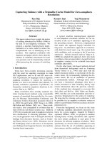
On physical examination, she was noted to have a soft,
skin-covered, midline mass, measuring 3 cm × 1 cm and
emanating from the perineum posterior to the vagina
(Figure 1). The child had normal external female genita-
lia and a sacral dimple but no anal opening. Her neuro-
logical examination was normal, as was the rest of her
physical examination. She did not pass meconium on
the first day of life. Ampicillin and gentamicin were
given for prophylaxis. On the second day of life, she
developed mild abdo minal distention and flecks of
meconium were noted in her diapers. Upon further
inspection, a small opening was noted in the posterior
aspect of the vaginal vestibule. This tract was probed
with a hemostat and meconium passed through this ves-
tibular fistula with partial decompression of the
abdomen.
Anteroposterior and lateral radiographs of the sacrum
and coccyx revealed normal bony structures without
adjacent soft tissue ca lcifications. A prone, cross-table,
lateral radio graph of the pelvis revealed a column of air
distending the rectum and sig moid colon, terminating
just below and anterior to the caudal aspect of the
sacrum.
An echocardiogram and ultrasound of the spine and
retroperitoneum were done i n order to assess the pre-
sence of VACTERL (see Abbreviations) anomalies
known to be associated with imperforate anus. These
studies were negative for cardiac, vertebral and renal
anomalies. The spinal cord terminated normally at L2
and there was normal motion of cauda equina nerve
roots. There was no tethered cord.
Ultrasound was done in order to further characterize
the perineal mass and it revealed a 2.5 cm × 3.6 cm ×
1.3 cm lesion with internal vascular flow and a possible
extension into the presac ral space. These findings were
suggestive of a presacral teratoma, which warranted
more urgent resection. The serum alpha-fetoprotein
levels were not inappropriately elevated f or a newborn.
Magnetic resonance imaging (MRI) revealed a peduncu-
lated mass with predominantly fat intensity and a single
central vessel (Figures 2 and 3). The structure did not
have a presacral component. The vagina and rectum
abutted one another and appeared to converge distally
just above the perineum.
Air and contrast enemas were performed through the
vestibular fistula in order to evaluate the distance
between the distal rectal pouch and the skin. The long,
narrow, fistulous tract connected to the distended color-
ectal pouch, which was approximately 3 to 3.5 cm from
the skin (Figure 4). The p atient underwent the creation
of a colosto my and mucous fistula to divert for a subse-
quent pull-through operation for impe rforate anus. She
recovered without complication and was discharged on
the sixth postoperative day.
Prior to her definitive surgical repair, a contrast enema
of the dis tal colon through the mucous fistula was done
in order to define the level of her rectum. Colostogram
revealed that the distance from the rectal pouch to the
perineal surface ranged from two to three vertebral
body heights. There were no strictures of the colon. At
six weeks of age, she underwent posterior sagittal anor -
ectoplasty and excision of the perineal mass. A midline
incision was made anterior and posterior to the perineal
mass, and the skin of the mass was surrounded circum-
ferentially. Upon palpation, the mass seemed to have a
Figure 1 Perineal mass. This photograph is taken from the inferior
aspect of the patient in the prone position and shows the perineal
mass extending from the sacrococcygeal region prior to surgical
repair.
Raines et al . Journal of Medical Case Reports 2010, 4:317
/>Page 2 of 5
central bony structure. The subcutaneous tissues sur-
rounding this structure were divided and the bony
structure was found to extend toward the sacrum, but
terminated prior to reaching it. Pathologic analysis
revealed a mass composed of benign skin including nor-
mal adnexal structures with underlying brown fat and a
central mature cartilage core, and it was designated a
pseudotail due to its cartilage component (Figure 5).
The patient recovered without complication and was
discharged two days postoperatively. She underwent
twice per day anal dilations until appropriate anal size
for age was attained. The 14-month old toddler is now
doing well with normal bowel function after colostomy
takedown.
Discussion
Dao and Netsky proposed the distinction between tails
and pseudot ails in 1984 [7]. A true tail is defined as the
remnant of the embryonic tail, w hich usually regresses
during the seventh and eighth weeks of gestation. A
pseudotail is a protrusion from the lumbosacrococcygeal
area that may be composed of normal or abnormal tis-
sue s but is not derived from the embryologic tail. Pseu-
dotails only resemble vestigial tails in location and can
represent an elongated coccyx, teratoma, lipoma, pygo-
melus, fibrolipoma or a prolonged sacrum [8]. True tails
are covered by skin and composed of muscle, adipose,
connective tissue, normal blood vessels and nerves,
which are components associated with the most distal
mesenchymal portion of the embryonic tail. The pre-
sence of bone, cartilage, notochord, or spinal cord tis-
sues exclude a tail from being classified as a true or
persistent vestigial tail.
This traditional classification has been recently chal-
lenged by some authors [9,10]. The designation of ‘true
tail’ by Dao and Netsky describes the most distal portion
of the embryonic tail which does not contain bone or
cartilage, but the embryonic ta il contains 10-12 caudal
vertebrae during the fifth and sixth weeks of develop-
ment [7]. Lu suggested that a tail should be classified
according to whether the anomaly is associated with
spinal dysraphism, since spinal dysraphism and tethered
cord are the most clinically significant associations due
to thei r propensity t o cause irrev ersible neurologic
sequelae [2].
Both human tails and imperforate anus are congenital
anomalies that may occur alone or be associated with a
wide range of abnormalities and syndromes. The differ-
ent ial diagnosis for a sacrococcygeal mass in a newbor n
Figure 2 Magnetic resonance imaging of pseudotail.ThisT1
coronal image through the sacrum demonstrates the fat containing
pseudotail (arrows).
Figure 3 Magnetic resonance imaging of pseudotail and
sacrum. This T2 fat-saturated coronal image demonstrates absence
of a presacral mass, relatively normal appearance of the sacrum, and
dilated rectum (R) in this child with known imperforate anus. The
fat containing pseudotail is incompletely included (arrow).
Raines et al . Journal of Medical Case Reports 2010, 4:317
/>Page 3 of 5
is extensive and includes teratoma, extra-spinal ependy-
moma, extremely rare malignancies such as chordoma,
benign entities such as lipoma and hemangioma, rectal
duplication and meningocele.
Optimal operative intervention for a patient with
imperforate anus and a sacrococcygeal mass requires a
thorough diagnostic evaluation to define the anatomic
relationships and to identify associated anomalies, such
as cardiac defects, which might limit the extent of inter-
vention that can be undertaken in the newborn period.
Suspected locally aggr essive tumors, such as chordomas
or lesions with an age-dependent likelihood of malig-
nancy, such as teratomas, mandate an urgent operative
intervention. The correction of associated anomalies,
such as spina bifida or cord tethering, can require neu-
rosurgical expertise.
This patient had an unusual combination of disorders
but she did not have significant comorbid anomalies.
Diversion with subsequent anoplasty was undertaken
because of the long fistulous tract which is needed to
mobilize the rectum and the additional perineal dissec-
tion required to resect the likely benign tail variant.
Conclusion
Anorectal malformations and skin-covered midline
masses represent challenging diagnostic and therapeutic
entities because of the exte nsive differential diagnoses
and the array of associated anomalies. This case presents
the unusual combination of imperforate anus with a rec-
tovestibular fistula and a human pseudotail in the
absence of associated anomalies or syndromes. It also
illustrates the necessity for a thorough preoperative eva-
luation and careful therapeutic decision making.
Figure 4 Fistulogram. This contrast study illustrates the long,
narrow fistulous tract and distended rectum above the expected
location of the anus.
Figure 5 Gross specimen.Photograph‘ A’ shows hair bearing skin with underlying fat and a firm, flexible circular center area loosely attached
to the fat. Representative image ‘B’ is the microscopic appearance of the pseudotail showing skin and adnexa with underlying brown fat and
mature cartilage in the center of the circular lesion (20× total magnification).
Raines et al . Journal of Medical Case Reports 2010, 4:317
/>Page 4 of 5
Consent
Written informed consent was obtained fr om the parent
of the described minor patient for publication of this
case report and accompanying images. A copy of the
written consent is available for review by the Editor-in-
Chief of this journal.
Abbreviations
VACTERL: an acronym representing an association of birth defects including
vertebral anomalies, anal atresia, cardiovascular anomalies,
tracheoesophageal fistula, esophageal atresia, renal or radial anomalies, and
limb defects; VATER: an acronym representing an association of birth defects
including vertebral anomalies, anal atresia, tracheoesophageal fistula,
esophageal atresia, renal or radial anomalies.
Acknowledgements
We thank Herman Kan MD for performing the magnetic resonance imaging
reconstructions and selecting the images for inclusion in this manuscript.
Author details
1
Vanderbilt University School of Medicine, 2134 Fairfax Avenue Apt C-3,
Nashville, TN 37212, USA.
2
Monroe Carell Jr. Children’s Hospital at Vanderbilt,
2200 Children’s Way, 11th Floor, Nashville, TN 37232, USA.
3
Monroe Carell Jr.
Children’s Hospital at Vanderbilt, 2200 Children’s Way, 7100 Doctor’s Office
Tower, Nashville, TN 37232, USA.
Authors’ contributions
MDR reviewed the literature and was primarily responsible for writing the
manuscript. MLW analysed the surgical specimen, reviewed the manuscript
and provided images of the specimen. GPJ oversaw the clinical care of this
patient, performed the operations and made revisions to the manuscript. All
authors have read and approved the final manuscript.
Competing interests
The authors declare that they have no competing interests.
Received: 24 September 2009 Accepted: 7 October 2010
Published: 7 October 2010
References
1. Amirjamshidi A, Abbassioun K, Shirani Bidabadi M: Skin-covered midline
spinal anomalies: a report of four rare cases with a discussion on their
genesis and milestones in surgical management. Childs Nerv Syst 2006,
22:460-465.
2. Lu FL, Wang PJ, Teng RJ, Yau KI: The human tail. Pediatr Neurol 1998,
19:230-233.
3. Dusmet M, Fete F, Crusi A, Cox JN: VATER association: report of a case
with three unreported malformations. J Med Genet 1988, 25:57-60.
4. Guven MA, Uzel M, Ceylaner S, Coskun A, Ceylaner G, Gungoren A: A
prenatally diagnosed case of sirenomelia with polydactyly and vestigial
tail. Genet Couns 2008, 19:419-424.
5. Parikh TB, Nanavati RN, Udani RH: Sirenomelia apus with vestigial tail.
Indian J Pediatr 2005, 72:367.
6. Kahler RJ, Merry GS: Spinal dysraphism and the Currarino triad. J Clin
Neurosci 1998, 5:339-342.
7. Dao AH, Netsky MG: Human tails and pseudotails. Hum Pathol 1984,
15:449-453.
8. Dubrow TJ, Wackym PA, Lesavoy MA: Detailing the human tail. Ann Plast
Surg 1988, 20:340-344.
9. Donovan DJ, Pedersen RC: Human tail with noncontiguous intraspinal
lipoma and spinal cord tethering: case report and embryologic
discussion. Pediatr Neurosurg 2005, 41:35-40.
10. Noack F, Reusche E, Gembruch U: Prenatal diagnosis of ‘true tail’ with
cartilage content? Fetal Diagn Ther 2003, 18:226-229.
doi:10.1186/1752-1947-4-317
Cite this article as: Raines et al.: Imperforate anus with a rectovestibular
fistula and pseudotail: a case report. Journal of Medical Case Reports 2010
4:317.
Submit your next manuscript to BioMed Central
and take full advantage of:
• Convenient online submission
• Thorough peer review
• No space constraints or color figure charges
• Immediate publication on acceptance
• Inclusion in PubMed, CAS, Scopus and Google Scholar
• Research which is freely available for redistribution
Submit your manuscript at
www.biomedcentral.com/submit
Raines et al . Journal of Medical Case Reports 2010, 4:317
/>Page 5 of 5