báo cáo khoa học: " Hypercalcemia and huge splenomegaly presenting in an elderly patient with B-cell non-Hodgkin’s lymphoma: a case report" potx
Bạn đang xem bản rút gọn của tài liệu. Xem và tải ngay bản đầy đủ của tài liệu tại đây (1.05 MB, 4 trang )
CASE REPO R T Open Access
Hypercalcemia and huge splenomegaly
presenting in an elderly patient with B-cell
non-Hodgkin’s lymphoma: a case report
Ali AM Ghazi
1
, Hamid Attarian
2
, Shirin Attarian
2*
, Abolghasem Abasahl
3
, Ebrahim Daryani
4
, Ebrahim Farasat
5
,
Marina Pourafkari
6
, Farrokh Tirgari
7
, Siavash M Ghazi
1
, Kalman Kovacs
8
Abstract
Introduction: Hypercalcemia is the major electrolyte abnormality in patients with malignant tumors. It can be due
to localized osteolytic hypercalcemia or elaboration of humoral substances such as parathyroid hormone-related
protein from tumoral cells. In hematological malignancies, a third mechanism of uncontrolled synthesis and
secretion of 1-25(OH)
2
D
3
from tumoral cells or neighboring macrophages may contribute to the problem.
However, hypercalcemia is quite unusual in patients with B-cell non-Hodgkin’s lymphoma.
Case presentation: An 85-year-old Caucasian woman presented with low grade fever, anorexia, abdominal
discomfort and fullness in her left abdomen for the last six months. She was mildly anemic and complained of
fatigability. She had huge splenomegaly and was hypercalcemic. After correction of her hypercalcemia, she had a
splenectomy. Microscopic evaluation revealed a malignant lymphoma. Her immunohistoche mistry was positive for
leukocyte common anti gen, CD20 and parathyroid hormone-related peptide.
Conclusion: Immunopositivity for parathyroid hormone-related peptide clearly demonstrates that hypersecretion of
a parathyroid hormone-like substance from the tumor had led to hypercalcemia in this case. High serum calcium is
seen in only seven to eight percent of patients with B-cell non-Hodgkin’s lymphoma, apparently due to different
mechanisms. Evaluation of serum parathyroid hormone -related protein and 1-25(OH)
2
D
3
can be helpful in
diagnosis and management. It should be noted that presentation with hypercalcemia has a serious impact on
prognosis and survival.
Introduction
Hypercalcemia is the major e lectrolyte abnormality in
patients with malignant tumors. It can be due to skeletal
invasion, known as localized osteolytic hypercalcemia or
elaboration of humoral substa nces such as para thyroid
hormone-related protein (PTHrP) from tumoral cells. In
hematological malignancies, a third mechanism, uncon-
trolled synthesis and secretion of 1-25(OH)
2
D
3
from
tumoral cells or neighboring macrophages, may contri-
bute to the problem [1,2].
Hypercalcemia is common in patients with hematolo-
gical malignancies. About 30% of patients with multiple
myeloma and 60% of patients with T-cell non-Hodgkin’s
lymphoma (NHL) experience hypercalcemia due to
osteolytic mechanisms or PTHrP hypersecretion respec-
tively. By contrast, hypercalcemia is seen in only seven
to eight percent of patients with B-cell NHL [3], most ly
due to uncontrolled endogenous production of 1-25
(OH)
2
D
3
from tumor cells. Hypercalcemia that is sec-
ondary to elaboration of PTHrP in patients with B-cell
NHL is quite unusual and, according to the best of our
knowledge, limited numbers of s uch patients have been
observed [3-7].
In our case report, we present the case of an 85-year-
old Iranian woman who had huge splenomegaly and
hypercalcemia. She was finally proven to have a PTHrP-
producing B-cell lymphoma of her spleen.
* Correspondence:
2
Department of Hematology and Oncology, Taleghani General Hospital,
Shahid Beheshti University of Medical Sciences, Tehran, Iran
Full list of author information is available at the end of the article
Ghazi et al. Journal of Medical Case Reports 2010, 4:330
/>JOURNAL OF MEDICAL
CASE REPORTS
© 2010 Ghazi et al; licensee BioMed Central Ltd. This is an Open Access article distributed under the terms of the Creative Commons
Attribution License ( which permits unrestricted use, di stribution, and reproduction in
any medium, provided the original work is properly cited.
Case presentation
An 85-year-old Iranian, Caucasian woman presented
with low grade fever, anorexia, abdominal discomfort
and fullness in her left abdomen for the last six months.
An abdominal computed tomography (CT) scan per-
formed six months previously revealed a filling defect in
her spleen, which was interpreted as a splenic cyst. No
specific treatment was done at that time.
On examination, she was mildly anemic and com-
plained of fatigability. On abdominal examination a
markedly enlarged spleen was palpated. No peripheral
lymphadenopathy was noted. Table 1 shows her labora-
tory data. Unfortunately, her PTHrP measurement was
not available to us. Her chest and mediastinal CT scan
was unremarkable exc ept for some fibrotic changes
compatible with her age. No mediastinal lymphadenopa-
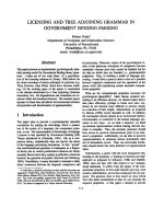
thy was seen. In her abdominal CT scan, it was noted
that her spleen was large and that it contained a definite
mass occupying about two thirds of the splenic space.
No abdominal or para-aortic lymph nodes were seen
(Figure 1).
Her serum calcium was gradually corrected by the use
of intravenous saline and furosemide over the next few
days. She did not receive any other specific treatment
for her hypercalcemia (such as calcitonin or bisphospho-
nates). On the fifth day of her admission, she underwent
a total splenectomy and a huge spleen measuring 22 ×
18 × 14 cm, weighing 1800 grams and harboring a firm
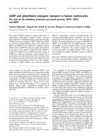
mass was extracted. Microscopic evaluation revealed a
high-grade malignant lymphoma with foci of necrosis
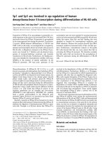
(Figure 2). Her immunohistochemistry was positive for
LCA, CD20, and PTHrP (Figure 3). After surgery her
serum calcium levels were 8.5-9.6 mg/dl but her low
grade fever and anorexia resumed. A bone marrow
biopsy was performed and t here was no bone marrow
involvement. Based on the lack of lytic bone lesions, no
bone marrow involvement, no plasmacytosis in her bone
marrow, and the lack of gammopathy in her serum pro-
tein electrophoresis, other hematological malignancies,
Table 1 Laboratory data of the patient on admission
Patient Normal range
Hb 11.4 12-14 g/dl
WBC 8.1*10/ml 4-10.8 × 10
3/
ml
Ca 13.3 8.5-10.3 mg/dl
P 3.5 2.5-4.5 mg/dl
Creatinin 1.9 0.5-1.2 mg/dl
PTH 15 15-65 pg/ml
LDH 936 <480 IU/L
25OH D
3
8.6 <30 ng/ml
1-25(OH)
2
D
3
12.7 20-70 pg/ml
24 h Urine Calcium 208 <120 mg/24 h
ESR 62 6-20 mm
Figure 1 An abdominal CT scan of the patient during the first
hospital admission.
Figure 2 PTHrP immunostaining.
Figure 3 H&E staining (hematoxylin and eosin staining).
Ghazi et al. Journal of Medical Case Reports 2010, 4:330
/>Page 2 of 4
including multiple myeloma, were ruled out. She was
treated with six courses of R-CHOP. Based on her age
(85-years-old), weight (70 kg), height (1.58 m) and body
surface area (1.7 m
2
), the dosage of the chemotherapy
regimen was as follows: 350 mg/m
2
of rituximab for a
total dose of 600 mg, 600 mg/m
2
of cyclophosphamide
for a total dose of 1000 mg, 40 mg/m
2
of Adriamycin
(doxorubicin) for a total dose of 70 mg, 1.4 mg/m
2
of
Oncovin (vincristine) for a total dose of 2 mg per injec-
tion and 75 mg of prednisolone daily for five days. After
the second course of chemotherapy, her general condi-
tion improved, her fever disappeared and her appetite
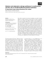
resumed. Five months after therapeutic courses, there
were no clinical or laboratory signs of disease. Figure 4
shows an abdominal CT sca n performed one year after
surgery.
Discussion
Immunohistochemistry immunopositivity for PTHrP
clearly demonstrates that hypersecretion o f the PTH-
like substance from the tumor had led to hypercalcemia
in this case. Contrary to Adult T-cell leukemia/lym-
phoma (ATLL) in which hypercalcemia is common and
almost always second ary to PTHrP hypersecretion, high
serum calcium is seen in only seven to eight percent of
patients with B-cell NHL, apparently due to different
mechanisms. Majumdar in his study on 112 patients
with B-cell NHL showed that eight patients (7.1%), six
with high grade and two with low grade disease had ele-
vated serum calcium levels [8]. Most patients had stage
3 and 4 (Stage 3: NHL is in two lymph node groups,
with/without partial involvement of an extran odal organ
or site above and below the diaphragm. Stage 4: NHL is
extensive (diffuse) in one organ or site, with/without
NHL in distant lymph nodes.) and survived between
two to 11 months after the appearance of hypercalce-
mia. No explanation about the etiology of hypercalcemia
was given in that paper.
The first study that linked e levated serum calcium to
hypersecretion of PT HrP from the tumoral cells belongs
to Wada et al [9]. In their study about a 40-year-old
man with B-cell NHL, the authors demonstrated not
only high serum levels of PTHrP, but also the parallel
changes in serum calcium and PTHrP d uring a course
of therapy. They also demonstrated the presence of
immunoreactive PTHrP in the tumor extract and proved
the bioactivity of the tumor extract producing C-AMP
in osteoblasts. Since that time, a limited number of
patients with hypercalcemic B-cell NHL secondary to
PTHrP have been reported [6,8-15]. Table 2 shows the
clinical and laboratory data of 10 such patients, includ-
ing ours. As shown in Table 2, the hypercalcemia was
severe and life-threatening and immediate therapeutic
modalities such as forced hydration and application of
Figure 4 A CT scan of the patient one year after surgery.
Table 2 Clinical and laboratory data of B-cell NHL patients with hypercalcemia due to PTHrP hypersecretion
Number Age (year) Gender Ca mg/dl PTHrP Pmol/L* LDH Iu/L 1-25(OH)
2
D
3
Pg/ml Outcome Author, Year
1 40 male 18.2 310
(21.8-44.8)
2349 41 died after 3 months Wada et al, 1992
2 64 female 16 151
(13.8-55.3)
1750 Normal Hamihara et al, 1996
3 70 female 26.3 10.3 (<2.5) - <20 Ranganath et al, 1998
4 49 female 16.2 52 (<16) 1795 20.5 died after 2 months Uno et al, 1998
5 73 male 17 1.3 (<0.5) - Normal partial improvement Daroszewski et al, 1999
6 52 male 18.6 8 (<0.8) - - partial improvement Knobel et al, 2001
7 93 female 16.6 5 (<0.6) - - died Ota, 2003
8 69 male 18.8 13 (<1.3) 356 47 died at hospital Schottker et al, 2006
9 50 female 18.3 6.2 (<0.6) 433 17 died at hospital Takasaki et al, 2006
10 85 female 13.3 NA 936 12.8 alive Ghazi et al, 2008
Ghazi et al. Journal of Medical Case Reports 2010, 4:330
/>Page 3 of 4
furosemide, calcitonin and pamidronate were underta-
ken to alleviate the problem.
Serum PTH and 1-25(OH)
2
D
3
were low in most cases
due to suppression of the parathyroid glands and renal
a-hydroxylase secondary to hypercalcemia.
It is also evident that hypercalcemia is a manifest ation
of advanced disease and, as with other cases of humoral
hypercalcemia of malignancy (HHM), points to a poor
prognosis. All the patients, except our patient who is
still in remission, died between two to 11 months after
the appearance of hypercalcemia.
Uncontrolled synthesis of 1-25(OH)
2
D
3
as the etiology
of hypercalcemia has also bee n described in patients
with B-cell NHL [6,16-18].
Conclusions
We conclude that a lthough hypercalcemia is rare in
patients with B-cell NHL, it should be properly diag-
nosed and urgently treated. The evaluation of serum
PTHrP and 1-25(OH)
2
D
3
can be helpful in diagnosis
and management. It sh ould also be noted that presenta-
tion with hype rcalcemia has a serious impact on prog-
nosis and survival.
Consent
Written informed consent was obtained from the patient
for publication of this case report and any accompany-
ing images. A copy of the written consent is available
for review by the Editor-in-Chief of this journal.
Author details
1
Research Institute for Endocrine Sciences, Taleghani General Hospital,
Shahid Beheshti University of Medical Sciences, Tehran, Iran.
2
Department of
Hematology and Oncology, Taleghani General Hospital, Shahid Beheshti
University of Medical Sciences, Tehran, Iran.
3
Department of Surgery, Imam
Khomeini Hospital, Tehran University of Medical Sciences, Tehran, Iran.
4
Department of Internal Medicine, Imam Khomeini Hospital, Tehran
University of Medical Sciences, Tehran, Iran.
5
Department of Cardiology, Sina
Hospital, Tehran University of Medical Sciences, Tehran, Iran.
6
Department of
Radiology, Taleghani General Hospital, Shahid Beheshti Universi ty of Medical
Sciences, Tehran, Iran.
7
Department of Pathology, Imam Khomeini Hospital,
Tehran University of Medical Sciences, Tehran, Iran.
8
Department of
Laboratory Medicine, Saint Michael’s Hospital, University of Toronto, Ontario,
Canada.
Authors’ contributions
AG analyzed and interpreted data regarding our patient’s endocrine disease
and hypercalcemia. HA analyzed and interpreted data regarding her
hematologic disease and performed her chemotherapy. SA carried out data
collection, was a major contributor in the writing of the manuscript and
coordinated all members of the group. AA performed splenectomy on our
patient. ED performed the gastrointestinal work up. EF undertook
cardiovascular management before the surgery. MP analyzed and
interpreted all X-rays and abdominal CT scans. FT perfomed, analyzed and
interpreted the pathological specimens resulting from her lymph node,
spleen, bone marrow, and all immunohistochemical studies. SG contributed
to writing the manuscript and the collection of data. KK undertook some
laboratory analysis and endocrine interpretation. All authors read and
approved the final manuscript.
Competing interests
The authors declare that they have no competing interests.
Received: 23 October 2009 Accepted: 19 October 2010
Published: 19 October 2010
References
1. Erban JK, Tang Z: A 54 year old man with hypercalcemia, renal
dysfunction and an enlarged liver. N Engl J Med 2004, 347:1952-1960.
2. Stewart AF: Hypercalcemia associated with cancer. N Engl J Med 2005,
352:373-379.
3. Cox M, Haddad JG: Lymphoma, hypercalcemia, and the sunshine vitamin.
Ann Intern Med 1994, 121:709-712.
4. Strewler GJ: The parathyroid hormone-related protein. Endocrinol Metab
Clin North Am 2000, 29:629-645.
5. Seymour JF, Gogel RF, Hagemeister FB, et al: Calcitriol production in
hypercalcemic and normocalcemic patients with non-Hodgkin
lymphoma. Ann Intern Med 1994, 121:638-640.
6. Uno H, Shima T, Maeda K, et al: Hypercalcemia associated with
parathyroid hormone-related protein produced by B-cell type primary
malignant lymphoma of the kidney. Ann Hematol 1998, 76:221-224.
7. Amezyane T, Lecoules S, Bordier L, Blade JS, Desrame J, Bechade D,
Coutant G, Algayres JP: Humoral hypercalcemia revealing a malignant
non Hodgkin lymphoma. Ann Endocrinol (Paris) 2008, 69(1):58-62.
8. Majumdar G: Incidence and prognostic significance of hypercalcemia in
B-cell non Hodgkin’s lymphoma. J Clin Pathol 2002, 55:637-638.
9. Wada S, Kitamura H, Matsaura Y, et al: Parathyroid hormone-related
protein as a cause of hypercalcemia in a B-cell type malignant
lymphoma. Internal Medicine 1992, 31:968-972.
10. Hanihara T, Takahashi T: Parathyroid hormone-related protein-associated
hypercalcemia in probable intravascular lymphoma of B-cell type. Am J
Hematol 1996, 53:144-146.
11. Ranganath L, Jamal H, Jones L, et al: Value of assessing parathyroid
hormone-like activity in a case of extreme hypercalcaemia. J Clin Pathol
1998, 51:257-258.
12. Daroszerski A, Bucknall RC, Chu P, et al: Severe hypercalcaemia in B-cell
lymphoma: combined effects of PTH-rP, IL-6 and TNF. Postgrad Med J
1999, 75:672-674.
13. Knobel B, Sommer I, Petchenko P, et al: Malignant humoral hypercalcemia
associated with angiotropic large B cell lymphoma. Harefuah 2001,
140:204-206.
14. Ota H, Azuma K, Horiuchi T, et al: An elderly case of non-Hodgkin’s
lymphoma with hypercalcemia. Nippon Ronen Igakkai Zasshi 2003,
40:167-171.
15. Schottker B, Heinz W, Weissinger F, et al: Parathyroid hormone related
protein-associated hypercalcemia in a patient with CLL type low grade
leukemic B cell lymphoma. Hematologica 2006, 91:119-122.
16. Takasaki H, Kanamori H, Takabayashi M, et al: Non-Hodgkin’s lymphoma
presenting as multiple bone lesions and hypercalcemia. Am J Hematol
2006, 81:439-442.
17. Klatte T, Said JW, Belldegrun AS, et al: Differential diagnosis of
hypercalcemia in renal malignancy. Urology 2007, 70:1790e7-1790e8.
18. Habra MA, Weaver EJ, Vance Prewitt P III: Primary cutaneous large B cell
lymphoma of the leg and acute hypercalcemia. J Clin Oncol 2007,
25:5825-5826.
doi:10.1186/1752-1947-4-330
Cite this article as: Ghazi et al.: Hypercalcemia and huge splenomegaly
presenting in an elderly patient with B-cell non-Hodgkin’s lymphoma: a
case report. Journal of Medical Case Reports 2010 4:330.
Ghazi et al. Journal of Medical Case Reports 2010, 4:330
/>Page 4 of 4